Original article (short paper)
Aerobic exercise training induces an
anti-apoptotic milieu in myocardial tissue
Eduardo Tadeu Santana Andrey Jorge Serra José Antonio Silva Junior
Nove de Julho University, Brazil
Danilo Sales Bocalini
São Judas University, Brazil
Valério Garrone Barauna José Eduardo Krieger
Institute of Heart of the University of São Paulo, Brazil
Paulo José Ferreira Tucci
Federal University of São Paulo, Brazil
Abstract—This study evaluated modulators of apoptosis in the myocardium of rats subjected to exercise training. Rats were assigned to non-trained and exercise-trained groups, respectively. The animals ran for 1 h per day, 6 times per week and, for a total of 13 weeks. The left ventricle was processed for analysis of gene and protein anti- (Bcl-2, c-IAP1, c-IAP2, Survivin, ILK, Akt and pAkt) and pro- (Bad) apoptotic expression by real-time PCR (except for Akt and pAkt) and Western blot, respectively. The Bad mRNA (p<0.05), but not the protein expression (p = 0.19), was signiicantly
lower after training. The exercise training signiicantly increased the gene and protein expression for all anti-apoptotic factors. However, a signiicant change in the c-IAP2 was seen only for gene expression (p<0.05). The present indings indicate that exercise can create a favorable milieu for the survival of cardiomyocytes when apoptosis is increased.
Keywords: apoptosis, exercise training, myocardial
Resumo—“Treinamento físico aeróbico induz ambiente anti-apoptótico em tecido miocárdico.” Este estudo analisou moduladores de apoptose no miocárdio de ratos submetidos a treinamento físico. Os ratos foram distribuídos nos se-guintes grupos, respectivamente: não treinados; treinados. Os animais realizaram exercício em esteira (60 min./dia; 6 x semana) por 13 semanas. O ventrículo esquerdo foi processado para análise da expressão gênica e protéica de fatores anti-apoptóticos (Bcl-2, c-IAP1, c-IAP2, Survivina, ILK, Akt e pAkt) e pro-apoptóticos (Bad) por PCR em tempo real (exceto Akt e pAkt) e Western blot, respectivamente. O teor de RNAm da Bad (p<0,05) foi signiicativamente reduzido após treinamento. Porém, a expressão protéica da Bad não foi diferente entre os grupos. A expressão gênica e proteica
de todos os fatores anti-apoptóticos foi signiicativamente aumentada com o treinamento. A exceção foi para c-IAP2,
que aumentou somente em nível transcripcional (p<0,05). Os achados deste estudo indicam que o exercício cria um ambiente favorável para sobrevivência dos cardiomiócitos a apoptose.
Palavras-chave:apoptose, treinamento físico, miocárdio
Resumen—“La práctica de ejercicio aeróbico induce un ambiente anti-apoptótico en tejido miocárdico.” El estudio analizó moduladores de la apoptosis en el miocardio de ratas entrenadas físicamente. Las ratas se dividieron en no en-trenado y entrenadas. Los animales se ha ejecutado (60 por día x 6 semanas) a las 13 semanas. El ventrículo izquierdo se procesan para el análisis de la expresión de sus genes y proteínas que inhiben (Bcl-2, c-IAP1, c-IAP2, survivina, ILK, Akt y pAkt) y causa (Bad) de la apoptosis por PCR en tiempo real (excepto Akt y pAkt) y Western blot. El nivel de ARNm de Bad (p<0,05) se redujo después de la entrenamiento, pero no era diferente de proteína. La expresión de
los inhibidores de la apoptosis fue signiicativamente mayor con la entrenamiento. La excepción fue para c-IAP2, que
aumentó sólo en el nivel transcripcional (p<0,05). Los resultados de este estudio indican que el ejercicio crea un entorno buen para la supervivencia de la apoptosis de los cardiomiocitos.
Introduction
Physically active lifestyle is recommended to maintain health and improve quality of life. In respect to the type of physical activity, aerobic exercise training (AET) is always recommen-ded, especially when the goal is to improve cardiovascular
itness (Garber et al., 2011). Thus, several lines of evidence
have shown that a regular AET program can reduce the risk for cardiovascular diseases (Bocalini, Santos, & Serra, 2008). Mo-reover, AET has been shown to be cardioprotective to different insults. In this issue, our group has shown cardioprotection to injury by ischemia and/or sustained sympathetic hyperactivity in rats submitted AET in pool and treadmill, respectively (Serra et al., 2008; Veiga et al., 2013).
An issue of interest is to identify the molecular mechanisms
for exercise-induced cardioprotective proile. Several potential
candidates have been previously evaluated in the myocardium, as follows: antioxidant defense (Pinho et al., 2012); increased
angiogenesis (Leosco et al., 2012); favorable inlammatory
status (Beavers, Brinkley, & Nicklas, 2010). There is a growing
body of evidence showing that AET is associated with an benei -cial modulation of programmed cell death (apoptosis) of the car-diomyocytes (Huang et al., 2012; Siu, Bryner, Martyn, & Alway, 2004). The apoptosis is a type of cellular death that allows the elimination of nonfunctional, abnormal, or damaged as well as harmful cells (Cohen, 1997; Thompson, 1995). However, when excessive (e.g. on pathological condition), apoptosis can lead to abnormal change in the structure and myocardial function.
The execution of an apoptotic program is principally
control-led under inluence of both endogenous pro- and antiapoptotic
factors. Thus, in this study, we examined the effects of a regular AET program on genes and proteins modulating apoptosis in the myocardial of rats. We tested the hypothesis that apoptotic suppressors (Bcl-2, c-IAP1, c-IAP2, Survivin, ILK and Akt) are upregulated after AET. We also tested the hypothesis that an apoptotic trigger (Bad) is downregulated post-exercise.
Methods
Animals and exercise training protocol
The research conforms to the Guide for the Care and Use of Laboratory Animals published by the US National Institutes of Health (NIH publication no. 85-23, revised 1996). The protocol was approved by the Institutional Research Ethics Committee of the Federal University of São Paulo, Brazil (No process:
1121/03). Twenty Wistar male rats, weighing 150-180 g, were assigned to one of two groups (n = 10 per group): non-trained rats (NT); exercise-trained rats (EXT). The animals were kept in plastic cages on an environment with temperature and light/ dark cycle controlled.
We take into account an aerobic exercise training protocol has been shown to be effective in improving myocardial per-formance and induce cardioprotection (Serra et al., 2008; Serra et al., 2010).The rats were subjected to run on a motor-driven treadmill for 1 h per day, 6 times per week, and for a total of 13 weeks. The animals were submitted to a pre-training
fami-liarization protocol of 12 days before the start of the formal exercise protocol. In this stage, running velocity was
progressi-vely increased by 3 m/min every 2 days until the inal velocity
of 18 m/min. The sessions initially lasted for 5 min and were increased by 5 min each day to reach 60 min on day 12. On main protocol, the speed race was 18 m/min for 30 min and 22 m/min for the remaining 30 min for each session.
Biological tissue preparation
After 24 hours of the last exercise session, the animals received a urethane overdose (4.8 g kg-1 i.p.) and hearts were
quickly removed and placed in 5% saline solution to remove excess blood. The left ventricle was removed, stored in cryo-genic tube and kept frozen in liquid nitrogen for later analysis of gene expression and protein.
Gene expression analysis
RNA Extraction
The left ventricle tissue was mixed with 1ml of TRIzol Reagent (Gibco BRL, Gaithersburg, USA) for isolation of the total RNA according to the manufacturer’s directions. The chlo-roform (200 µl) was placed to the homogenate and the mixture was stirred for 15s. Then, mixture was kept at room temperature for 5 min and centrifuged for 15min (12,000 x g at 4° C). The aqueous layer of samples was transferred to a 1.5 ml Eppendorf sterile tube and 500 µl of isopropanol were added. After 10 min at room temperature, the samples were again centrifuged (12 000 x g / 4° C / 10 min) and supernatants were removed. The RNA pellets were washed with 1ml of 75% ethanol (prepared with water treated with diethylpyrocarbonate, DEPC, 0.01%) and samples were once again centrifuged (12.000xg/4°C/5 min) and the supernatants were discarded. The pellets were dried in the open air, and then resuspended with 50 µl of DEPC water. The RNAs were measured in spectrophotometer (Eppendorf)
on a ilter of 260 nm. The integrity of the RNAs was determined
on gel electrophoresis in 1% agarose stained with ethidium bromide. The RNA samples with integrity of the subunits 18S and 28S of the RNA ribosomal were used in the experiments.
Total RNA
To eliminate the contamination by genomic DNA, 1 µg of total RNA was incubated with 1 unit of DNAse I / RNAse free (Invitrogen, USA), 0.5 unit of RNase OUT (Invitrogen, USA), 0.5 µl of MgCl2 (50 mM) and DEPC water, in a reaction of 13.5 µl. The samples were incubated at 37°C for 15 min, and 95°C per 5 min to inactivate the DNAse I.
Reverse transcription (RT)
(Invitrogen, USA), 0.5 unity of RNase-OUT (Invitrogen, USA), 1 µl of dideoxinucleotideos (dATP, dCTP, dGTP, dTTP (dNTPs, 10 mM, Amersham Biosciences, USA), 50 ng of a mixture of primers (Random Primer Hexamer, RH, Amersham Biosciences, USA) and 1 µl of dithiothreitol (10 mM LGC Biotechnology,
Brazil) in a reaction of inal volume of 200 µl. Then, samples
were incubated: 20°C per 10 min, 42°C per 45 min, 95°C per 5 min. The complementary DNA (cDNA) samples were kept
at -20° C. To ensure the eficacy of the treatment with DNAseI,
RT reactions were performed without the reverse transcriptase enzyme, as negative control, RNAs treated as described above.
Polymerase Chain Reaction in real time (RT-PCR)
The RT-PCR was done with a ABI 7500 (Applied Biosystems, USA) using a SYBRGreen core reaction kit (Applied Biosystems, USA). The RT- PCR reaction was performed in the following manner: 0.5 µl of sense primer (10 uM), 0.5 ul of antisense primer (10 µM), 7.5 µl of buffer 2 x Master Mix (Applied Biosystems, USA) and enough water to 15 µl of reaction were added to 1 µl of cDNA. The samples were incubated at 95°C for 10 min, and underwent 40 thermal cycles at 95°C for 15s, 60°C for 30s and 72°C for 90s. All experiments were performed in triplicate. Two
parameters were taken into account for control of speciicity: pre
-sence of only one peak in the luorescence dissociation protocol; absence of ampliication in the reactions with negative control
for RT. Table 1 illustrates all primers set used.
Quantiication of mRNA expression
The quantiication of mRNA was performed as a value relative
to an internal reference for Glyceraldehyde phosphate dehydroge-nase (GAPDH). The mRNA values were obtained from an arbitrary
threshold of luorescence, analyzed in the exponential phase of the ampliication curve. Thus, the cycle threshold (Ct) was calculated
for each sample, reaction and gene. The average Ct was calculated and relative expression was done by subtracting this average Ct
gene from the average Ct gene for GAPDH, obtaining the ΔCt. Sin-ce it is unusual to use the relative expression of a given gene in ΔCt values due to their logarithmic characteristics, the 2-∆∆Ct parameter was used in order to analyze the relative expression of mRNA.
Western blot analysis
The frozen myocardial tissue was homogenized as previously described (Serra et al., 2010). Samples containing 30 μg of the homogenate were subjected to SDS-PAGE (10% polyacrylamide gels) and separated proteins were transferred onto Hydrophobic Polyvinylidene membranes (Hybond-P, Amersham Biosciences; Piscataway, NJ, USA). The membrane was soaked in a blocking bu-ffer (5% non-fat dry milk, 10mM Tris–HCl, pH7.6, 150mM NaCl and 0.1% Tween 20) for 2 h at room temperature and then incubated
overnight at 4◦C with Rabbit anti-Bad (1:200 dilution; Santa Cruz
Biotechnology, INC.), Mouse anti-Bcl-2 (1:200 dilution; Santa Cruz Biotechnology, INC), Rabbit anti c-IAP1, (1:200 dilution; Santa Cruz Biotechnology, INC), Rabbit anti c-IAP2 (1:200 dilu-tion; Santa Cruz Biotechnology, INC), Mouse anti-Survivin (1:200 dilution; Santa Cruz Biotechnology, INC), Rabbit anti-ILK (1:200 dilution; Santa Cruz Biotechnology, INC), Rabbit anti-Akt (1:200 dilution; Santa Cruz Biotechnology, INC), Rabbit anti-phospho -Akt (Ser 473) (1:200 dilution; Santa Cruz Biotechnology, INC). After overnight incubation, membranes were washed three times and then incubated for 1 h at room temperature with horseradish peroxidase-conjugated secondary antibodies (1:5000 dilution; Zymed, San Franscisco, CA, USA). Detection was performed with enhanced chemiluminescence reagents (Amersham Biosciences). The GAPDH expression was used to normalize the results.
Statistical analysis
The Kolmogorov-Smirnov test was used to verify normal statistic distributions. The Student or Mann-Whitney tests were applied when appropriate. The results are shown as mean ±
standard error and signiicance level was set at p≤0.05.
F, forward primer; R, reverse primer. Gene GenBank Forward
Bad NM_022698 F: 5′-CAGTGATCTGCTCCACATTC-3′ (sense)
R: 5′-ATATT TCCAGCTAGGATGATAGGAC-3′ (antisense)
Bcl-2 NM_016993 F: 5′-GCTACGAGTGGGATACTGG-3′ (sense)
R: 5′-GTGTGCAGATGCCGGTTCA-3′ (antisense)
c-IAP1 AF183430 F: 5’ -CTCCAGCCTTTCTCCAAACCC-3’(sense) R: 5’-CCAGTTACTGAGCTTCCCACCAC-3’ (antisense)
c-IAP2 AF183431 F: 5’-TCCATCAAATCCTGTAAACTCC-3’(sense) R: 5’-AGCAAGCCACTCTGTCTCC-3’ (antisense)
Survivin AF276775 F: 5’-GACCACCGCATCTCTACATTCAAG-3’(sense) R: 5’-AAGGAAAGCGCAACCGGAC-3’ (antisense)
ILK NM_133409 F: 5’- ACCCAACCCTCATCACACACT-3’ (sense) R: 5’- GCCTCTTGCCATGTCCAAA-3’ (antisense)
Results
The AET protocol was well tolerated, thereby all animals completed the study without presenting any complication. Thus, no animal of the non-trained or exercise-trained group was discharged.
The transcriptional results for modulators of apoptosis are summarized in Table 2. As shown by quantitative RT-PCR
analysis, the AET over a 13 week period resulted in signiicant
reduction in the pro-apoptotic Bad mRNA content. More im-portantly, this exercise training protocol was effective to induce
a signiicant increase in all modulators well known to have
anti-apoptotic actions in myocardial tissue.
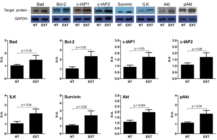
In respect to protein analysis conducted with Western blot
approach, the results were wispy different to the indings in
transcriptional level. As illustrated in Figure 1, the
pro-apop-totic Bad expression was not signiicantly different between
NT and EXT groups. This condition was also observed for the
anti-apoptotic c-IAP2 protein, in which there was no signiicant
trend of increase caused by exercise (p= 0.06). On the other hand, several proteins that restrict myocardial apoptosis were affected by training. Therefore, out to Bad, all anti-apoptotic
proteins were signiicantly increased in myocardial as in gene
expression analysis. To expand our investigation (Figure 1), we also analyzed the expression of another molecule well known to promote cell survival, the Akt, in which the AET shown to
indu-ce a signiicant increase. To date, the active Akt form was also
affected by training. Thus, the phosphorylated Akt expression
was signiicantly higher in EXT group compared with NT group.
Discussion
The present study provides interesting evidence to a benei -cial exercise training role on apoptosis in postmitotic myocytes. We showed that rats subjected to chronic AET for 13 wk
exhibi-ted a signiicant increase in several anti-apoptotic factors when
measurements are taken 24 h after the last exercise bout. It is important to note that the several anti-apoptotic factors were increased in transcriptional and translational level.
In respect to Bad, a well-known trigger for apoptosis, we saw a distinct expression pattern. The AET decreased the Bad
Gene NT EXT p value
Bad (a.u.) 2.12±0.18 1.46±0.3 0.001
Bcl-2 (a.u.) 0.12±0.02 0.35±0.11 0.0006
c-IAP1 (a.u.) 0.23±0.04 0.43±0.08 0.0005
c-IAP2 (a.u.) 0.25±0.12 0.45±0.09 0.04
Survivin (a.u.) 0.27±0.05 0.39±0.05 0.007
ILK (a.u.) 1.36±0.14 2.31±0.36 0.003 Table 2. Gene expression by real-time RT-PCR in myocardium of non-trained (NT) and exercise-trained (EXT) rats.
Figure 1. The protein expression for pro- (Bad) and anti (Bcl-2, c-IAP1, c-IAP2, ILK, Survivin, Akt and pAkt) - apoptotic factors by Western blot in myocardium. The upper panel is a representative Western blot for target proteins. All values were normalized for levels of GAPDH. The
References
Beavers, K. M., Brinkley, T. E., & Nicklas, B. J. (2010). Effect
of exercise training on chronic inlammation. Clinica Chimica Acta, 411, 785-793.
Bocalini, D. S., Santos, L., & Serra, A. J. (2008). Physical exercise improves the functional capacity and quality of life in patients with heart failure. Clinics, 63, 437-442.
Cohen, G. M. (1997). Caspases: the executioners of apoptosis. Bio-chemistry Journal, 15, 1516-1526.
DeBosch, B., Sambandam, N., Weinheimer, C., Courtois, M., & Muslin, A. J. (2006). Akt2 regulates cardiac metabolism and cardiomyocyte survival. Journal Biological Chemitry, 281, 32841-32851. Delchev, S. D., Georgieva, K. N., Koeva, Y. A., & Atanassova, P. K.
(2006). Bcl-2/Bax ratio, mitochondrial membranes and aerobic enzyme activity in cardiomyocytes of rats after submaximal trai-ning. Folia Medica (Plovdiv), 48, 50-56.
Fujio, Y., Nguyen, T., Wencker, D., Kitsis, R. N., & Walsh, K. (2000). Akt promotes survival of cardiomyocytes in vitro and protects against ischemia-reperfusion injury in mouse heart. Circulation, 101, 660-667.
Garber, C. E., Blissmer, B., Deschenes, M. R., Franklin, B. A., Lamonte, M. J., Lee, I-M., … & Swain, D. P. (2011). Quantity and quality of exercise for developing and maintaining cardiorespiratory,
musculoskeletal, and neuromotor itness in apparently healthy
adults: Guidance for prescribing exercise. Medicine Science Sports Exercise, 43, 1334-1359.
Gu, R., Bai, J., Ling, L., Ding, L., Zhang, N., Ye, J., … & Xu, B. (2012). Increased expression of integrin-linked kinase improves cardiac function and decreases mortality in dilated cardiomyopathy model of rats. PLoS One, 7, e31279. doi: 10.1371/journal.pone.0031279 Huang, C. Y., Yang, A. L., Lin, Y. M., Wu, F. N., Lin, J. A., Chan, Y. S., … & Lee, S. D. (2012). Anti-apoptotic and pro-survival effects of exercise training on hypertensive hearts. Journal Applied Phy-siology, 112, 883-891.
Lee, R. T. (1993). Interleukin-33 prevents apoptosis and improves survival after experimental myocardial infarction through ST2 signaling. Circulation Heart Failure, 2, 684-691.
Leosco, D., Rengo, G., Iaccarino, G., Golino, L., Marchese, M., For-tunato, F., … &
Pei, Z. H., Chen, B. Y., Tie, R., Zhang, H. F., Zhao, G., Qu, P, … Yu, J. (2011) Infrasound exposure induces apoptosis of rat cardiac myo-cytes by regulating the expression of apoptosis-related proteins. Cardiovascular Toxicology, 11, 341-346.
Pinho, C. A., Tromm, C. B., Tavares, A. M., Silva, L. A., Silveira, P. C., Souza, C. T., … & Pinho, R. A. (2012). Effects of different phy-sical training protocols on ventricular oxidative stress parameters in infarction-induced rats. Life Science, 90, 553-559.
Rengo, F. (2008). Exercise promotes angiogenesis and improves be-ta-adrenergic receptor signalling in the post-ischaemic failing rat heart. Cardiovascular Research, 78, 385-394.
Seki, K., Sanada, S., Kudinova, A. Y., Steinhauser, M. L., Handa, V., Gannon, J., & Lee, R. T. (1993). Interleukin-33 prevents apoptosis and improves survival after experimental myocardial infarction through ST2 signaling. Circulation Heart Failure, 2, 684-691. Serra, A. J., Higuchi, M. L., Ihara, S. S., Antônio, E. L., Santos, M. H.,
Bombig, M. T., & Tucci, P. J. (2008). Exercise training prevents beta-adrenergic hyperactivity-induced myocardial hypertrophy and lesions. European Journal Heart Failure, 10, 534-539.
Serra, A. J., Santos, M. H. H., Bocalini, D. S., Antônio, E. L., Levy, R. F., Santos, A. A., ... & Tucci, P. J. F. (2010). Exercise training
inhibits inlammatory cytokines and more than prevents myocar -dial dysfunction in rats with sustained β-adrenergic hyperactivity.
expression in transcript level, but the protein Bad content
was similar to that observed in untrained rats. These indings
suggest that may occur post-transcriptional changes to protein Bad expression also not be increased as in transcript level. Our
indings for a similar protein Bad level are consistent with the
idea that myocardial adaptation to AET is not associated with cardiac apoptosis. A study on the cardiac repercussions of treadmill exercise for 13 wk demonstrated that there was no positive cardiomyocytes for staining with digioxigenin-dUTP terminal dexytransferase - a method to indicate apoptosis. Siu,
Bryner, Martyn, and Always (2004) investigated the inluence of
treadmill exercise for 13 wk on apoptosis in myocardial. They found that the proteases activity of caspase-3 was not altered for similar exercise intensity as used in our study.
In respect to analysis for factors inhibiting the execution of
apoptosis in myocardial, we have conirmed results of previous
studies in which the Bcl-2 was up-regulated after AET (Delchev, Georgieya, Koeya, & Atanassova, 2006; Siu, Bryner, Martyn, & Always, 2004). In our line of investigation, the effects of
exer-cise training were wider and we demonstrate for the irst time a
relationship between regular AET and increased transcriptional and translational levels for other members of anti-apoptotic IAP family, which included c-IAP1 and Survivin (Pei et al., 2011; Seki et al., 2009). Moreover, we also observed that the c-IAP2
mRNA expression was signiicantly increased and there was a
pronounced tendency to increase the protein content in trained animals (p=0.06). The extent for AET effects on this family of factors suppressing apoptosis was previously reported by Siu, Bryner, Murlasits, and Always (2005). The authors showed a 14% increase in the myocardial XIAP content for animals that were trained by running 5 days weekly for 8 weeks.
This study was also designed to investigate the AET effects on myocardial ILK and Akt signaling expression. The ILK is a multifunctional kinase linking the extracellular matrix to intra-cellular signaling pathways, whose activation in the heart gives rise to a number of functional results (Gu et al., 2012). Of our interest are the cardioprotective effects for apoptosis associated with ILK. This issue was evident in a recent study conducted by Gu et al. (2012). In a experimental model of doxorubicin-in-duced cardiomyopathy, the authors observed that the treatment with adeno-ILK was associated with a reduction in apoptosis of
cardiomyocytes. These indings are particularly important when
considering that we showed a myocardial ILK increase with exercise. We have found that Akt content and its active form were
also increased with AET. These indings are important because
the Akt is involved in diverse cellular processes, including the promotion of cell survival and inhibition of apoptosis (DeBos-ch, Sambandam, Weinheimer, Courtois, & Muslin, 2006; Fujio, Nguyen, Wencker, Kitsis, & Walsh, 2000; Zhang, Xia, La Cour, 7 Ren, 2011). Since ILK overexpression can promote activation of Akt (White et al., 2006), we can assume that ILK exerts these
be-neicial effects through increasing the phosphorylation and hence
activation of Akt. In fact, this issue may be acceptable since we have found a similar increase in ILK and Akt in the present study.
Journal of Physiology, 588, 2431-2442.
Siu, P. M., Bryner, R. W., Martyn, J. K., & Always, S. E. (2004). Apoptotic adaptations from exercise training in skeletal and cardiac muscles. FASEB Journal, 18, 1150-1152.
Siu, P. M., Bryner, R. W., Murlasits, Z., & Always, S. E. (2005). Res-ponse of XIAP, ARC, and FLIP apoptotic suppressors to 8 wk of treadmill running in rat heart and skeletal muscle. Journal Applied Physiology, 99, 204-209.
Thompson, C. B. (1995). Apoptosis in the pathogenesis and treatment of disease. Science, 267,1456-1462.
Veiga, E. C., Portes, L. A., Bocalini, D. S., Antonio, E. L., dos Santos, A. A., Santos, M. H., ... & Tucci. P. J. (2013). Cardiac implications after myocardial infarction in rats previously undergoing physical exercise. Arquivos Brasileiros de Cardiologia, 100, 37-43. White, D. E., Coutu, P., Shi, Y. F., Tardif, J. C., Nattel, S., St Arnaud
R, … & Muller W. J. (2006). Targeted ablation of ILK from the murine heart results in dilated cardiomyopathy and spontaneous heart failure. Genes Development., 20, 2355-2360.
Zhang, Y., Xia, Z., La Cour, K. H., & Ren, J. (2011). Activation of Akt rescues endoplasmic reticulum stress-impaired murine cardiac contractile function via glycogen synthase kinase-3β-mediated su-ppression of mitochondrial permeation pore opening. Antioxidants & Redox Signaling, 15, 2407-2424.
Authors’ note
Eduardo Tadeu Santana and José Antonio Silva Junior are afiliated
with the graduate program in rehabilitation sciences, University Nove de Julho.
Andrey Jorge Serra is afiliated with the graduate program in reha -bilitation sciences and the undergraduate physical education course, University Nove de Julho.
Danilo Sales Bocalini is afiliated with the undergraduate physical
education course, University Nove de Julho.
Valério Garrone Barauna and José Eduardo Krieger are afiliated with
Laboratory of genetics and molecular cardiology, Institute of Heart, College of Medicine, University of São Paulo.
Paulo José Ferreira Tucci is afiliated with the laboratory of physiology
and cardiac physiopathology of the Federal University of São Paulo.
Acknowledgments
This study was supported by grants from Conselho Nacional de
De-senvolvimento Cientiico e Tecnológico and Fundação de Amparo à
Pesquisa do Estado de São Paulo.
This article is the full version of the abstract presented at the 2013 International Congress of Human Movement Sciences and the São Paulo Symposium of Physical Education.
Corresponding author
Andrey Jorge Serra
Rua Araurari 159, Postal Code: 03650-040 - São Paulo, SP, Brazil E-mail address: [email protected]
Tel-Fax: (5511) 20230381
Manuscript received on March 26, 2013 Manuscript accepted on May 4, 2014