https://doi.org/10.1590/0004-282X20170156
ARTICLE
High expression of XIAP and Bcl-2 may inhibit
programmed cell death in glioblastomas
Alta expressão de XIAP e Bcl-2 pode inibir morte celular programada em glioblastomas
Daniela Pretti da Cunha Tirapelli1, Isis Lacrose Lustosa1, Sarah Bomfim Menezes1, Indira Maynart Franco1, Andressa Romualdo Rodrigues1,
Fernanda Maris Peria
2, Alexandre Magno da Nóbrega Marinho3, Luciano Neder Serafini4, Carlos Gilberto Carlotti Jr1, Luís Fernando Tirapelli1
Despite recent advances in the understanding of high-grade gliomas, they are among the most malignant of
all cancers, with dismal patient outcomes. he classiication
of gliomas follows the World Health Organization (WHO)
classiication, which is based on knowledge of cytologic fea
-tures and degrees of malignancy. he most aggressive form of glioma, the glioblastoma (GBM), represents 29% of primary brain tumors and about 55% of all gliomas1,2,3. In spite of
stan-dard treatment with surgery, chemotherapy with
temozolo-mide, and radiotherapy, glioblastomas are always fatal with
a median survival rate of less than a year and a ive-year sur
-vival rate of less than 10% of the cases4,5,6,7.
he glioblastomas are characterized by complex hetero
-geneity. A new deinition of this heterogeneity was recently proposed based on genomic, transcriptomic and epigenomic studies carried out by he Cancer Genome Atlas Network. hrough the latter’s analyses, the GBMs were clustered into four subgroups: proneural, neural, classical and mesenchy
-mal, which correlate with biological properties of the tumors
and measures of clinical outcome8,9.
1. Universidade de São Paulo, Departamento de Cirurgia e Anatomia, Ribeirão Preto SP, Brasil;
2. Universidade de São Paulo, Departamento de Medicina Interna, Ribeirão Preto SP, Brasil;
3. Universidade Federal de Campina Grande, Unidade Acadêmica de Ciências Médicas, Campina Grande PB, Brazil;
4. Universidade de São Paulo, Faculdade de Medicina de Ribeirão Preto, Departamento de Patologia e Medicina Legal, Ribeirão Preto SP, Brasil.
Correspondence: Daniela Pretti da Cunha Tirapelli; Departamento de Cirurgia e Anatomia da USP; Avenida dos Bandeirantes, 3900; 14049-900 Ribeirão Preto
SP, Brasil; E-mail: lab.biomol.cirurgia@fmrp.usp.br
Conflict of interest: There is no conflict of interest to declare.
Received 11 August 2016; Received in final form 02 August 2017; Accepted 14 August 2017. ABSTRACT
Glioblastoma (GBM) is the most malignant glioma and represents 29% of all brain tumors. Tumorigenesis is intimately connected with characteristics acquired in the physiologic pathway of cellular death. Objective: In the present study, the expression of anti-apoptotic (XIAP and Bcl-2) and apoptotic (cytochrome C, caspase 9, APAF-1), caspase 3 and the Smac/DIABLO genes related to the apoptosis pathway were evaluated in 30 samples of glioblastoma. Methods: The gene expression was evaluated in 30 glioblastomas (WHO grade IV) and compared to 10 white matter control samples with real-time PCR. Results and Conclusion: There were higher expressions of XIAP (p = 0.0032) and Bcl-2 (p = 0.0351) in the glioblastoma samples compared to the control samples of normal brain. These results raise the question of whether Bcl-2 and XIAP genes can be responsible for the inhibition of programmed cell death in glioblastomas. Moreover, they provide additional information capable of allowing the development of new target therapy strategies.
Keywords: glioblastoma; apoptosis; X-linked inhibitor of apoptosis protein; B-cell lymphoma 2.
RESUMO
O glioblastoma (GBM) é o glioma mais maligno e representa 29% de todos os tumores cerebrais. A tumorigênese está intimamente ligada à características adquiridas na via fisiológica de morte celular. Objetivo: Avaliar a expressão de genes anti-apoptóticos (XIAP e Bcl-2) e apoptóticos (citocromo C, a caspase 9, APAF-1), caspase 3 e SMAC/DIABLO, relacionados à apoptose, em 30 amostras de tecido de pacientes com glioblastoma. Métodos: A expressão gênica foi avaliada em trinta glioblastomas e comparada a dez amostras controles de substância branca por PCR em tempo real. Resultados e Conclusão: Houve maior nível de expressão de XIAP (p = 0,0032) e Bcl-2 (p = 0,0351) em comparação com as amostras controle, de cérebro normal. Estes resultados levantam a questão de que os genes Bcl-2 e XIAP podem ser responsáveis pela inibição da morte celular programada em glioblastomas, além disso, proporcionam informação adicional capaz de permitir o desenvolvimento de novas estratégias de terapia alvo.
Tumorigenesis is intimately connected with heredity or acquired defects in physiological pathways of cellular death. Defects may include loss of a tumor suppressor gene
such as the TP53, enhanced expression of anti-apoptotic genes or reduction of the expression of apoptotic gene products such as Bcl-2 and BAX, respectively4. Knowing
the mechanisms of apoptosis makes it possible to support
the development of new drugs that target specific apop-totic pathways or genes10.
The intrinsic pathway of cellular apoptosis is trig-gered in response to a wide range of death stimuli that are generated within the cell. The intrinsic pathway is
mediated by mitochondria and, in response to apoptotic
stimuli, several proteins are released from the
intermem-brane space of mitochondria into the cytoplasm. The
release of mitochondrial proteins (such as cytochrome
C) is mediated by BAK and BAX, which make up part of the multidomain pro-apoptotic members of Bcl-2 fam
-ily proteins. Some of the well-described proteins include cytochrome C, second mitochondria-derived activator of caspase (Smac)/direct inhibitor of apoptosis (DIABLO),
and apoptosis-inducing factor. The most intriguing of the
pro-apoptotic proteins is cytochrome C, which binds to
and activates the protein APAF-1 in the cytoplasm. The
binding of cytochrome C to APAF-1 induces a conforma
-tional change that allows APAF-1 to bind to ATP/dATP
resulting in the apoptosome molecule, which mediates
activation of caspase-9, thereby triggering a cascade of
caspase activation11,12.
The inhibition of cytochrome C release and the pre
-vention of binding to APAF-1 are carried out by Bcl-2 and Bcl-XL molecules, both anti-apoptotic Bcl-2 pro
-teins located in the external mitochondrial membrane. Other proteins, besides the cytochrome C, can medi
-ate the apoptotic process, such as Smac/DIABLO, apop
-tosis inducing factor, endonuclease G and OMI/HTRA2 (high-temperature-requirement protein A2)11.
he X-linked inhibitor of apoptosis (XIAP) is a protein whose activity mediates apoptosis resistance, its efects being mediated by the ability to directly suppress the cas
-pases. It has recently been pursued as a new therapeutic
target in solid tumors and is associated with poor survival
among GBM patients13,14,15.
In order to know more about the mechanism of apopto
-sis in GBM, this study was developed to evaluate the gene expression of anti-apoptotic (XIAP and Bcl-2) and apop
-totic (cytochrome C, caspase 9, APAF-1), caspase 3 and the Smac/DIABLO genes of the intrinsic pathway of apopto
-sis. his could contribute to a better understanding of the expression of these proteins in human glioblastoma, and pos
-sibly provide additional prognostic information for the devel -opment of new target therapies associated with the glioma apoptosis pathway, resulting in an improvement in treat-ment responses and patients outcomes.
METHODS
Patient samples
For analysis of this study, we used 30 consecutive samples diagnosed with glioblastoma, collected from 2006 to 2007
from adult patients (men and women), with a mean age of
45 years (average age with a histopathologic diagnosis based
on WHO criteria)1,2, who were treated by the neurosurgery
team of the Ribeirão Preto medical school clinical hospital, University of São Paulo. In addition, 10 white matter samples
from patients with no tumor, with a drug-resistant epilepsy diagnosis who underwent surgery, were used as controls.
All frozen tumor and control specimens were submitted to a
microdissection process.
his study was approved by the Research Ethics Committee of Ribeirão Preto Medical School at the University of São Paulo. he committee is in agreement with the Helsinki Declaration requirements for research carried out
on humans. Informed consent was also received from each patient (or their legal representative) involved in this project.
RNA extraction and cDNA synthesis
Total RNA was extracted with Trizol reagent (Applied Biosystems, USA) according to the manufacturer’s instruc
-tions. For veriication of the integrity of the RNA obtained, each sample was subjected to electrophoresis on agarose gel 1% RNA and through the spectrophotometer (Nanodrop 2000) that provided the RNA concentration in a sample of 1μl to 2μl. In addition to the concentration, this device provided
us with values of a reference for the integrity of the samples
(260/280 ratio). he ideal range to be obtained was 1.7 to 1.9.
In preparation of real-time polymerase chain
reac-tion (PCR), reverse transcripreac-tion (RT-PCR) of RNA samples was performed using the High-Capacity cDNA kit (Applied Biosystems, USA).
Analysis of gene expression patterns by RQ-PCR
For the quantitative analysis of the genes being stud
-ied, caspase 9 (Assay ID Hs00154260_m1), cytochrome C (Assay ID Hs01588973_m1), APAF-1 (Assay ID Hs00559441_ m1), caspase 3 (Assay ID Hs00234385_m1), Smac/DIABLO (Assay ID 00219876_m1), XIAP (Assay AI 00236913_m1) and Bcl-2 (Assay ID 00608023_m1), the commercially-available system TaqMan Assay-on-demand was used (Applied Biosystems, Foster City, CA, USA). Reverse transcrip
-tion was performed using 1 μg total RNA for each sample in 20 μl of the total reaction mixture. he cDNA obtained was diluted 1:10 and 4.5 μl was used for each 10 μl of the real-time quantitative RT-PCR (RQ-PCR) mixture using the TaqMan Master Mix (Applied Biosystems).
All reactions were carried out in duplicate and
standard deviation between duplicates was 10%. he total RNA absorbed was normalized on the basis of the Ct value
for β-actin gene (Hs0099999_m1). he variation of expres
-sion among samples was calculated by the 2-∆∆Ct (cycle) method, with the mean delta Ct value of the control group being used as a calibrator. To obtain the Ct values, a thresh
-old of 0.1 was established and the PCR conditions were: pre
-heating at 50° for 2 min, denaturation at 95° for 10 min and 50 cycles of ampliication and quantiication (15 sec. at 95° and 1 min. at 60°).
Statistical analysis
Comparisons of gene expression of the tumor and control samples were performed by nonparametric Mann-Whitney tests. he level of signiicance was set at p<0.05 in all anal
-yses. he tests were accomplished with the support of the GraphPad Prism-version 4.0 program (GraphPad Software Inc, San Diego, CA, USA).
RESULTS
Comparison of apoptotic and anti-apoptotic genes mRNA levels in high grade gliomas and normal tissue
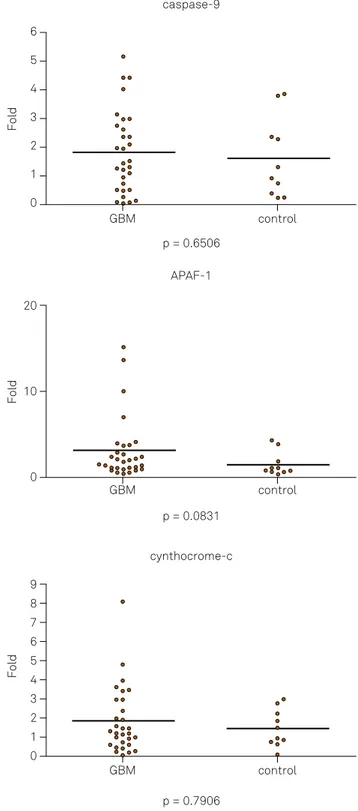
he expression of apoptotic (cytochrome C, APAF-1, cas
-pase 9, cas-pase 3, Smac/DIABLO) and anti-apoptotic genes (XIAP and Bcl-2) were analyzed using relative quantiication of mRNA levels in normal brain and glioblastomas. Among the apoptotic genes (cytochrome C, p = 0.7906; APAF-1, p = 0.0831; caspase 9, p = 0.6506; caspase 3, p = 0.1743 and Smac/DIABLO, p = 0.4630), no signiicant diferences in gene expression between high-grade glioma tumor and normal tis
-sue were observed (Figure 1).
However, the anti-apoptotic genes (XIAP and Bcl-2) were expressed at higher levels in GBMs when compared to con
-trol samples (p < 0.05; Figure 2).
DISCUSSION
Recent studies analyzing gene expression have shown that the treatment of glioblastoma cells with retinamide (apopto
-sis inducer) resulted in decreased levels of Bcl-2 and Bcl-X in glioblastoma cells. When associated with carmustine (a che
-motherapy agent), it leads to a downregulation of Bcl-2 and Bcl-X proteins in these cells and increases the apoptosis inci
-dence in the U87 glioblastoma cellular lineage. In this way, it was demonstrated that the modulation of Bcl-2 and Bcl-X
anti-apoptotic proteins levels may raise the sensitivity of the
glioblastomas to chemotherapy16. In another study, Tirapelli
et al.17, through immunohistochemical analysis of GBM sam
-ples, found high expression of Bcl-2, providing molecular evi
-dence that the protein Bcl-2 can reduce cell death in GBM. he upregulation of the anti-apoptotic Bcl-2 and Bcl-XL and
downregulation of the pro-apoptotic Bax have been detected in recurrent GBMs, showing, in part, the resistance of GBM to TRAIL-directed treatments18. Our results support this
hypoth-esis, demonstrating a signiicant diference in the expression of XIAP and Bcl-2, both anti-apoptotic genes, in glioblasto
-mas, and it can suggest that the cellular death triggered by the
GBM control
p = 0.7906 cynthocrome-c
GBM control
p = 0.0831 APAF-1
GBM control
p = 0.6506 caspase-9
Fold
9
7
5
3
1 0 8
6
4
2
Fold
20
0 10
Fold
5
3
1
0 6
4
2
apoptosis intrinsic pathway was inhibited in these tumors. Strik et al., also showed that the changes in Bcl-2 family pro
-tein expression can result from radiochemotherapy, but also relect the natural course of disease19.
Several proteins are released from the mitochondrial
intermembrane space into the cytoplasm, including cyto
-chrome C and Smac/DIABLO. Cyto-chrome C binds and activates the APAF-1 in the cytoplasm. he binding of cyto
-chrome C to APAF-1 induces conformational changes that allow APAF-1 to bind ATP/dATP and form the apoptosome that activates caspase 9. In this way, it triggers a caspase
activation cascade11. he glioblastoma samples showed low
expression levels of mRNA of cytochrome C, APAF-1 and cas
-pase 9 in our analysis, which suggests decreased apoptotic activity in these cells. hese data are in accordance with a study carried out by Watanabe et al., who observed, through immunohistochemistry and RT-PCR, the low expression of loss of heterozygosity of 12q22-23 chromosome in 70% of 33 glioblastomas and the consequent inactivation of APAF-1 in the glioblastomas20.
he resistance to apoptosis in cells of human astrocy
-toma was also examined by Ceruti et al. using the induction of mitochondria-damaging agents. hey reported that the
resistance to the apoptosis was due to an intrinsic defect of
caspase 9, leading to inhibition of enzyme activation and/or deteriorated interaction with proteins released by depolar -ization of the mitochondria21. he caspase 3 has been iden
-tiied as an apoptosis key mediator. Once activated, it is responsible for proteolytic cleavage of a broad spectrum of
cellular targets, leading to cell death22. he degradation of the
PARP (polyADP-ribose polymerase), actin and SREBP (ste
-rol regulatory element binding protein) related to the apop -tosis23, conirms the activation of caspase 3 and suggests
an irreversible phase in the death process by apoptosis in tumors. Ray et al.24 showed that the increase of the expression
of the pro-apoptotic genes, Bax, CALPAIN (cysteine protease dependent) and caspase 3 associated with DNA fragmenta -tion, indicates that the apoptosis is a mechanism that causes the death of malignant tumor cells and increasing the apop-tosis in the therapeutic process may result in a reduction of
GBM control
p = 0,0032 XIAP
Fold
20
0 30
10
GBM control
p = 0,0351 Bcl-2
Fold
20
0 30
10
GBM control
p = 0,1743 caspase-3
Fold
4
0 6
2
GBM control
p = 0,4630 SMAC
Fold
10
0 15
5 3
5
1
tumor size24. In this study, there was no signiicant diference
in the expression of caspase 3 between glioblastoma and nor
-mal brain tissue samples, providing evidence of an unlikely
occurrence of apoptosis in these tumors.
he Smac/DIABLO that binds to XIAP, and possibly to other IAPs, is released by the mitochondria, in a manner that keeps caspases away from XIAP. herefore, the Smac/ DIABLO is an IAP negative regulator and, consequently, a
molecule that increases apoptosis12. In the present study,
there were no signiicant changes in Smac/DIABLO expres
-sion, suggesting the connection of the XIAP and caspase 9, and, in this way, the inhibition of apoptosis. hese data are compatible with the results of Rajalingan et al.25, who, in a
study with HeLa cells, could not detect any compensation for Smac loss as the formation of P17 active fragment of caspase 3 and induction of apoptosis were clearly Smac dependent25.
Smac deicient cells are protected from apoptosis or another form of programmed cellular death. he XIAP is a potent
inhibitor of cell death that has been attributed largely to its ability to suppress speciic caspases12. he interaction with
the caspase 9 and 3 allows the XIAP to block the caspases activities eiciently, preventing, in this way, the proteolytic
cascade. If the apoptotic stimulus persists, the progressive generation of activated caspases, together with a
competi-tion among the mitochondrial proteins that block the XIAP activity, may overwhelm the protective efect of the XIAP26.
In conclusion, despite the limitations of our study due to
the small number of samples, the results demonstrated that there are diferences in the expressions of Bcl-2 and XIAP genes in glioblastomas and this can contribute for the inhi
-bition of programmed cell death, thus explaining one of the mechanisms of resistance of this type of tumor. Moreover, our results may provide additional information to enable the development of new therapeutic strategies capable of specii -cally targeting the apoptotic pathway in gliomas, aiming for
better treatment responses and patient outcomes.
References
1. Louis DN, Ohgaki H, Wiestler OD, Cavenee WK, Burger PC, Jouvet A et al. The 2007 WHO classification of tumours of the central nervous system. Acta Neuropathol. 2007;114(2):97-109. https://doi.org/10.1007/s00401-007-0243-4
2. Louis DN, Perry A, Reifenberger G, Deimling A, Figarella-Branger D, Cavenee WK et al. The 2016 World Health organization classification of tumors of the central nervous system: a summary. Acta Neuropathol. 2016;131(6):803-20. https://doi.org/10.1007/s00401-016-1545-1
3. Ceccarelli M, Barthel FP, Malta TM, Sabedot TS, Salama SR, Murray BA et al. Molecular profiling reveals biologically discrete subsets and pathways of progression in diffuse glioma. Cell. 2016;164(3):550-63. https://doi.org/10.1016/j.cell.2015.12.028
4. Frei K, Ambar B, Adachi N, Yonekawa Y, Fontana A. Ex vivo malignant glioma cells are sensitive to Fas (CD95/APO-1) ligand-mediated apoptosis. J Neuroimmunol. 1998;87(1-2):105-13. https://doi.org/10.1016/S0165-5728(98)00065-4
5. Stupp R, Hegi ME, Gilbert MR, Chakravarti A.
Chemoradiotherapy in malignant glioma: standard of care and future directions. J Clin Oncol. 2007;25(26):4127-36. https://doi.org/10.1200/JCO.2007.11.8554
6. Ohgaki H, Kleihues P. Genetic pathways to primary and secondary glioblastoma. Am J Pathol. 2007;170(5):1445-53. https://doi.org/10.2353/ajpath.2007.070011
7. Griguer CE, Oliva CR. Bioenergetics pathways and therapeutic resistance in gliomas: emerging role of mitochondria. Curr Pharm Des. 2011;17(23):2421-7. https://doi.org/10.2174/138161211797249251
8. Network CG. Comprehensive genomic characterization defines human glioblastoma genes and core pathways. Nature. 2008;455(7216):1061-8. https://doi.org/10.1038/nature07385
9. Verhaak RG, Hoadley KA, Purdom E, Wang V, Qi Y, Wilkerson MD et al. Integrated genomic analysis identifies clinically relevant subtypes of glioblastoma characterized by abnormalities in PDGFRA, IDH1, EGFR, and NF1. Cancer Cell. 2010;17(1):98-110. https://doi.org/10.1016/j.ccr.2009.12.020
10. Goldar S, Khaniani MS, Derakhshan SM, Baradaran B. Molecular mechanisms of apoptosis and roles in cancer development and treatment. Asian Pac J Cancer Prev. 2015;16(6):2129-44. https://doi.org/10.7314/APJCP.2015.16.6.2129
11. Riedl SJ, Shi Y. Molecular mechanisms of caspase regulation during apoptosis. Nat Rev Mol Cell Biol. 2004;5(11):897-907. https://doi.org/10.1038/nrm1496
12. Salvesen GS, Duckett CS. IAP proteins: blocking the road to death’s door. Nat Rev Mol Cell Biol. 2002;3(6):401-10. https://doi.org/10.1038/nrm830
13. Emery IF, Gopalan A, Wood S, Chow KH, Battelli C, George J et al. Expression and function of ABCG2 and XIAP in glioblastomas. J Neurooncol. 2017;133(1):47-57. https://doi.org/10.1007/s11060-017-2422-z
14. Vellanki SH, Grabrucker A, Liebau S, Proepper C,
Eramo A, Braun V et al. Small-molecule XIAP inhibitors enhance gamma-irradiation-induced apoptosis in glioblastoma. Neoplasia. 2009;11(8):743-52.
15. Lee FA, Zee BC, Cheung FY, Kwong P, Chiang CL, Leung KC et al. Randomized Phase II study of the X-linked Inhibitor of Apoptosis (XIAP) antisense AEG35156 in combination with sorafenib in patients with advanced hepatocellular carcinoma (HCC). Am J Clin Oncol. 2016;39(6):609-13. https://doi.org/10.1097/COC.0000000000000099
16. Lytle RA, Jiang Z, Zheng X, Higashikubo R, Rich KM.
Retinamide-induced apoptosis in glioblastomas is associated with down-regulation of Bcl-xL and Bcl-2 proteins. J Neurooncol. 2005;74(3):225-32. https://doi.org/10.1007/s11060-005-7305-z
17. Tirapelli LF, Bolini PH, Tirapelli DP, Peria FM, Becker AN,
Saggioro FP et al. Caspase-3 and Bcl-2 expression in glioblastoma: an immunohistochemical study. Arq Neuropsiquiatr. 2010;68(4):603-7. https://doi.org/10.1590/S0004-282X2010000400023
18. Krakstad C, Chekenya M. Survival signalling and apoptosis resistance in glioblastomas: opportunities for targeted therapeutics. Mol Cancer. 2010;9:135. https://doi.org/10.1186/1476-4598-9-135
19. Strik H, Deininger M, Streffer J, Grote E, Wickboldt J, Dichgans J et al. BCL-2 family protein expression in initial and recurrent glioblastomas: modulation by radiochemotherapy. J Neurol Neurosurg Psychiatry. 1999;67(6):763-8.
https://doi.org/10.1136/jnnp.67.6.763
21. Ceruti S, Mazzola A, Abbracchio MP. Resistance of human astrocytoma cells to apoptosis induced by mitochondria-damaging agents: possible implications for anticancer therapy. J Pharmacol Exp Ther. 2005;314(2):825-37. https://doi.org/10.1124/jpet.105.085340
22. Shi Y. Mechanical aspects of apoptosome assembly. Curr Opin Cell Biol. 2006;18(6):677-84. https://doi.org/10.1016/j.ceb.2006.09.006
23. Konstantinidou AE, Givalos N, Gakiopoulou H, Korkolopoulou P, Kotsiakis X, Boviatsis E et al. Caspase-3 immunohistochemical expression is a marker of apoptosis, increased grade and early recurrence in intracranial meningiomas. Apoptosis. 2007;12(4):695-705. https://doi.org/10.1007/s10495-006-0001-4
24. Ray SK, Patel SJ, Welsh CT, Wilford GG, Hogan EL, Banik NL. Molecular evidence of apoptotic death in malignant brain tumors including glioblastoma multiforme: upregulation of calpain and caspase-3. J Neurosci Res. 2002;69(2):197-206. https://doi.org/10.1002/jnr.10265
25. Rajalingam K, Oswald M, Gottschalk K, Rudel T. Smac/DIABLO is required for effector caspase activation during apoptosis in human cells. Apoptosis. 2007;12(8):1503-10.
https://doi.org/10.1007/s10495-007-0067-7