Submitted 17 July 2015 Accepted 5 October 2015 Published22 October 2015
Corresponding author
Willias Masocha, [email protected]
Academic editor
Jafri Abdullah
Additional Information and Declarations can be found on page 10
DOI10.7717/peerj.1350
Copyright
2015 Masocha
Distributed under
Creative Commons CC-BY 4.0
OPEN ACCESS
Astrocyte activation in the anterior
cingulate cortex and altered glutamatergic
gene expression during paclitaxel-induced
neuropathic pain in mice
Willias Masocha
Department Pharmacology and Therapeutics, Faculty of Pharmacy, Kuwait University, Safat, Kuwait
ABSTRACT
Spinal astrocyte activation contributes to the pathogenesis of paclitaxel-induced neuropathic pain (PINP) in animal models. We examined glial fibrillary acidic protein (GFAP; an astrocyte marker) immunoreactivity and gene expression of GFAP, glutamate transporters and receptor subunits by real time PCR in the anterior cingulate cortex (ACC) at 7 days post first administration of paclitaxel, a time point when mice had developed thermal hyperalgesia. The ACC, an area in the brain involved in pain perception and modulation, was chosen because changes in this area might contribute to the pathophysiology of PINP. GFAP transcripts levels were elevated by more than fivefold and GFAP immunoreactivity increased in the ACC of paclitaxel-treated mice. The 6 glutamate transporters (GLAST, GLT-1 EAAC1, EAAT4, VGLUT-1 and VGLUT-2) quantified were not significantly altered by paclitaxel treatment. Of the 12 ionotropic glutamate receptor subunits transcripts analysed 6 (GLuA1, GLuA3, GLuK2, GLuK3, GLuK5 and GLuN1) were significantly up-regulated, whereas GLuA2, GLuK1, GLuK4, GLuN2A and GLuN2B were not significantly altered and GLuA4 was lowly expressed. Amongst the 8 metabotropic receptor subunits analysed only mGLuR8 was significantly elevated. In conclusion, during PINP there is astrocyte activation, with no change in glutamate transporter expression and differential up-regulation of glutamate receptor subunits in the ACC. Thus, targeting astrocyte activation and the glutamatergic system might be another therapeutic avenue for management of PINP.
Subjects Molecular Biology, Neuroscience, Anaesthesiology and Pain Management, Pharmacology
Keywords Neuropathic pain, Anterior cingulate cortex, Astrocyte, Paclitaxel, Glutamate, Glutamate receptors, Glutamate transporters
INTRODUCTION
The anterior cingulate cortex (ACC) is a cortical area in the brain that has been described to be involved with pain, possibly including both perception and modulation (Vogt, 2005;
et al., 2005;Sewards & Sewards, 2002;Vogt, 2005). Neuroimaging studies have shown increased activity in the ACC during chronic pain, including neuropathic pain (Hsieh et al., 1995;Peyron, Laurent & Garcia-Larrea, 2000;Tseng et al., 2013). Neurophysiological and molecular changes have also been observed in the ACC during chronic or neuropathic (Wrigley et al., 2009;Xu et al., 2008;Yamashita et al., 2014).
One of the changes that has been observed in the ACC during chronic or neuropathic pain is the activation of astrocytes or astrogliosis (Chen et al., 2012;Kuzumaki et al., 2007;Lu, Zhu & Gao, 2011;Narita et al., 2006;Yamashita et al., 2014). Astrocytes are the most numerous non-neuronal cells in the brain involved in modulation of neuronal activities e.g., extracellular and synaptic cleft neurotransmitter level regulation, release of neuroactive molecules amongst other activities (Maragakis & Rothstein, 2006;Seifert, Schilling & Steinhauser, 2006). Astrocytes express transporters which remove neuro-transmitters such asγ-aminobutyric acid (GABA) and glutamate from the extracellular space or synaptic cleft (Conti et al., 1998;Danbolt, 2001;Gosselin, Bebber & Decosterd, 2010;Minelli et al., 1995;Wang & Bordey, 2008). Astrocyte activation has been linked with increase in transporters for GABA and a decrease in transporters for glutamate resulting in a more excitatory state in the brain (Gosselin, Bebber & Decosterd, 2010;Maragakis & Rothstein, 2006). Recently, we observed an increase in the transcripts of GABA transporter 1 (GAT-1) in a rodent model of paclitaxel-induced neuropathic pain (PINP) (Masocha, 2015). However, it is not known whether paclitaxel induces astrocyte activation in the ACC although it has been shown to induce astrocyte activation in the spinal cord (Peters et al., 2007;Zhang, Yoon & Dougherty, 2012). Paclitaxel is a chemotherapeutic agent that causes dose-dependent neuropathic pain in some patients (Scripture, Figg & Sparreboom, 2006;Wolf et al., 2008). In the rodent models, we observed that the PINP is linked with disturbances in the GABAergic system (Masocha, 2015) resulting in increased excitability of the ACC to electrophysiological stimulation (H Nashawi, IO Edafiogho, SB Kombian, W Masocha, 2015, unpublished data). GABA is the major inhibitory neurotransmitter while glutamate is the major stimulatory neurotransmitter in the brain (Meldrum, 2000;
Petroff, 2002). It is not known whether paclitaxel causes any changes in the glutamatergic system in the ACC, although it has been shown to decrease the expression of glutamate transporters such as GLAST and GLT-1 in the spinal cord (Weng et al., 2005;Zhang, Yoon & Dougherty, 2012). There are 8 known glutamate transporters, which are excitatory amino acid transporter 1 (EAAT1; referred to as GLAST in rodents), EAAT2 (GLT-1), EAAT3 (EAAC1), EAAT4, EAAT5, vesicular glutamate transporter 1 (VGLUT1), VGLUT2, and VGLUT3 (Danbolt, 2001;Shigeri, Seal & Shimamoto, 2004). Of the transporters, GLAST and GLT-1 are expressed on astrocytes (Danbolt, 2001) and play an important role in removal of glutamate from the synaptic cleft and extracellular space (Danbolt, 2001;
Shigeri, Seal & Shimamoto, 2004); if their expression is down-regulated, this results in increased levels of glutamate and excitotoxicity (Danbolt, 2001;Rothstein et al., 1996;
(NMDA) receptors which have 18 subunits GLuA1 to 4, GLuK1-5 and GLuN1, GLuN2A to D, GLuN3A and B, and GLuD1 and 2 (Collingridge et al., 2009). There are 8 subunits of the metabotropic receptors mGLUR1to8(Conn & Pin, 1997;Niswender & Conn, 2010).
Astrocyte activation, which has been observed in the ACC in models of chronic and neu-ropathic pain (Chen et al., 2012;Kuzumaki et al., 2007;Lu, Zhu & Gao, 2011;Narita et al., 2006;Yamashita et al., 2014), might occur in the ACC during PINP together with molecular changes in the glutamatergic system contributing to the pathogenesis or maintenance of PINP. Thus, in this study, astrocyte activation and the gene expression of molecules of the astrocyte marker (glial fibrillary acidic protein (GFAP), glutamate transporters and receptors in the ACC were evaluated in mice at a time point when the mice had paclitaxel-induced thermal hyperalgesia (Nieto et al., 2008;Parvathy & Masocha, 2013).
MATERIALS AND METHODS
AnimalsNinety eight female BALB/c mice (8–12 weeks old) supplied by the Animal Resources Cen-tre (ARC) at the Health Sciences Center (HSC), Kuwait University were used. The animals were housed and handled in compliance with the Kuwait University, HSC, ARC guidelines and published ethical guidelines for research in experimental pain with conscious animals (Zimmermann, 1983). All animal experiments were approved by the Ethical Committee for the use of Laboratory Animals in Teaching and in Research, HSC, Kuwait University.
Paclitaxel administration
Paclitaxel (Cat. No. 1097; Tocris, Bristol, UK) was dissolved in a solution made up of 50% Cremophor EL and 50% absolute ethanol to a concentration of 6 mg/ml and then diluted in normal saline (NaCl 0.9%), to a final concentration of 0.2 mg/ml just before administration. The vehicle for paclitaxel, thus, constituted of about 1.7% Cremophor EL and 1.7% ethanol in normal saline. Paclitaxel 2 mg/kg or its vehicle were administered to mice intraperitoneally (i.p.), daily for 5 consecutive days. This treatment regimen has been reported to produce painful neuropathy and thermal hyperalgesia in mice (Nieto et al., 2008;Parvathy & Masocha, 2013).
Hot plate test
Reaction latencies to hot plate test were measured before (baseline latency) and on day 7 after first administration of paclitaxel. Briefly, mice were placed on a hot plate (Panlab SL, Barcelona, Spain) with the temperature adjusted to 55±1◦C. The time to the first sign of
nociception, paw licking or flinching, was recorded and the animal immediately removed from the hot plate. A cut-offperiod of 20 s was maintained to avoid damage to the paws.
ACC tissue preparation
Table 1 PCR primer sequences of cyclophilin, GFAP and glutamatergic system molecules.
Gene Polarity
Sense
Sequence 5′–3′
Anti-sense Sequence 5′–3′
Cyclophilin GCTTTTCGCCGCTTGCT CTCGTCATCGGCCGTGAT
GFAP ACAGCGGCCCTGAGAGAGAT CTCCTCTGTCTCTTGCATGTTACTG
GLAST ACCAAAAGCAACGGAGAAGAG GGCATTCCGAAACAGGTAACTC
GLT-1 ACAATATGCCCAAGCAGGTAGA CTTTGGCTCATCGGAGCTGA
EAAC1 CTTCCTACGGAATCACTGGCT CGATCAGCGGCAAAATGACC
EAAT4 AGCAGCCACGGCAATAGTC ATGCCAAGCTGACACCAATGA
VGLUT-1 GGTGGAGGGGGTCACATAC AGATCCCGAAGCTGCCATAGA
VGLUT-2 CCCTGGAGGTGCCTGAGAA GCGGTGGATAGTGCTGTTGTT
GLuA1 CCGTTGACACATCCAATCAGTTT GTCGATAATGCTAATGAGAGCTTCCT
GLuA2 AAATTGCCAAACATTGTGG ATGGAGCCATGGCAATATCA
GLuA3 ACACCATCAGCATAGGTGGA TCAGTGGTGTTCTGGTTGGT
GLuA4 TTGGAATGGGATGGTAGGAG TAGGAACAAGACCACGCTGA
GLuK1 TCACACCCTACGAGTGGTATAAC AGCTCCAACGCCAAACCAG
GLuK2 ATCGGATATTCGCAAGGAACC CCATAGGGCCAGATTCCACA
GLuK3 AGGTCCTAATGTCACTGACTCTC GCCATAAAGGGTCCTATCAGAC
GLuK4 CCAAGGTCGAAGTGGACATCT CTGGGGTGAAGGTTCAGGG
GLuK5 ATAGTCGCCTTCGCCAATCC GTGTCCGTGGTCTCGTACTG
GLuN1 GGCATCGTAGCTGGGATCTTC TCCTACGGGCATCCTTGTG
GLuN2A GTTTGTTGGTGACGGTGAGA AAGAGGTGCTCCCAGATGAA
GLuN2B ATGTGGATTGGGAGGATAGG TCGGGCTTTGAGGATACTTG
mGluR1 TGTCATCAACGCCATCTATGC CCCACGTAGCCAGGACATAGAG
mGluR2 CGCTCTCTGCACGCTCTATG GATGAACTTGGCCTCGTTGAA
mGluR3 AAGCCATCGCCTGTCATCTG GGAGGTCCCAAGCCCAAGT
mGluR4 GATGCTCTACATGCCCAAAGTCTAC CGGTGACAACGGCTTTGAG
mGluR5 TGACCCTGAGCCCATTGC AACGAAGAGGGTGGCTAGCA
mGluR6 TCATGGCCACCACAACTATCA CAGAGGCGCGGACTATGG
mGluR7 AAGCCTGGGCAGAGGAAGA TCCATCACAGGGCTCACAAG
mGluR8 CAGCATCTGTCTGCAGCCTG CGGTTTTCTTCCTCTCCCCA
Real time RT-PCR
Gene transcripts of the astrocyte marker GFAP, 6 glutamate transporters (GLAST, GLT-1, EAAC1, EAAT4, VGLUT1, VGLUT2), 12 ionotropic glutamate receptor subunits (GLuA1 to 4, GLuK1 to 5, GLuN1, GLuN2A and GLuN2B) and 8 metabotropic glutamate subunits (mGluR1to8) were quantified in the ACC of vehicle-treated or paclitaxel-treated by real
at 50◦C for 2 min, a second hold at 95◦C for 2 min followed by 40 cycles at 95◦C for 15 s and 63◦
C for 1 min. Threshold cycle (Ct) values for all cDNA samples were obtained and the amount of mRNA of individual animal sample (n=6–24 per group) was normalized to
cyclophilin (housekeeping gene) (ΔCt). The relative amount of target gene transcripts was calculated using the 2−ΔΔCtmethod as described previously (Livak & Schmittgen, 2001).
Immunohistochemistry
Fresh-frozen brains were cut on a cryostat into 25µm thick sections and thaw-mounted on chrome-alum gelatin–coated slides. The sections at a level of the lateral ventricles and the ACC were fixed in 4% formalin and 14% picric acid in PBS for 30 s at 4◦
C, rinsed in PBS, fixed in acetone for 30 s at−20◦C, and then rinsed in PBS. All sections were preincubated
with 1% bovine serum albumin and 0.3% Triton X-100 in PBS (solution used as diluent for primary and secondary antibodies) for 30 min at room temperature. Sections were incubated with rabbit anti- GFAP (1:100; DAKO, Glostrup, Denmark) for 2 h at room temperature to immunostain astrocytes. Sections were then rinsed in PBS and incubated with DyLight 594-conjugated Affinipure donkey Anti-rabbit IgG (H+L) (1:100, Jackson
ImmunoResearch Laboratories, West Grove, Pennsylvania, USA) for 1 h. The sections were rinsed in PBS and mounted in ProLong®Gold antifade reagent (Invitrogen, USA). Sections were examined and analysed using a LSM 700 laser scanning confocal microscope. Images were taken from the ACC using an Axio imager (Carl Zeiss MicroImaging GmbH, Oberkochen, Germany).
Statistical analyses
Statistical analyses were performed using unpaired two-tailed Student’st-test using Graph Pad Prism software (version 5.0). The differences were considered significant atp<0.05. The results in the text and figures are expressed as the means±S.E.M.
RESULTS
Paclitaxel-induced thermal hyperalgesia
Mice developed thermal hyperalgesia on day 7 after first administration of paclitaxel as we previously described (Masocha, 2014;Parvathy & Masocha, 2013) i.e., paclitaxel-treated mice had significant reduction in response latency time in the hot plate test on day 7 compared to the baseline latency and vehicle-treated animals (6.23±0.28 s compared to
9.66±0.16 s and 9.00±0.38 s, respectively;n=10 vehicle-treated mice and 16 paclitaxel
treated-mice;p<0.05 for both comparisons).
Astrocyte activation in the ACC at 7 days after paclitaxel administration
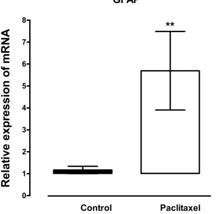
Figure 1 Effects of paclitaxel on glial fibrillary acidic protein (GFAP) transcript levels in the anterior cingulate cortex (ACC).Relative GFAP mRNA expression in the ACC of BALB/c mice on day 7 after first administration of the drug or its vehicle. Each point represents the mean±S.E.M of the values obtained from 21 vehicle-treated control mice and 24 paclitaxel-treated mice.∗∗p<0.01 compared to vehicle-treated control mice.
in the ACC compared to vehicle-treated controls (Fig. 2). However, the change in GFAP immunoreactivity in paclitaxel-treated animals varied across the ACC and animals i.e., it was not robust in all animals and did not cover most of the ACC.
Expression of transcripts of glutamate transporters in the ACC at 7 days after paclitaxel administration
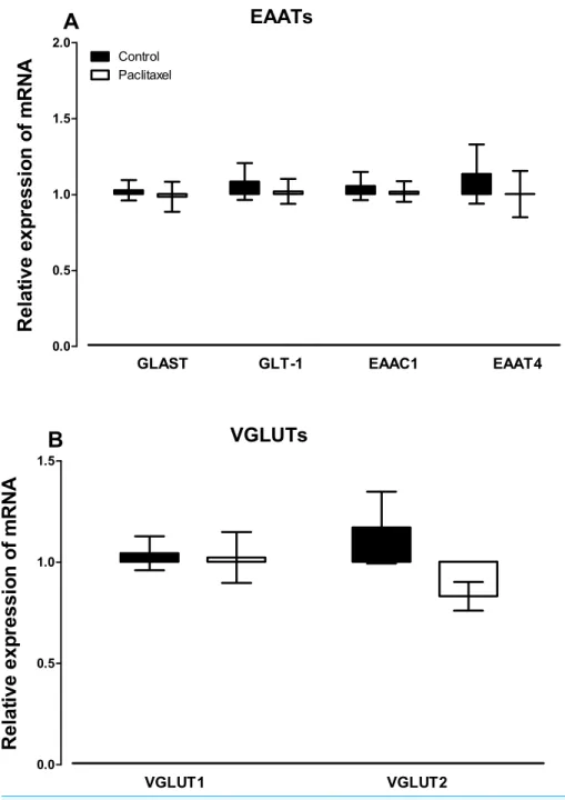
There were no differences observed in the transcript levels of all the six glutamate transporters analysed (Fig. 3) in the ACC of paclitaxel-treated mice compared to vehicle-treated mice. Using the unpaired two-tailed Student’st-test thepvalues obtained are: 0.7243 for GLAST, 0.6608 for GLT-1, 0.7575 for EAAC1, 0.5925 for EAAT4, 0.8885 for VGLUT-1 and 0.0858 for VGLUT-2.
Expression of transcripts of glutamate receptors in the ACC at 7 days after paclitaxel administration
Figure 2 Effects of paclitaxel on glial fibrillary acidic protein (GFAP) immunoreactivity in the an-terior cingulate cortex (ACC).GFAP immunoreactivity in the ACC of BALB/c mice on day 7 after first administration of the drug or its vehicle. GFAP immunoreactivity in astrocytes is increased in 3 paclitaxel-treated mice (D–F) compared to 3 vehicle-treated control mice (A–C) in the ACC. Note that in a paclitaxel-treated mouse (D) increased immunoreactivity of GFAP appears to be along a blood vessel: Scale bar: 50µm.
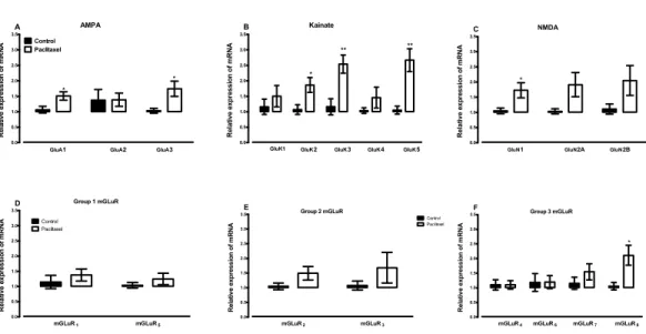
significantly increased the expression of GLuA1 (p=0.0166) and GLuA3 (p=0.0243) subunits compared to vehicle-treated controls (Fig. 4A).
Amongst the 5 kainate receptor subunits analysed, treatment with paclitaxel signifi-cantly increased the expression of the 3 subunits GluK2 (p=0.0136), GluK3 (p=0.0026) and GluK5 (p=0.0011), but not 2 subunits GluK1 (p=0.4367) and GluK4 (p=0.2785), compared to vehicle-treated controls (Fig. 4B).
Amongst the 3 NMDA receptor subunits analysed, treatment with paclitaxel signifi-cantly increased the expression of GluN1 (p=0.0209) only, but not 2 subunits GluN2A (p=0.0612) and GluN2B (p=0.1105), compared to vehicle-treated controls (Fig. 4C).
Of all the eight metabotropic glutamate receptors subunits quantified, only mGLuR8
was significantly altered (p=0.0144) in the ACC by treatment with paclitaxel compared to treatment with vehicle (Figs. 4Eand4F). Using the unpaired two-tailed Student’st-test, thepvalues obtained are: 0.4439 for mGLuR1, 0.1340 for mGLuR2, 0.3201 for mGLuR3,
0.9971 for mGLuR4, 0.3375 for mGLuR5, 0.9693 for mGLuR6and 0.2780 for mGLuR7.
DISCUSSION
Figure 3 Effects of paclitaxel on glutamate transporters transcript levels in the anterior cingulate cortex (ACC).Relative mRNA expression of (A) excitatory amino acid transporters GLAST, GLT-1, EAAC1, EAAT4, and (B) vesicular glutamate transporters VGLUT1 and VGLUT2 in the ACC of BALB/c mice on day 7 after first administration of the drug or its vehicle. Each point represents the mean±S.E.M of the values obtained from 11 to 15 vehicle-treated control mice and 13–15 paclitaxel-treated mice.
modulation (Vogt, 2005;Xie, Huo & Tang, 2009;Zhuo, 2008), during paclitaxel-induced neuropathic pain (PINP).
Figure 4 Effects of paclitaxel on glutamate receptors transcript levels in the anterior cingulate cortex (ACC).Relative mRNA expression of (A) AMPA receptor subunits GLuA1 to 3, (B) kainate receptor sub-units GLuK1 to 5, (C) NMDA receptor subsub-units GLuN1, GLuN2A and GLuN2B, and (D-F) metabotropic glutamate receptors mGLuR1 to8 in the ACC of BALB/c mice on day 7 after first administration of the drug or its vehicle. Each point represents the mean± S.E.M of the values obtained from 6 to 15 vehicle-treated control mice and 8–16 paclitaxel-treated mice.∗p<0.05,∗∗p<0.01 compared to vehicle-treated control mice.
Gao, 2011). Astrocyte activation has also been observed in the ACC in other models of neuropathic pain (Xu et al., 2008;Yamashita et al., 2014) but had not been reported in PINP. However, astrocyte activation in the spinal cord has been reported to contribute to PINP in rodents (Ruiz-Medina et al., 2013;Zhang, Yoon & Dougherty, 2012). In the current study, the expression of GFAP transcripts and immunoreactivity in the ACC was increased in mice treated with PINP. During peripheral nerve injury neurons have been reported to release neurotransmitters such substancePand glutamate and neuronal chemokines that cause astrocyte activation in the CNS (Milligan & Watkins, 2009;Wang et al., 2009;
Watkins et al., 2007). Activated astrocytes in turn release molecules that contribute to the pathophysiology of pain through modulation of neuronal functioning (Milligan & Watkins, 2009;Wang et al., 2009;Watkins et al., 2007). Thus, the current results suggest that astrocyte activation in the ACC might also contribute to the pathophysiology of PINP.
the ACC during neuropathic pain in both humans and animal models (Hsieh et al., 1995;
Peyron, Laurent & Garcia-Larrea, 2000;Tseng et al., 2013;Wrigley et al., 2009;Xu et al., 2008;Yamashita et al., 2014).
Although we did not observe any changes in the glutamate transporters in the ACC, we observed that transcripts of various glutamate receptors and receptor subunits were elevated in the ACC of mice with PINP. The increased expression of some of the glutamate receptors and receptor subunits could have been linked to astrocyte activation since all of the up-regulated receptors are expressed on astrocytes (Geurts et al., 2005;Mart´ınez-Lozada & Ortega, 2015). Several receptors have been reported to be differentially expressed in the ACC in rodent models of PINP. We observed an increase in the expression of various GABA receptors in the ACC during PINP (Masocha, 2015).Ortega-Legaspi et al. (2011)
andOrtega-Legaspi et al. (2010). reported a differential expression of muscarinic-1 and
−2 receptors and dopamine D1 and D2 receptors in the ACC of rodents with PINP.
The increased expression glutamate receptors in the ACC also suggest a role of the glutamatergic system in the pathogenesis of PINP.
CONCLUSIONS
In conclusion, the results of this study show that animals with paclitaxel-induced neuropathic pain (PINP) have increased transcripts and immunoreactivity of the astrocyte marker GFAP and transcripts of some glutamate receptors and receptor subunits, but not glutamate transporters, in the ACC. In a previous study, transcripts of a GABA transporter GAT-1, whose increase has been associated with astrocyte activation in the spinal cord of rodents with PINP (Yadav et al., 2015), was found increased in the ACC of mice with PINP (Masocha, 2015). Thus, inhibition of astrocyte activation and GAT-1 activity and/or antagonism of specific glutamate receptors could be therapeutic modalities of managing PINP and possibly other types on chemotherapy-induced neuropathic pain.
ACKNOWLEDGEMENTS
I am grateful to Dr. Subramanian S Parvathy, Ms. Salini Soman from the Department of Pharmacology and Therapeutics, Faculty of Pharmacy, and Ms. Jucy Gabriel from the Research Core Facility, HSC, Kuwait University for their technical assistance and to the staff from the Animal Resources Centre, HSC, Kuwait University for their support.
ADDITIONAL INFORMATION AND DECLARATIONS
Funding
This study was supported by grants PT01/09 and SRUL02/13 from Kuwait University Research Sector. The funders had no role in study design, data collection and analysis, decision to publish, or preparation of the manuscript.
Grant Disclosures
Competing Interests
The authors declare there are no competing interests.
Author Contributions
• Willias Masocha conceived and designed the experiments, performed the experiments,
analyzed the data, contributed reagents/materials/analysis tools, wrote the paper, prepared figures and/or tables, reviewed drafts of the paper.
Animal Ethics
The following information was supplied relating to ethical approvals (i.e., approving body and any reference numbers):
All animal experiments were approved by the Ethical Committee for the use of Laboratory Animals in Teaching and in Research, HSC, Kuwait University.
Supplemental Information
Supplemental information for this article can be found online athttp://dx.doi.org/ 10.7717/peerj.1350#supplemental-information.
REFERENCES
Aldskogius H, Kozlova EN. 1998.Central neuron-glial and glial–glial interactions following axon injury.Progress in Neurobiology55:1–26DOI 10.1016/S0301-0082(97)00093-2.
Chen FL, Dong YL, Zhang ZJ, Cao DL, Xu J, Hui J, Zhu L, Gao YJ. 2012.Activation of astrocytes in the anterior cingulate cortex contributes to the affective component of pain in an inflamma-tory pain model.Brain Research Bulletin87:60–66DOI 10.1016/j.brainresbull.2011.09.022.
Collingridge GL, Olsen RW, Peters J, Spedding M. 2009.A nomenclature for ligand-gated ion channels.Neuropharmacology56:2–5DOI 10.1016/j.neuropharm.2008.06.063.
Conn PJ, Pin JP. 1997.Pharmacology and functions of metabotropic glutamate receptors.Annual Review of Pharmacology and Toxicology37:205–237DOI 10.1146/annurev.pharmtox.37.1.205.
Conti F, Melone M, De Biasi S, Minelli A, Brecha NC, Ducati A. 1998.Neuronal and glial localization of GAT-1, a high-affinity gamma-aminobutyric acid plasma membrane transporter, in human cerebral cortex: with a note on its distribution in monkey cortex.Journal of Comparative Neurology396:51–63
DOI 10.1002/(SICI)1096-9861(19980622)396:1<51::AID-CNE5>3.0.CO;2-H.
Danbolt NC. 2001.Glutamate uptake.Progress in Neurobiology65:1–105
DOI 10.1016/S0301-0082(00)00067-8.
Geurts JJ, Wolswijk G, Bo L, Redeker S, Ramkema M, Troost D, Aronica E. 2005.Expression pat-terns of Group III metabotropic glutamate receptors mGluR4 and mGluR8 in multiple sclerosis lesions.Journal of Neuroimmunology158:182–190DOI 10.1016/j.jneuroim.2004.08.012.
Gosselin RD, Bebber D, Decosterd I. 2010.Upregulation of the GABA transporter GAT-1 in the gracile nucleus in the spared nerve injury model of neuropathic pain.Neuroscience Letters 480:132–137DOI 10.1016/j.neulet.2010.06.023.
Hsieh JC, Belfrage M, Stone-Elander S, Hansson P, Ingvar M. 1995.Central representation of chronic ongoing neuropathic pain studied by positron emission tomography.Pain63:225–236
Kuzumaki N, Narita M, Hareyama N, Niikura K, Nagumo Y, Nozaki H, Amano T, Suzuki T. 2007.Chronic pain-induced astrocyte activation in the cingulate cortex with no change in neural or glial differentiation from neural stem cells in mice.Neuroscience Letters415:22–27
DOI 10.1016/j.neulet.2006.12.057.
Livak KJ, Schmittgen TD. 2001.Analysis of relative gene expression data using real-time quantitative PCR and the 2(-Delta Delta C(T)) Method. Methods 25:402–408
DOI 10.1006/meth.2001.1262.
Lu Y, Zhu L, Gao YJ. 2011.Pain-related aversion induces astrocytic reaction and proinflammatory cytokine expression in the anterior cingulate cortex in rats.Brain Research Bulletin84:178–182
DOI 10.1016/j.brainresbull.2010.12.007.
Maragakis NJ, Rothstein JD. 2006.Mechanisms of Disease: astrocytes in neurodegenerative disease.Nature Clinical Practice Neurology2:679–689DOI 10.1038/ncpneuro0355.
Mart´ınez-Lozada Z, Ortega A. 2015.Glutamatergic transmission: a matter of three.Neural Plasticity2015:787396DOI 10.1155/2015/787396.
Masocha W. 2009.Systemic lipopolysaccharide (LPS)-induced microglial activation results in different temporal reduction of CD200 and CD200 receptor gene expression in the brain.
Journal of Neuroimmunology214:78–82DOI 10.1016/j.jneuroim.2009.06.022.
Masocha W. 2014.Paclitaxel-induced hyposensitivity to nociceptive chemical stimulation in mice can be prevented by treatment with minocycline. Scientific Reports
4:6719DOI 10.1038/srep06719.
Masocha W. 2015.Comprehensive analysis of the GABAergic system gene expression profile in the anterior cingulate cortex of mice with Paclitaxel-induced neuropathic pain.Gene Expression 16:145–153DOI 10.3727/105221615X14181438356337.
Meldrum BS. 2000.Glutamate as a neurotransmitter in the brain: review of physiology and pathology.Journal of Nutrition130:1007S–1015S.
Milligan ED, Watkins LR. 2009.Pathological and protective roles of glia in chronic pain.Nature Reviews Neuroscience10:23–36DOI 10.1038/nrn2533.
Minelli A, Brecha NC, Karschin C, DeBiasi S, Conti F. 1995.GAT-1, a high-affinity GABA plasma membrane transporter, is localized to neurons and astroglia in the cerebral cortex.Journal of Neuroscience15:7734–7746.
Narita M, Kuzumaki N, Narita M, Kaneko C, Hareyama N, Miyatake M, Shindo K, Miyoshi K, Nakajima M, Nagumo Y, Sato F, Wachi H, Seyama Y, Suzuki T. 2006.Chronic pain-induced emotional dysfunction is associated with astrogliosis due to cortical delta-opioid receptor dysfunction.Journal of Neurochemistry97:1369–1378DOI 10.1111/j.1471-4159.2006.03824.x.
Nieto FR, Entrena JM, Cendan CM, Pozo ED, Vela JM, Baeyens JM. 2008.Tetrodotoxin inhibits the development and expression of neuropathic pain induced by paclitaxel in mice.Pain 137:520–531DOI 10.1016/j.pain.2007.10.012.
Niswender CM, Conn PJ. 2010.Metabotropic glutamate receptors: physiology, pharmacology, and disease.Annual Review of Pharmacology and Toxicology50:295–322
DOI 10.1146/annurev.pharmtox.011008.145533.
Ortega-Legaspi JM, De Gortari P, Garduno-Gutierrez R, Amaya MI, Leon-Olea M, Coffeen U, Pellicer F. 2011.Expression of the dopaminergic D1 and D2 receptors in the anterior cingulate cortex in a model of neuropathic pain.Molecular Pain7:97DOI 10.1186/1744-8069-7-97.
Ortega-Legaspi JM, Leon-Olea M, De Gortari P, Amaya MI, Coffeen U, Simon-Arceo K, Pellicer F. 2010.Expression of muscarinic M1 and M2 receptors in the anterior cingulate cortex associated with neuropathic pain.European Journal of Pain14:901–910
Parvathy SS, Masocha W. 2013.Matrix metalloproteinase inhibitor COL-3 prevents the development of paclitaxel-induced hyperalgesia in mice.Medical Principles and Practice 22:35–41DOI 10.1159/000341710.
Peters CM, Jimenez-Andrade JM, Kuskowski MA, Ghilardi JR, Mantyh PW. 2007.An evolving cellular pathology occurs in dorsal root ganglia, peripheral nerve and spinal cord following intravenous administration of paclitaxel in the rat.Brain Research1168:46–59
DOI 10.1016/j.brainres.2007.06.066.
PetroffOA. 2002.GABA and glutamate in the human brain. Neuroscientist 8:562–573
DOI 10.1177/1073858402238515.
Peyron R, Laurent B, Garcia-Larrea L. 2000.Functional imaging of brain responses to pain. A review and meta-analysis (2000).Neurophysiologie Clinique30:263–288
DOI 10.1016/S0987-7053(00)00227-6.
Rothstein JD, Dykes-Hoberg M, Pardo CA, Bristol LA, Jin L, Kuncl RW, Kanai Y, Hediger MA, Wang Y, Schielke JP, Welty DF. 1996.Knockout of glutamate transporters reveals a major role for astroglial transport in excitotoxicity and clearance of glutamate.Neuron16:675–686
DOI 10.1016/S0896-6273(00)80086-0.
Ruiz-Medina J, Baulies A, Bura SA, Valverde O. 2013.Paclitaxel-induced neuropathic pain is age dependent and devolves on glial response.European Journal of Pain17:75–85
DOI 10.1002/j.1532-2149.2012.00172.x.
Scripture CD, Figg WD, Sparreboom A. 2006.Peripheral neuropathy induced by paclitaxel: recent insights and future perspectives. Current Neuropharmacology 4:165–172
DOI 10.2174/157015906776359568.
Seifert G, Schilling K, Steinhauser C. 2006.Astrocyte dysfunction in neurological disorders: a molecular perspective.Nature Reviews Neuroscience7:194–206DOI 10.1038/nrn1870.
Senapati AK, Lagraize SC, Huntington PJ, Wilson HD, Fuchs PN, Peng YB. 2005.Electrical stimulation of the anterior cingulate cortex reduces responses of rat dorsal horn neurons to mechanical stimuli.Journal of Neurophysiology94:845–851DOI 10.1152/jn.00040.2005.
Sewards TV, Sewards MA. 2002.The medial pain system: neural representations of the motiva-tional aspect of pain.Brain Research Bulletin59:163–180DOI 10.1016/S0361-9230(02)00864-X.
Shigeri Y, Seal RP, Shimamoto K. 2004.Molecular pharmacology of glutamate transporters, EAATs and VGLUTs.Brain Research Reviews45:250–265
DOI 10.1016/j.brainresrev.2004.04.004.
Tseng MT, Chiang MC, Chao CC, Tseng WY, Hsieh ST. 2013. fMRI evidence of
degeneration-induced neuropathic pain in diabetes: enhanced limbic and striatal activations.
Human Brain Mapping34:2733–2746DOI 10.1002/hbm.22105.
Vogt BA. 2005.Pain and emotion interactions in subregions of the cingulate gyrus.Nature Reviews Neuroscience6:533–544DOI 10.1038/nrn1704.
Wang DD, Bordey A. 2008. The astrocyte odyssey.Progress in Neurobiology86:342–367
DOI 10.1016/j.pneurobio.2008.09.015.
Wang W, Wang W, Mei X, Huang J, Wei Y, Wang Y, Wu S, Li Y. 2009.Crosstalk between spinal astrocytes and neurons in nerve injury-induced neuropathic pain.PLoS ONE4:e6973
DOI 10.1371/journal.pone.0006973.
Watkins LR, Hutchinson MR, Ledeboer A, Wieseler-Frank J, Milligan ED, Maier SF. 2007.
Norman Cousins Lecture. Glia as the “bad guys”: implications for improving clinical pain control and the clinical utility of opioids.Brain, Behavior, and Immunity21:131–146
Weng HR, Aravindan N, Cata JP, Chen JH, Shaw AD, Dougherty PM. 2005.Spinal glial glutamate transporters downregulate in rats with taxol-induced hyperalgesia.Neuroscience Letters386:18–22DOI 10.1016/j.neulet.2005.05.049.
Wolf S, Barton D, Kottschade L, Grothey A, Loprinzi C. 2008.Chemotherapy-induced peripheral neuropathy: prevention and treatment strategies.European Journal of Cancer44:1507–1515
DOI 10.1016/j.ejca.2008.04.018.
Wrigley PJ, Press SR, Gustin SM, Macefield VG, Gandevia SC, Cousins MJ, Middleton JW, Henderson LA, Siddall PJ. 2009.Neuropathic pain and primary somatosensory cortex reorganization following spinal cord injury.Pain141:52–59DOI 10.1016/j.pain.2008.10.007.
Xie YF, Huo FQ, Tang JS. 2009.Cerebral cortex modulation of pain.Acta Pharmacologica Sinica 30:31–41DOI 10.1038/aps.2008.14.
Xu H, Wu LJ, Wang H, Zhang X, Vadakkan KI, Kim SS, Steenland HW, Zhuo M. 2008.
Presynaptic and postsynaptic amplifications of neuropathic pain in the anterior cingulate cortex.Journal of Neuroscience28:7445–7453DOI 10.1523/JNEUROSCI.1812-08.2008.
Yadav R, Yan X, Maixner DW, Gao M, Weng HR. 2015.Blocking the GABA transporter GAT-1 ameliorates spinal GABAergic disinhibition and neuropathic pain induced by paclitaxel.Journal of Neurochemistry133:857–869DOI 10.1111/jnc.13103.
Yamashita A, Hamada A, Suhara Y, Kawabe R, Yanase M, Kuzumaki N, Narita M, Matsui R, Okano H, Narita M. 2014.Astrocytic activation in the anterior cingulate cortex is critical for sleep disorder under neuropathic pain.Synapse68:235–247DOI 10.1002/syn.21733.
Yi JH, Pow DV, Hazell AS. 2005.Early loss of the glutamate transporter splice-variant GLT-1v in rat cerebral cortex following lateral fluid-percussion injury.Glia49:121–133
DOI 10.1002/glia.20099.
Zhang H, Yoon SY, Dougherty PM. 2012.Evidence that spinal astrocytes but not microglia contribute to the pathogenesis of Paclitaxel-induced painful neuropathy.The Journal of Pain 13:293–303DOI 10.1016/j.jpain.2011.12.002.
Zhuo M. 2008.Cortical excitation and chronic pain. Trends in Neurosciences31:199–207
DOI 10.1016/j.tins.2008.01.003.