Riparin A, a compound from
Aniba riparia
, attenuate the inflammatory
response by modulation of neutrophil migration
Renan O. Silva
a, Samara R.B. Damasceno
a, Irismara S. Silva
b, Valdelânia G. Silva
b, Camila F.C. Brito
b,
Antônio Éder A. Teixeira
a, Geandra B.L. Nunes
c, Celso A. Camara
d, José Maria B. Filho
e,
Stanley J.C. Gutierrez
f, Ronaldo A. Ribeiro
a, Marcellus H.L.P. Souza
a, André L.R. Barbosa
b,
Rivelilson M. Freitas
c, Jand Venes R. Medeiros
b,⇑aDepartment of Physiology and Pharmacology, Federal University of Ceará, Fortaleza, CE, Brazil bPost-Graduation Program in Biotechnology, Federal University of Piauí, Parnaíba, PI, Brazil
cLaboratory for Research in Experimental Neurochemistry, Federal University of Piauí, Teresina, PI, Brazil dMolecular Sciences Department, Federal Rural University of Pernambuco, Recife, PE, Brazil
eLaboratory of Pharmaceutical Technology, Federal University of Paraiba, João Pessoa, PB, Brazil
fLaboratory Chemistry of Bioactive Natural and Synthetic Products, Federal University of Piauí, Teresina, PI, Brazil
a r t i c l e
i n f o
Article history:
Received 22 November 2014
Received in revised form 9 January 2015 Accepted 24 January 2015
Available online 3 February 2015
Keywords:
Inflammation Neutrophil Riparin Oxidative stress
a b s t r a c t
Inflammation is a local tissue response to attacks characterized by vascular and cellular events, including intense oxidative stress. Riparin A, a compound obtained fromAniba riparia, has been shown to have anti-oxidant activity and cytotoxicityin vitro. This study was aimed at evaluating the anti-inflammatory effect of riparin A against acute inflammation. The results of our evaluations in various experimental models indicated that riparin A reduced paw edema induced by carrageenan, compound 48/80, histamine, and serotonin. Furthermore, it decreased leukocyte and neutrophil counts, myeloperoxidase activity, thiobar-bituric acid reactive substance (TBARS) levels, and cytokine (tumor necrosis factor-aand interleukin-1b) levels increased by carrageenan-induced peritonitis, and reversed glutathione levels. Riparin A also reduced carrageenan-induced adhesion and rolling of leukocytes on epithelial cells and did not produce gastric-damage as compared with indomethacin. In conclusion, the data show that riparin A reduces inflammatory response by inhibiting vascular and cellular events, modulating neutrophil migration, inhibiting proinflammatory cytokine production, and reducing oxidative stress.
Ó2015 Elsevier Ireland Ltd. All rights reserved.
1. Introduction
Inflammatory responses are a series of well-coordinated events that depend on an increase in vascular permeability and sequential release of inflammatory mediators, leading to edema and the arri-val of inflammatory leukocytes at the site of inflammation[1]. The drugs commonly used to treat inflammatory conditions, such as non-steroidal anti-inflammatory drugs (NSAIDs), are associated with low therapeutic efficacy. In addition, these drugs have major adverse effects, such as gastrointestinal ulcers, and bleeding and renal disorders[2].
Natural products are an important source for research aimed at the discovery of new substances with pharmacological activity
[3,4]. Secondary metabolites of natural products, such as lactones [5], alkaloids[6], and terpenoids[7]have attracted the attention of many researchers because of their anti-inflammatory activities. In our study, alkamide alkaloids were isolated from the green fruit of Aniba riparia (Nees) Mez (Lauraceae), a typical plant of the Amazon Region, where it is popularly known as ‘‘louro’’ [8]. After structural elucidation of one of its compounds,O-methyl-N -(2-hydroxybenzoyl) tyramine[9], new riparin derivatives were iso-lated and designated riparin A–F[10]. Riparin A,N-(2-phenylethyl) benzamide, (Fig. 1) the target of this study, is obtained from the coupling of a methyl ester of tyramine with a benzoyl chloride [11]. Previously, riparin A has been shown to have antioxidant activity[8]and fewer toxic effects than classical medicines [12]. However, there are no studies about the possible anti-inflamma-tory properties of riparin A.
Thus, considering that many studies have been conducted on alkaloids, and based on the fact that these molecules have impor-tant pharmacological activities with great therapeutic potential, in
http://dx.doi.org/10.1016/j.cbi.2015.01.029
0009-2797/Ó2015 Elsevier Ireland Ltd. All rights reserved.
⇑ Corresponding author at: BIOTEC/LAFFEX/UFPI, Av. São Sebastião, n°2819, CEP 64202-020, Parnaíba, PI, Brazil. Tel.: +55 86 9986 2374/3323 4750; fax: +55 86 3323 5406.
E-mail address:[email protected](J.V.R. Medeiros).
Contents lists available atScienceDirect
Chemico-Biological Interactions
the current study, we investigated the anti-inflammatory proper-ties of riparin A, a semisynthetic derivative isolated fromA. riparia, in mice.
2. Materials and methods
2.1. Preparation
Riparin A was prepared using as method described previously [13]. The purification were performed by column chromatography using a vertical glass column (silica gel 60 (SiO2) 70–230 Mesh; SysCroma) and a mixture of organic solvents in increasing order of polarity (hexane – hexane:dichloromethane (1:1) – dichloro-methane and dichlorodichloro-methane:methanol 90:10) and the determi-nation of the melting point by MQAPF-302 digital apparatus model manufactured by Micro Chemical[10].
2.2. Obtention and identification of riparin A (N-phenethylbenzamide)
Riparin A was obtained through Schotten–Bauman reaction. In a 50 ml flask 0.41 ml of benzoyl chloride and 0.89 ml of 2-phenyleth-ylamine were mixed. The reaction mixture without solvent was left in magnetic stirring for 30 min at room temperature. After purification by column chromatography it was obtained 0.68 g of
N-phenethylbenzamide (riparin A) with melting point of 115°C and yield of 84%. The identification of the riparin A was performed by analyzing1H and13C NMR spectral data consistent with litera-ture values[10].
2.3. Drugs and reagents
k-Carrageenan, compound 48/80, serotonin and histamine were purchased from Sigma Chemical (St. Louis, MO, USA). Heparin was provided by Merck (Brazil). All drugs were dissolved in saline. Rip-arin A was dissolved in 2% DMSO. All other chemicals were of ana-lytical grade and obtained from standard commercial suppliers.
2.4. Animals
Male Swiss mice (25–30 g) were randomly housed in appropri-ate cages with free access to food and wappropri-ater. All procedures were in accordance with the Guide for Care and Use of Laboratory Animals (National Institute of Health, Bethesda, MD, USA) and experimental protocols were approved by the Ethics Committee in Research of the Federal University of Piauí (protocol No 068/14).
2.5. Effect of riparin A on carrageenan-induced paw edema
Initially, mice received riparin A (1, 3, and 10 mg/kg, i.p.), after 30 min carrageenan (500
l
g/paw, 50l
l) was administered into the right hind paw. The control untreated received only 2% DMSO. The doses of riparin A were selected based on previous studies in our research group. Paw volume was measured in a plethysmometer (Panlab, Barcelona, Spain) before (V0; time zero) at 1, 2, 3, and 4 h after carrageenan treatment (Vt), as previously described[14]. In the 4th hour, samples of paw were removed and fixed in 10% formalin for subsequent histopathological analysis. Additionally,paw edema was also measured 24, 48, 72 and 96 h after carra-geenan (500
l
g/paw) administration [15]. Data were expressed as mean ± SEM of 5–6 animals per group.2.6. Effect of riparin A in paw edema induced by various agents
Mice were pretreated with riparin A (10 mg/kg, i.p.) 30 min before stimuli. Next, compound 48/80 (12
l
g/paw), serotonin (5-HT; 1% w/v) or histamine (Hist; 100l
g/paw) was administered into the right hind paw[6]. The control untreated received only 2% DMSO. Right paw volume was measured before (V0; time zero) at 30, 60, 90 and 120 min after stimuli (Vt). Data were expressed as mean ± SEM of 5–6 animals per group.2.7. Histological evaluation
For histological evaluation, the paw samples were embedded in paraffin and sectioned; 4-
l
m-thick sections were deparaffinized, stained with hematoxylin and eosin (H & E), and then examined under a light microscope by an experienced pathologist without knowledge of the treatments.2.8. Carrageenan-induced peritonitis
Mice were pretreated with riparin A (Rip A: 10 mgkg, i.p.) or indomethacin (10 mg/kg, i.p.) 30 min before carrageenan (500
l
g/cavity) administration. After 4 h, animals were sacrificed and peritoneal cavity washed with heparinized PBS. Total cell counts were performed in a Neubauer chamber, and differential cell counts (100 cells total) in slides stained with hematoxylin and eosin (H & E). Results are expressed as number of leukocytes or neutrophils per milliliter of peritoneal exudate. Aliquots of per-itoneal exudates were collected for analysis of glutathione (GSH) levels, TBARS levels, myeloperoxidase (MPO) activity and cytokine (TNF-a
and IL-1b) levels. Data were expressed as mean ± SEM of 5– 6 animals per group.2.9. Myeloperoxidase (MPO) activity
MPO activity of peritoneal exudate was assayed by measuring the change in absorbance at 450 nm using o-dianisidine and 1% H2O2[17]. Results are expressed as MPO units per milliliter of per-itoneal exudate (UMPO/ml).
2.10. TNF-
a
and IL-1blevelsThe concentrations of tumor necrosis factor-
a
(TNF-a
), interleu-kin-1b(IL-1b) were measured by ELISA, using commercially avail-able kits and according to instructions supplied by the manufacturer (DuoSet ELISA Development kit R & D Systems, Min-neapolis, MN, USA). Results are expressed as pictograms of cyto-kines per milliliter of peritoneal exudate (pg/ml).2.11. Glutathione (GSH) levels
Initially, 400
l
l of each supernatant of peritoneal exudate was mixed with 800l
l of 0.4 M Tris buffer and 20l
l of 0.01 M DTNB. The samples were stirred for 3 min and read on a spectrophotom-eter at 412 nm[18]. Results are expressed as micrograms of NPSH per milliliter of peritoneal exudate (l
g/ml).2.12. Malondialdehyde (MDA) concentration
diately in an ice water bath followed by the addition of 4 ml ofn -butanol. This mixture was shaken and the butanol layer was sepa-rated by centrifugation at 1200 rpm for 15 min and read on a spec-trophotometer to be 535 and 520 nm[19]. Results are expressed as nanomoles per milliliter of peritoneal exudate (nmol/ml).
2.13. Intravital microscopy of leukocytes to assess rolling and adhesion to the mesentery
Intravital microscopy was used to establish leukocyte rolling and adhesion to mesenteric microcirculation and examined as pre-Fig. 2.Riparin A reduces carrageenan-induced paw edema. Mice were pretreated with 2% DMSO or riparin A (1, 3, and 10 mg/kg, i.p.). After 30 min carrageenan (500lg/paw) was administrated. Each line represents the mean ± SEM of 5–6 animals per group.#p< 0.05versuscontrol group;⁄p< 0.05versuscarrageenan group. One-way analysis of
variance (ANOVA) followed by Newman–Keuls test.
Fig. 3.Photomicrographs from mice paw stained with hematoxylin and eosin (Panel A, C and E: 40magnification; Panel B and D: 10magnification). Panel A: 2% DMSO
viously described[16]. Briefly, mice were pretreated with riparin A (10 mg/kg) 30 min before carrageenan (500
l
g/cavity) administra-tion. After 4 h, mice were anesthetized with a mixture of ketamine (100 mg/kg, i.p.)/xylazine (10 mg/kg, i.p.) and mesenteric tissue was exteriorized for microscopic examinationin situ. Animals were maintained on a special board thermostatically controlled at 37°C, with a transparent platform on which the tissue to be transillumi-nated was placed. Vessels selected for study were third-order ven-ules, defined according to their branch-order location within the microvascular network. These vessels corresponded to postcapil-lary venules, with a diameter of 10–15l
m. Leukocyte interaction with the luminal surface of the venular endothelium was studied in a vessel segment. Rolling leukocytes were defined as white blood cells that move at a lower velocity than erythrocytes in the same stream. The number of rolling leukocytes was determined at 10-min intervals. These leukocytes moved at a sufficiently slow pace to be individually visible and were counted as they rolled past a 10l
m length of venule. Leukocyte was considered to be adherent to the venular endothelium if it remained stationary for 30 s. The number of adherent cells (stickers) was expressed as the number per 10l
m length of venule. Cells were counted in the recorded image, using 3–4 different fields for each animal to avoid sampling variability. Data were expressed as mean ± SEM of 5–6 animals per group.2.14. Evaluation of gastric damage
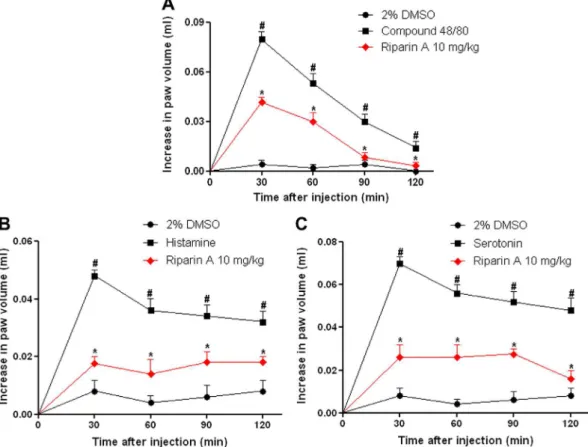
Mice received 2% DMSO, riparin A (10 and 100 mg/kg, p.o.) or indomethacin (20 mg/kg p.o.). After 7 h, the animals were sacri-ficed; the stomach was located, removed and opened along the greater curvature. The gastric damage macroscopic was measured Fig. 4.Riparin A reverse paw edema induced by various stimuli. Mice were pretreated with 2% DMSO or riparin A (10 mg/kg, i.p.). After 30 min the paw edema was induced by compound 48/80 (12lg/paw; A), histamine (100lg/paw; B), or serotonin (1% w/v; C). Each line represents the mean ± SEM of 5–6 animals per group.#p< 0.05versus control group;⁄p< 0.05versusstimuli. One-way analysis of variance (ANOVA) followed by Newman–Keuls test.
Fig. 5.Riparin A decrease cell migration in carrageenan-induced peritonitis. Mice were pretreated with 2% DMSO or riparin A (10 mg/kg, i.p.). After 30 min carrageenan (500lg/cavity) was administered. Cell migration was evaluated 4 h later. Leukocyte (A) and neutrophil (B) counts are shows. Each column represents the mean ± SEM of 5–6 animals per group.#p< 0.05versuscontrol group;⁄p< 0.05
using digital caliper (MitutoyoÒ) and calculated as sum of the
lengths of all linear erosions. In addition, samples were collected for assay myeloperoxidase (MPO) activity as described above.
2.15. Statistical analysis
Results are expressed as mean ± SEM of 5–6 animals per group and statistical analysis was performed using one-way analysis of variance (ANOVA) followed by the Newman–Keuls post hoc test, when appropriate. Statistical significance was set atp< 0.05.
3. Results
3.1. Effect of riparin A on carrageenan-induced paw edema
Fig. 2shows that carrageenan administration into the right hind paw promoted the formation of edema, peaking at 4 h (0.14 ± 0.01 ml) and 72 h (0.15 ± 0.02 ml), and then declining at 96 h (0.10 ± 0.01 ml). Pretreatment with riparin A (1, 3, and 10 mg/kg) markedly reduced the edematogenic response induced by the administration of carrageenan in dose-dependent manner.
The maximal effect was observed at a dose of 10 mg/kg riparin, which significantly (p< 0.05) reduced at all time points evaluated, including peaking at 4 h (0.06 ± 0.01 ml; 57% reduction) and 72 h (0.04 ± 0.01 ml; 73% reduction). There was not increased anti-inflammatory effect with doses of 30 and 60 mg/kg riparin A (data not shown). Because a riparin A dose of 10 mg/kg afforded the most antiedematogenic activity, this dose was selected for the study of the possible mechanisms of action involved in riparin A-mediated antiinflammatory effects.
3.2. Histological evaluation
The paw tissue of the control animals had a normal histological appearance, with dispersed cell infiltration and preserved mem-branes (Fig. 3A). However, 4 h after carrageenan administration, the paw tissue showed marked cell inflammatory infiltrates, char-acterized mainly by neutrophils and accompanied by intense edema (Fig. 3B and C). Furthermore, it was observed that pretreat-ment with riparin A (10 mg/kg) decreased neutrophilic infiltration (Fig. 3D and E).
3.3. Effect of riparin A on paw edema induced by various stimuli
Fig. 4shows paw edema induced by compound 48/80 (Fig. 4A: 0.08 ± 0.01 ml), serotonin (Fig. 4B: 0.07 ± 0.01 ml), and histamine (Fig. 4C: 0.05 ± 0.01 ml), with the peak occurring at 30 min after the administration of the phlogistic agent. Riparin A (10 mg/kg) sig-nificantly reversed all edema, including peaking (0.04 ± 0.01 ml for compound 48/80, 50% inhibition; 0.02 ± 0.01 ml for serotonin, 71% inhibition; 0.01 ± 0.01 ml for histamine, 80% inhibition).
3.4. Effect of riparin A on carrageenan-induced peritonitis
As shown inFig. 5, leukocyte (4.83 ± 0.99103cells/ml;Fig. 5A) and neutrophil (3.55 ± 0.32103cells/ml;Fig. 5B) counts in the peritoneal cavity significantly (p< 0.05) increased 4 h after carra-geenan administration, compared to corresponding counts in the control group (0.25 ± 0.11103cells/ml and 0.16 ± 0.15103 cells/ml, respectively). However, pretreatment with riparin A (10 mg/kg) considerably reduced these counts (1.97 ± 0.36103 cells/ml and 0.68 ± 0.11103cells/ml, respectively). Similarly, when compared to carrageenan treatment, pretreatment with indomethacin produced a reduction (2.34 ± 0.43103cells/ml) in total leukocyte count and a reduction (0.63 ± 0.09103cells/ml) in neutrophil migration to the peritoneal cavity.
3.5. Effect of riparin A on myeloperoxidase (MPO) activity
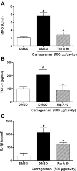
Carrageenan (7.69 ± 0.80 U/ml of peritoneal exudate) adminis-tration produced a significant (p< 0.05) increase in MPO activity in the peritoneal exudates, which is an important marker of neutr-ophilic infiltration (Fig. 6A). Pretreatment with riparin A (10 mg/ kg) reduced MPO (2.80 ± 0.75 U/ml of peritoneal exudate) levels to values similar to those in the control group (2.14 ± 0.24 U/ml of peritoneal exudate).
3.6. Effect of riparin A on TNF-
a
and IL-1blevelsAs shown inFig. 6, carrageenan administration increased TNF-
a
(208.7 ± 39.2 pg/ml; Fig. 6B) and IL-1b (1073.0 ± 180.1 pg/ml; Fig. 6C) levels in peritoneal fluid, compared to the corresponding levels in the control group (100.7 ± 15.5 pg/ml and 176.9 ± 9.5 pg/ml, respectively). Pretreatment with riparin A (10 mg/kg) sig-nificantly (p< 0.05) reduced TNF-
a
(90.4 ± 32.8 pg/ml) and IL-1b (635.2 ± 65.9 pg/ml) levels.Fig. 6.Riparin A reduces myeloperoxidase (MPO) activity and cytokine (TNF-aand IL-1b) levels in carrageenan-induced peritonitis. Mice were pretreated with 2% DMSO or riparin A (10 mg/kg, i.p.). After 30 min carrageenan (500lg/cavity) was administrated, and analyses realized after 4 h. (A) MPO activity; (B) TNF-a; and 6C: IL-1b. Each column represents the mean ± SEM of 5–6 animals per group.#p< 0.05
versus control group;⁄p< 0.05 versuscarrageenan group. One-way analysis of
3.7. Effect of riparin A on carrageenan-induced leukocyte rolling and adhesion
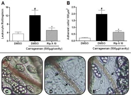
Fig. 7shows that riparin A (10 mg/kg) administration signifi-cantly (p< 0.05) reduced leukocyte rolling and adherence in the mesentery microcirculation, compared to those in the carrageenan group (5.09 ± 1.14versus 12.27 ± 2.33 leukocyte rolling (Fig. 7A) and 0.65 ± 0.09versus 1.98 ± 0.29 adhering leukocytes (Fig. 7B)). In addition,Fig. 7shows a typical video image of the control group (Panel C); the leukocyte-endothelium interaction increased after administration of carrageenan (Panel D) and decreased with ripa-rin A (Panel E) treatment.
3.8. Effect of riparin A on glutathione (GSH) levels
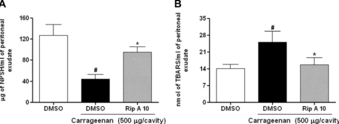
Fig. 8A shows that carrageenan (44.2 ± 8.8
l
g/ml) administra-tion reduced GSH levels, compared to those in the control group (127.6 ± 20.1l
g/ml). Furthermore, pretreatment with riparin A (10 mg/kg) reverted the effect of the carrageenan, significantly (p< 0.05) increasing GSH (95.6 ± 9.7l
g/ml) levels.3.9. Effect of riparin A on thiobarbituric acid reactive substance (TBARS) levels
Carrageenan (25.1 ± 4.4 nmol/ml) administration promoted a significant (p< 0.05) increase in TBARS levels in the peritoneal exu-date, compared to that in the control group (12.1 ± 1.7 nmol/ml). However, pretreatment with riparin A (10 mg/kg) inhibits this
effect of carrageenan, significantly reducing TBARS
(15.7 ± 2.8 nmol/ml) levels to values similar to those in the control group (Fig. 8B).
3.10. Evaluation of gastric damage
This experiment was performed for evaluate the gastric toxicity of riparin A and compare with indomethacin (20 mg/kg, standard drug).Fig. 9shows that indomethacin promoted gastric damage (9.84 ± 2.37 mm,Fig. 9A) and increased MPO activity (8.01 ± 1.53 UMPO/mg of gastric tissue,Fig. 9B) significantly (p< 0.05), when compared to the DMSO group (0.95 ± 0.36 UMPO/mg of tissue Fig. 9B). In addition, the treatment with riparin (10 or 100 mg/ kg) did not promote gastric damage or change in MPO activity (0.41 ± 0.16 and 0.44 ± 0.30 UMPO/mg of tissue, respectively, Fig. 9B).
4. Discussion
Natural products, particularly those obtained from medicinal plants, are commonly used for the treatment of inflammatory dis-eases and are considered an important source of molecules with potential therapeutic efficacy[20]. In this study, we show that rip-arin A, a compound obtained fromA. riparia, could reduce inflam-matory responses in classic experimental models of inflammation; the underlying mechanism involves inhibition of neutrophil migra-tion to the damaged site, accompanied by the reducmigra-tion of proin-flammatory cytokine levels and oxidative stress.
Initially, carrageenan-induced paw edema model, which is widely used for the evaluation of anti-inflammatory effects of mol-ecules with pharmacological potential [21,22], was established. The method was biphasic, with the initial phase involving the release of inflammatory mediators such as histamine and seroto-nin, followed by intense infiltration of polymorphonuclear (PMN) cells and a peak occurring at the 4th hour after carrageenan admin-istration[23]. The second phase was observed after 72 h, and was characterized by the release of TNF-
a
[15]and monocyte migration Fig. 7.Riparin A decrease rolling and adhesion of leukocyte in the mesentery microcirculation mice. The animals were initially treated with 2% DMSO or riparin A (10 mg/kg, i.p.) and then administrated carrageenan (500lg/cavity). The rolling (A) and adhesion (B) of leukocytes were evaluated in the mesentery microcirculation by intravital microscopy, 4 h after inflammatory stimuli. Video photomicrographs displaying leukocyte-endothelium interaction is shown in Panel C, D and E. Panel C: 2% DMSO group, show leukocyte rolling basal. Panel D: carrageenan group, showing leukocyte rolling significantly increased on the vascular endothelium. Panel E: riparin A (10 mg/kg, i.p.) group, showing significantly reduction of leukocyte rolling in the mesenteric microvasculature. Each column represents the mean ± SEM of 5–6 animals per group.#p< 0.05[24]. Our results showed that riparin A inhibited carrageenan paw edema in all time points evaluated. This suggested that the anti-inflammatory effects are associated with the inhibition of vascular events, such as the reduction of release and/or production of vaso-active mediators and inhibition of cell migration.
In order to prove this mechanism, paw edema was induced using different agents (compound 48/80, histamine, and seroto-nin). Compound 48/80 administration is known to promote edema through potent induction of mast cell degranulation, followed by a massive release of vasoactive amines, such as histamine and sero-tonin, which greatly contributes to the vascular events of the inflammatory response[25,26]. In this study, riparin A markedly reduced compound 48/80-induced paw edema, which could be associated with downregulation of mast cell degranulation, through the potential stabilization of its outer membrane[6]. This point was confirmed by the reduction of histamine- and serotonin-induced paw edema. In addition, this corroborates with the inhib-itory effect of riparin A on the early phase of the carrageenan-induced paw edema.
The acute inflammatory response also consists of a cellular component, primarily represented by neutrophils [27]. In this study, using histopathological analysis, we demonstrated that cell recruitment in the inflamed paw tissue 4 h after carrageenan administration markedly reduced in mice pretreated with riparin A. Furthermore, our results showed that riparin A inhibited leuko-cyte and neutrophil migration in the carrageenan-induced perito-nitis. Corresponding to these findings, riparin A also reduced MPO activity in the peritoneal fluid. MPO is an enzyme present in the azurophilic granules of neutrophils, which is released during
the process of migration into the inflammatory site[6,28]and is determined to be an index of neutrophil infiltration[15].
Several studies have consistently shown that activated neutro-phils may synthesize and release an arsenal of molecules, such as cytokines TNF-
a
, IL-1b, and IL-6, which can subsequently activate new neutrophils. In addition, they can activate endothelial cells that lead to neutrophil interaction culminating in the sequential events of rolling, adhesion, and transmigration[29,30]. Cytokines are protein messengers released in the circulation, or directly into the tissue, and are responsible for cell localization through interac-tion with specific receptors[31]. Overproduction, or inappropriate production, of proinflammatory cytokines can result in conditions such as septic shock [32] and rheumatoid arthritis [33], which involve tissue destruction and exacerbation of inflammation.Similar to previous studies, our study showed that carrageenan administration induced an increase in TNF-
a
and IL-1blevels in the peritoneal fluid[6,14]. Supporting the effects of riparin A in the experimental models above, which demonstrated inhibition of cell migration to the inflamed site, we observed that pretreatment with riparin A promoted a reduction of cytokine levels to levels similar to those in the control group. Considering that the effects of carra-geenan administration on neutrophil migration significantly atten-uated by pretreatment with riparin A, we suggesting that this compound can modulate the process of leukocyte recruitment.Next, to elucidate this mechanism we pretreated mice with rip-arin A and evaluated carrageenan-induced leukocyte rolling and adhesion in mesenteric postcapillary venules by using an intravital microscopy system. The neutrophil migration from the circulation to the site of inflammation is controlled by interactions with the Fig. 8.Riparin A reverse glutathione (GSH) and TBARS levels in carrageenan-induced peritonitis. Mice were pretreated with 2% DMSO or riparin A (10 mg/kg, i.p.). After 30 min carrageenan (500lg/cavity) was administrated, and biochemical parameters measured 4 h after carrageenan injection. (A) GSH levels; (B) TBARS levels. Each column represents the mean ± SEM of 5–6 animals per group.#p< 0.05versuscontrol group;⁄p< 0.05versuscarrageenan group. One-way analysis of variance (ANOVA) followed by
Newman–Keuls test.
vascular endothelium[34]. Interestingly, our results demonstrated that riparin A significantly inhibited leukocyte rolling and adhe-sion. Riparin A treatment also decreased TNF-
a
and IL-1blevels. Considering the important role of these cytokines in neutrophil recruitment and their involvement of mechanism dependent of molecular events among leukocytes and endothelial cells, our results suggest that riparin A reduces the inflammatory response by mechanisms dependent, at least in part, on the inhibition of pro-inflammatory cytokine production at inflammatory sites, which culminate in a decrease in leukocyte rolling and adhesion in the endothelial cells.Neutrophils represent the body’s primary line of defense in the inflammatory process [35]. They play an important role in the migration events to the inflammation site by releasing proteases contained within their azurophilic granules, such as myeloperoxi-dase, elastase, and proteinase-3[36]. They can generate reactive oxygen species (ROS) that cause toxic tissue damage that has been observed in inflammatory diseases[37].
Our results showed a decrease in GSH concentration and an increase in TBARS levels in the peritoneal fluid in carrageenan-induced peritonitis. This confirms that neutrophil migration to the inflamed site produces reactive metabolites that exacerbate the inflammatory process [6,38]. On the other hand, mice pre-treated with riparin A showed a marked reduction in these param-eters. In cases of ROS involvement, the GSH levels decreased, while the TBARS concentration, an important marker of lipid peroxida-tion, increased. These data reinforce the results obtained by other authors that show that riparin A has direct antioxidant activity, being capable of decreasing the levels of free radicals even in low concentrations[39,40]. This effect, probably is related to the pres-ence of unstable hydrogen in riparin A that is captured by the free radicals, inactivating them.
Nonsteroidal anti-inflammatory drugs (NSAIDs) are widely pre-scribed for treatment of pain and inflammation. However, these drugs are associated with low efficacy and specificity and impor-tant side effects, such as bleeding and perforation in gastrintestinal
tissues[41]. For this reason, our study is conducted to identify a novel therapeutic option to develop and introduce new drugs with greater safety and efficacy. We confirm that indomethacin pro-moted gastric damage and increased MPO activity. Therefore, at the tested doses, the treatment with riparin A did not promote gas-tric damage. The present data provide evidence that riparin A is devoid of side effects on gastric mucosa.
This study demonstrated the anti-inflammatory potential of riparin A, a compound obtained fromA. riparia, in experimental models of acute inflammation. This compound promotes its effect through mechanisms dependent on the inhibition of vascular events and neutrophil migration to the damaged site. In addition, riparin A reduced the production of proinflammatory cytokines (TNF-
a
and IL-1b) and oxidative stress. Fig. 10summarizes the modulatory effects of riparin A on inflammatory response. In con-clusion, the results of this study suggest that riparin A is a major target for future therapeutic approaches for the treatment of inflammatory diseases.Conflict of Interest
The authors declare that there are no conflicts of interest.
Transparency Document
TheTransparency documentassociated with this article can be found in the online version.
Acknowledgments
The authors gratefully acknowledge the financial support from National Counsel of Technological and Scientific Development – CNPq (Brazil) and Research Foundation for the State of Piauí – FAPEPI.
References
[1]R. Medzhitov, Origin and physiological roles of inflammation, Nature 454 (2008) 428–435.
[2]L.J. Quintans-Júnior, A.G. Guimarães, M.T. Santana, B.E.S. Araújo, F.V. Moreira, L.R. Bonjardim, A.A.S. Araújo, J.S. Siqueira, A.R. Antoniolli, M.A. Botelho, J.R.G.S. Almeida, M.R.V. Santos, Citral reduces nociceptive and inflammatory response in rodents, Rev. Bras. Farmacogn. 21 (2011) 497–502.
[3]M.S. Butler, The role of natural product chemistry in drug discovery, J. Nat. Prod. 67 (2004) 2141–2153.
[4]U.P. Albuquerque, N. Hanazaki, As pesquisas etnodirigidas na descoberta de novos fármacos de interesse médico e farmacêutico: fragilidades e perspectivas, Rev. Bras. Farmacogn. 16 (2006) 678–689.
[5]D.A. Valério, T.M. Cunha, N.S. Arakawa, H.P. Lemos, F.B. Da Costa, C.A. Parada, S.H. Ferreira, F.Q. Cunha, W.A. Verri Jr., Anti-inflammatory and analgesic effects of then sesquiterpene lactone budlein A in mice: inhibition of cytokine production-dependent mechanism, Eur. J. Pharmacol. 562 (2007) 155–163. [6]V.G. Silva, R.O. Silva, S.R.B. Damasceno, N.S. Carvalho, R.S. Prudêncio, K.S.
Aragão, M.A. Guimarães, S.A. Campos, L.M. Véras, M. Godejohann, J.R.A. Leite, A.L.R. Barbosa, J.V. Medeiros, Anti-inflammatory and antinociceptive activity of epiisopiloturine, an imidazole alkaloid isolated fromPilocarpus microphyllus, J. Nat. Prod. 76 (2013) 1071–1077.
[7]J. Gershenzon, N. Dudareva, The function of terpene natural products in the natural world, Nat. Chem. Biol. 3 (2007) 408–414.
[8]A.M.R. Carvalho, N.F.M. Rocha, L.F. Vasconcelos, E.R.V. Rios, M.L. Dias, M.I. Silva, M.M. França Fonteles, J.M. Filho, S.J. Gutierrez, F.C. Sousa, Evaluation of the anti-inflammatory activity of riparin II (O-methil-N-2-hidroxi-benzoyl tyramine) in animal models, Chem. Biol. Interact. 205 (2013) 165–172. [9]R.M.R. Catão, J.M.B. Barbosa-Filho, E.O. Lima, M.S.V. Pereira, M.A.R. Silva, T.A.
Arruda, R.M.P. Antunes, Evaluation of the antimicrobial activity and biological effect by riparins about elimination the resistance of drugs in samples of Staphylococcus aureus, RBAC 42 (2010) 9–14.
[10]G.B.L. Nunes, P.R. Policarpo, L.M. Costa, T.G. Silva, G.C.G. Militão, C.A. Câmara, J.M. Barbosa Filho, S.J. Gutierrez, M.T. Islam, R.M. Freitas,In vitroantioxidant and cytotoxic activity of synthetic compounds derived from riparins, Molecules 19 (2013) 4595–4607.
[11]J.M. Barbosa-Filho, E.C. Silva, J. Bhattacharyya, Synthesis of several new phenylethylamides of substituted benzoic acids, Quim. Nova 13 (1990) 332– 334.
[12]F.C.F. Sousa, C.T.V. Melo, A.P. Monteiro, V.T. Lima, S.J.C. Gutierrez, B.A. Pereira, Antianxiety and antidepressant effects of riparin III fromAniba riparia(Nees) Mez (Lauraceae) in mice, Pharmacol. Biochem. Behav. 78 (2004) 27–33. [13]R.T. Morrison, R.N. Boyd, Organic Chemistry, sixth ed., Prentice Hall, New
Jersey, 1992.
[14]L.S. Chaves, L.A.D. Nicolau, R.O. Silva, F.C. Barros, A.L. Freitas, K.S. Aragão, R.A. Ribeiro, M.H. Souza, A.L. Barbosa, J.V. Medeiros, Anti-inflammatory and antinociceptive effects in mice of a sulfated polysaccharide fraction extracted from the marine red algaeGracilaria caudata, Immunopharmacol. Immunotoxicol. 35 (2013) 93–100.
[15]I. Posadas, M. Bucci, F. Roviezzo, A. Rossi, L. Parente, L. Sautebin, G. Cirino, Carrageenan-induced mouse paw oedema is biphasic, age-weight dependent and displays differential nitric oxide cyclooxygenase-2 expression, Br. J. Pharmacol. 142 (2004) 331–338.
[16]Z.B. Fortes, S.P. Farsky, M.A. Oliveira, J. Garcia-Leme, Direct vital microscopic study of defective leukocyte–endothelial interaction in diabetes mellitus, Diabetes 40 (1991) 1267–1273.
[17]P.P. Bradley, D.A. Priebat, R.D. Christensen, G. Rothstein, Measurement of cutaneous inflammation: estimation of neutrophil content with an enzyme marker, J. Invest. Dermatol. 78 (1982) 206–209.
[18]J. Sedlak, R.H. Lindsay, Estimation of total, protein-bound, and nonprotein sulfhydryl groups in tissue with Ellman’s reagent, Anal. Biochem. 25 (1968) 192–205.
[19]M. Mihara, M. Uchiyama, Determination of malonaldehyde precursor in tissues by thiobarbituric acid test, Anal. Biochem. 86 (1978) 271–278. [20]J.H. Yim, O.H. Lee, U.K. Choi, Y.C. Kim, Antinociceptive and anti-inflammatory
effects of ethanolic extracts ofGlycine max(L.) Merr andRhynchosia nulubilis seeds, Int. J. Mol. Sci. 10 (2009) 4742–4753.
[21]G. Trevisan, M.F. Rossato, C. Hoffmeister, L.G. Müller, C. Pase, M.M. Córdova, F. Rosa, R. Tonello, B.S. Hausen, A.A. Boligon, R.N. Moresco, M.L. Athayde, M.E.
Burguer, A.R. Santos, J. Ferreira, Antinociceptive and antiedematogenic effect of pecan (Carya illinoensis) nut shell extract in mice: a possible beneficial use for a product of the nut industry, J. Basic Clin. Physiol. Pharmacol. 27 (2014) 1– 10.
[22]M. Fezai, L. Senovilla, M. Jemaà, M. Ben-Attia, Analgesic, anti-inflammatory and anticancer activities of extra virgin olive oil, J. Lipid. 2013 (2013) 1–7. [23]C.J. Morris, Carrageenan-induced paw edema in the rat and mouse, Methods
Mol. Biol. 225 (2003) 115–121.
[24]P. Ghiara, M. Bartalini, A. Tagliabue, D. Boraschi, Anti-inflammatory activity of IFN-beta in carrageenan-induced pleurisy in the mouse, Clin. Exp. Immunol. 66 (1986) 606–614.
[25]R.O. Silva, F.B. Sousa, S.R. Damasceno, N.S. Carvalho, V.G. Silva, F.R. Oliveira, D.P. Sousa, K.S. Aragão, A.L. Barbosa, R.M. Freitas, J.V. Medeiros, Phytol, a diterpene alcohol, inhibits the inflammatory response by reducing cytokine production and oxidative stress, Fundam. Clin. Pharmacol. 28 (2014) 455–464. [26] Subhashini, P.S. Chauhan, S. Kumari, D. Dash, R. Singh, Curcumin inhibits compound 48/80 induced systemic anaphylaxis, Am. J. Life Sci. 1 (2013) 165– 170.
[27]M.N. Ajuebor, A. Singh, J.L. Wallace, Cyclooxygenase-2-derived prostaglandin D(2) is an early anti-inflammatory signal in experimental colitis, Am. J. Physiol. Gastrointest. Liver Physiol. 279 (2000) 238–244.
[28]S.J. Klebanoff, Myeloperoxidase, Proc. Assoc. Am. Physicians 111 (1999) 383– 389.
[29]S. Fujishima, A.R. Hoffman, T. Vu, K.J. Kim, H. Zheng, D. Daniel, Y. Kim, E.F. Wallace, J.W. Larrick, T.A. Raffin, Regulation of neutrophil interleukin 8 gene expression and protein secretion by LPS, TNF-alpha, and IL-1 beta, J. Cell. Physiol. 154 (1993) 478–485.
[30] J.M. Hallett, A.E. Leitch, N.A. Riley, R. Duffin, C. Haslett, A.G. Rossi, Novel pharmacological strategies for driving inflammatory cell apoptosis and enhancing the resolution of inflammation, Trends Pharmacol. Sci. 29 (2008) 250–257.
[31]D. Figarella-Branger, M. Civatte, C. Bartoli, J.F. Pellissier, Cytokines, chemokines and cell adhesion molecules in inflammatory myopathies, Muscle Nerve 28 (2003) 659–682.
[32]F.Q. Cunha, J. Assreuy, D.W. Moss, D. Rees, L.M. Leal, S. Moncada, M. Carrier, C.A. O’Donnell, F.Y. Liew, Differential induction of nitric oxide synthase in various organs of the mouse during endotoxaemia: role of TNF-alpha and IL-1-beta, Immunology 81 (1994) 211–215.
[33]I.B. McInnes, G. Schett, Cytokines in the pathogenesis of rheumatoid arthritis, Nat. Rev. Immunol. 7 (2007) 429–442.
[34]D. Dal Secco, A.P. Moreira, A. Freitas, J.S. Silva, M.A. Rossi, S.H. Ferreira, F.Q. Cunha, Nitric oxide inhibits neutrophil migration by a mechanism dependent on ICAM-1: role of soluble guanylate ciclase, Nitric Oxide 15 (2006) 77–86. [35]C. Summers, S.M. Rankin, A.M. Condliffe, N. Singh, A.M. Peters, E.R. Chilvers,
Neutrophil kinetics in health and disease, Trends Immunol. 31 (2010) 318– 324.
[36]B. Korkmaz, M.S. Horwitz, D.E. Jenne, F. Gauthier, Neutrophil elastase, proteinase 3, and cathepsin G as therapeutic targets in human diseases, Pharmacol. Rev. 62 (2010) 726–759.
[37]H.L. Wright, R.J. Moots, R.C. Bucknall, S.W. Edwards, Neutrophil function in inflammation and inflammatory diseases, Rheumatology 49 (2010) 1618– 1631.
[38]H. Uzkeser, E. Cadirci, Z. Halici, F. Odabasoglu, B. Polat, T.N. Yuksel, S. Ozaltin, F. Atalay, Anti-inflammatory and antinociceptive effects of salbutamol on acute and chronic models of inflammation in rats: involvement of an antioxidant mechanism, Mediators Inflamm. 2012 (2012) 1–10.
[39]G.B.L. Nunes, L.M. Costa, S.J.C. Gutierrez, P. Satyal, R.M. Freitas, Behavioral tests and oxidative stress evaluation in mitochondria isolated from the brain and liver of mice treated with riparin A, Life Sci. 121 (2015) 57–64.
[40] G.B.L. Nunes, P.R. Policarpo, L.M. Costa, T.G. Silva, G.C.G. Militão, C.A. Camara, J.M. Barbosa Filho, S.J.C. Gutierrez, M.T. Islam, R.M. Freitas, In vitro antioxidant and cytotoxic activity of some synthetic riparin-derived compounds, Molecules 19 (2014) 4595–4607.