R E S E A R C H A R T I C L E
Physicochemical and rheological characterization of
different Carnoy
0
s solutions applied in oral and
maxillofacial surgery
Francisco Samuel Rodrigues Carvalho
1,2| Victor Pinheiro Feitosa
3|
Said Gonçalves da Cruz Fonseca
4| Thiago Dias de Vasconcelos Araújo
5|
Eduardo Costa Studart Soares
6| Cristiane Sá Roriz Fonteles
7| Thyciana Rodrigues Ribeiro
7|
Fábio Wildson Gurgel Costa
81
Division of Oral and Maxillofacial Surgery, Federal University of Ceará, Fortaleza, Brazil
2
Division of Oral and Maxillofacial Morphology and Oral and Maxillofacial Surgery, UNIFOR, Fortaleza, Brazil
3
Paulo Picanço School of Dentistry, Fortaleza, Ceará, Brazil
4Division of Pharmacology Sciences, Federal
University of Ceará, Fortaleza, Brazil
5
Division of Pharmacology, Federal University of Ceará, Fortaleza, Brazil
6Division of Oral and Maxillofacial
Surgery, Walter Cantídio University Hospital, Federal University of Ceará, Fortaleza, Brazil
7
School of Dentistry, Federal University of Ceará, Fortaleza, Brazil
8Division of Oral Radiology, Walter
Cantídio University Hospital, Federal University of Ceará, Fortaleza, Brazil
Correspondence
Francisco Samuel Rodrigues Carvalho, Division of Oral and Maxillofacial Surgery, Federal University of Ceará, Rua Monsenhor Furtado, Rodolfo Teófilo, Fortaleza, Ceará CEP: 60.430‐350, Brazil. Email: [email protected]; [email protected]
Abstract
Carnoy0s solution has been widely undertaken as adjunctive therapy for benign
odontogenic tumors. Although its outcomes were highly investigated, little information is available regarding its mechanism of action and the role of chlo-roform in composition. The aim of this study was to characterize Carnoy0s
solu-tion (with and without chloroform) in order to survey its physicochemical and pharmacological properties. UV/Vis spectroscopy, Raman microspectroscopy, viscosity assessment, pH analysis, and ion (Fe3+) concentration were studied. All solutions were prepared by the same operator and assayed at each 7 days within a 4‐week period. All solutions depicted acidic pH whereas the viscosity was increased by the addition of chloroform. Starting in the seventh day after preparation, solutions with chloroform showed iron precipitation when nonfiltered. Carnoy0s solution without chloroform was stable during the survey
period as evidenced by Raman spectroscopy. In conclusion, Carnoy0s solution
possesses an acidic characteristic, it is stable when stored at room temperature, and precipitates iron salts when in the presence of chloroform in a nonfiltered solution. The chloroform increases the viscosity of the solution and makes its pH slightly more acidic when compared to the Carnoy0s solution without
chlo-roform. Raman spectroscopy favored the evaluation of components0stability in
solutions by using the spectra analysis and the established correlations between the peaks in different periods.
K E Y W O R D S
Carnoy0s solution, chloroform, oral surgery, Raman spectroscopy
1
|
I N T R O D U C T I O N
The maxillofacial region may be affected by a variety of benign lesions but locally invasive with a high incidence
of recurrences such as ameloblastoma, odontogenic myx-oma, and keratocystic odontogenic tumor. Conservative treatment might lead to recurrence, and radical approach could result in aesthetic‐functional sequels.[1,2] Thus,
DOI: 10.1002/jrs.5227
complementary therapies to the surgery are largely employed in the literature in order to reduce recurrence rate and potential damages.[2]Within the different modal-ities of surgery treatments, there is no method associated with zero recurrence rate.[3] Regarding these therapies, Carnoy0s solution has been highlighted principally for
keratocystic odontogenic tumor,[1–17] once lowest recur-rence rates were found for surgery resection method followed by enucleation with adjunctive use of Carnoy0s
solution.[3]
In 1886, Jean Baptiste Carnoy improved the fixation solution of Clarke (1851) for botanic investigations, thereby proposing a mixture of absolute ethanol, chloro-form, and glacial acetic acid in 6:3:1 ratio, respectively. This solution is a fixation agent of rapid activity, useful for visualization of cell core, proteins, and glycogen but unsuitable for lipids.[18,19] After almost half century from this invention, the first use in surgery occurred by Zollinger and Moritz.[20]Authors surveyed the effect of different necrobiotic agents for cysts in dog brains. Carnoy0s solution with ferric chloride addition was
assessed in order to enclose hemostatic features for the treatment.[20] This combination provided moderate tis-sue penetration, rapid local fixation of cystic surround-ing cells and optimal hemostasis. In 1933, Cutler and Zollinger employed this solution for clinical treatment of patients with cysts and fistulas.[21] Voorsmit et al.[4] published the first report of the adjunctive usage of Carnoy0s solution for surgical treatment of odontogenic
keratocyst, which presented, until that period, high inci-dence of recurrence.[4] According to the authors, the application of Carnoy0s solution is based on three
aspects for such odontogenic lesions: Cyst epithelium is fragile and its remnants might remain in bone cavity; the occurrence of remaining microcysts or epithelium islands, which were on the walls of initial cyst; and development of new keratocysts arising from derivations from basal layer of oral epithelium.[4] Therefore, Carnoy0s solution warrants reduction of lesions, less
fra-gility, improvement of complete enucleation, and ade-quate local hemostasis.[22]
A wide variety of treatments has been suggested for the therapy of odontogenic tumors, encompassing surgi-cal approaches (enucleation, curettage, peripheral osteotomy, cryosurgery, etc.) isolated or combined with conventional Carnoy0s solution or the modified one
(with-out chloroform).[4,12,23–25]It has been demonstrated that the usage of Carnoy0s solutions (with and without
chloro-form) induces lower recurrence rate when compared with further treatments.[12,24–26]
In 1992, American Food and Drug Administration (FDA) Compliance Policy Guide (chapter 4 subchapter 460)[27] prohibited pharmaceutical compositions of
therapeutic agents containing chloroform, as considered a carcinogenic compound.[24,25] Indeed, this guidance promoted the replacement of chloroform and Carnoy0s
solution changed to the composition: 9‐ml absolute eth-anol, 3‐ml acetic acid, and 1‐g ferric chloride.[28] Never-theless, both Carnoy0s solutions (with and without
chloroform) are currently used in clinical practice as adjunctive for several surgical interventions, and the principal parameters evaluated are the recurrence of lesions.[1–17,24] However, two aspects of such agent were not previously surveyed: (a) physicochemical properties of the solution and (b) effects of storage period on the features and formation of by‐products.
In a recent systematic review, Al‐Moraissi et al.[3] found in 2,287 non‐syndromic keratocystic odontogenic tumors recorded in 35 studies that relapse rates associ-ated with treatment of these lesions were enucleation alone (23.1%), enucleation with peripheral ostectomy (17.4%), enucleation and Carnoy0s solution (11.5%),
enucleation plus liquid nitrogen cryotherapy (14.6%), marsupialization alone (32.3%), decompression followed by residual cystectomy (14.6%), and resection (8.4%). Indeed, this highlights the role of Carnoy0s solution as
one of the main less invasive treatments.[3]
Physicochemical characterization of modified Carnoy0s solution has never been realized up to this
inves-tigation. Although Puchtler et al.[29]had investigated the chemical components of Carnoy0s solution in 1968, that
study did not consider the presence of ferric chloride as well as the presence/absence of chloroform, substances well known by maxillofacial surgeons.[25,26]The isolated compounds featured in the present investigation depicted fundamental aspects to perceive the mechanism of action of Carnoy0s solution in the treatment of maxillary
aggres-sive odontogenic lesions.
Ethanol (CH2H5OH) presents the properties of a
coagulative fixative that denature insoluble proteins in water at room temperature and extract phospholipids from cells without affecting the carbohydrates,[29,30] principally when in major concentration in the solution (described as solvent). Chloroform (CHCl3) enhances
and accelerates ethanol penetration in the tissue. It is a compound with lipophilic nature that improves the dehydration of tissues by dissolving the lipids in mem-branes, favoring the action of ethanol in the pro-cess.[31–33] Indeed, such feature may likely attain lower efficacy of Carnoy0s solution without chloroform. This
corroborates with the findings of Dashow0s studies[17,24]
that demonstrated higher recurrence rates in patients submitted to adjunct therapy with Carnoy0s solution
allow the use of Carnoy0s solution with chloroform for
clinical trials, because, in histology, chloroform is a sol-vent for lipids relatively inert in other tissues.[29] The limit of occupational exposure to chloroform in an 8‐hr shift is 2 ppm (9.9 mg/m3), and for buffered neutral formalin, it is 1 ppm (1.2 mg/m3). Moreover, formalin is known as a carcinogenic agent, whereas chloroform is classified as a likely carcinogenic compound.[32] Such aspects may justify the use of chloroform within Carnoy0s solution in relation to the possibility of the
reduction in recurrence rates[26] and lower occupational risk.[32]
Glacial acetic acid (CH3COOH) penetrates rapidly in
tissues and has the role of preserving chromosomes through the coagulation of nucleic acids.[19]Furthermore, this compound brakes the cross‐links between proteins and releases hydrophilic radicals, swelling the tissue, thereby preventing the excessive shrinking and stiffening promoted by ethanol action.[34]
Ferric chloride (FeCl3) is a brownish chemical agent
with acidic and protein coagulating properties, which pro-vides its characteristics as strong hemostatic agent. By the contrary of further known hemostatic agents, ferric chlo-ride displays its effects through the chemical reaction with blood, regardless the action of normal hemostatic system. As most topical hemostatic agents are employed to control superficial bleeding, requiring normal hemostatic func-tions to achieve the effects, the addition of a local hemo-static agent that exempts the need for a normal hemostatic system may indeed improve the coagulative effect.[35]
In 1928, the physicist Sir Chandrasekhara Venkata Raman observed and interpreted the phenomenon of inelastic scattering of light through the matter using a microspectroscopy technique, currently denominated as Raman effect.[36]Raman spectroscopy is a valuable ana-lytic technique able to assay the chemical composition of a variety of samples, such as fluids, cells, and tissues. Furthermore, it may detect a molecular “fingerprint”of different substrates useful for diagnosis and assessment of new therapies, thereby providing qualitative and quan-titative information in a noninvasive high‐resolution (1μm) experiment.[36,37]
In this regard, the aim of this study was to characterize two formulations of Carnoy0s solution with ferric chloride
(with and without chloroform) employed in oral maxillo-facial surgery, in order to survey their physicochemical and pharmacological properties, and further characterize the Raman spectra of these solutions along the time. To date, no investigations in our knowledge have character-ized Carnoy0s solution or demonstrated physicochemical
alterations of such solution in presence or absence of chloroform over time.
2
|
M A T E R I A L S A N D M E T H O D S
2.1
|
Preparation of solutions
The Carnoy0s solutions used in this investigation were
prepared by the same trained pharmaceutical, by using 6‐ml absolute ethanol, 3‐ml chloroform, 1‐ml acetic acid and 1‐g FeCl3.6H2O (ferric chloride hexahydrate) to
obtain an approximately 10‐ml final solution.[4,21] To obtain Carnoy0s solution without chloroform, the 3 ml of
chloroform was replaced by 3‐ml absolute ethanol.[17,23,28] The solutions produced, as described above, have supersaturated concentration relative to the ferric chlo-ride hexahydrate salt. We proceeded with the filtration of the samples using a 0.45‐μm syringe filter, which
allowed to obtain solutions of Carnoy0s with or without
chloroform filtered. Thus, four types of solution were ana-lyzed in the present study: unfiltered Carnoy0s solution
with chloroform, filtered Carnoy0s solution with
chloro-form, unfiltered Carnoy0s solution without chloroform,
and filtered Carnoy0s solution without chloroform.
2.2
|
Pharmacological characterization
(density, pH, viscosity, and Fe
3+concentration)
The pharmacological properties of Carnoy0s solutions
with and without chloroform determined were mass den-sity and relative denden-sity (pycnometer, Prolab, Materials for Laboratory, São Paulo, Brazil), viscosity (viscometer, Viscotester 6L Thermo Haake—Thermo Fisher Scientific Inc., Germany), and pH (Benchtop pHmeter DM23, Digimed, Campo Grande, Brazil).[38] Determination of Fe3+concentration was undertaken by means of ultravio-let/visible (UV/Vis) spectroscopy (GENESYS 20 UV/Vis Spectrophotometer, Thermo Fisher Scientific Inc., Germany) by the reaction with potassium thiocyanate in aqueous solutions.[39]
2.3
|
Raman spectroscopy
Spatial distribution of organic and inorganic components was evaluated through the relative intensities of peaks obtained in a Raman microspectrophotometer (Xplora, Horiba Scientific, Paris, France). An argon laser with 638‐nm wavelength and 3.2‐mW power were used along with 10× magnification lens (Olympus American Inc., London, UK) to obtain the focus. Solutions were adapted in optical buckets for standardization. Raman spectra were attained in the range between 200 and 3,500 cm−1
spectra was conducted in the same temperature and pres-sure (22 °C and 1 atm) conditions. Ten milliliter of each solution was surveyed.
All components were assayed separately as well as possible combinations, in order to facilitate the process of identifying the peaks in the spectrum of the solution. Ferric chloride hexahydrate was dissolved in absolute ethanol, as the common solvent. All analyses were realized once a week for 4 weeks in order to detect possible alterations over time. The Raman spectra were acquired in LabSpec 6 software (Horiba) and further manipulated in OriginPro 9.0 software (OriginLab Co., Northampton, USA). They were processed with baseline correction, smoothing by polynomial method (Savitzk‐ Golay) and peaks position/intensity identification by Gaussian and Lorentzian methods to ensure characteri-zation and deconvolution of graphs.[40]
2.4
|
Statistical analysis
The data were tabulated in Microsoft Excel®, and the rela-tive expressions were calculated as a percentage of the height, width, and area of each peak in relation to the total of existing peaks. Area/width ratios were used to standard-ize these measurements.After that, the values obtained were exported to the Statistical Packing for Social Sciences (SPSS®) version 17.0 software with a 95% confidence inter-val for the design of the multivariate analysis model (ANOVA‐3‐way followed by the posttest Bonferroni) to evaluate the interaction between the time, filtration, and
chloroform factors in the heights, widths, areas, and rela-tive area/width ratio of each of the observed peaks. For the graphical representation of significant associations, the means and standard deviations were depicted.
3
|
R E S UL T S
3.1
|
Pharmacological characterization
Relative density and viscosity of Carnoy0s solution were
influenced by the presence of chloroform, with higher den-sity and viscoden-sity than water when with chloroform. In its absence, lower viscosity was attained (Table 1). The pH of solutions was highly acidic during the evaluation. Carnoy0s
solution with chloroform (pH = 0.12) was more acid than without chloroform (p= .27). Turbidity was more observed in the presence of chloroform than without, starting from the seventh day after manipulation. Iron precipitation was detected in all nonfiltered solutions.
The assessment of Fe3+concentration by UV/Vis spec-troscopy exhibited a slight increase in ferric ions concen-tration over the 4 weeks survey. Besides, higher concentrations were attained in nonfiltered solutions (Table 2).
3.2
|
Raman spectroscopy
The spectra of isolated compounds are described in Figure 1. They were organized in order to identify peaks of each component of Carnoy0s solutions with and
with-out chloroform. The relative intensity of peaks was established in relation to the major peak of the com-pound, thereby determining very weak (vw), weak (w), medium (m), strong (s), and very strong (vs) peaks as well as shoulders (Table 3). Based on the identified peaks of isolated solutions as described, one may highlight charac-teristic peaks of the components in solutions with and without chloroform, depicted in Figure 2.
Thus, in Carnoy0s solutions, the main peaks were
absolute ethanol (433, 879, 1,047, 1,091, 1,273, and TABLE 1 Characterization of viscosity of Carnoy0s solutions with
and without chloroform in relation to water
Mean spreading time (s)
Density (g/ml)
Viscosity (mPa·s)
Water 7.24 0.99704 1
Carnoy0s solution 7.36 1.0805 1.1017
Carnoy0s solution
without chloroform
7.72 0.8862 0.9478
TABLE 2 Concentration of [Fe3+] by UV/Vis calculated by the line equation in calibration curve (Abs = 0.1521.[Fe3+] + 0.0077)
Week 1 Week 2 Week 3 Week 4
[Fe3+] (μg/ml) [Fe3+] (μg/ml) [Fe3+] (μg/ml) [Fe3+] (μg/ml)
Mean (1, 1F, 2, and 2F) 7.67 7.75 7.70 8.27
Mean 1 and 2 7.70 7.70 7.69 7.95
Mean 1F and 2F 7.64 7.81 7.72 8.60
Standard deviation 0.090 0.290 0.016 0.611
1,452 cm−1), acetic acid (614 cm−1), ferric chloride
(326 cm−1), and chloroform (257, 362, 664, and 756 cm−1;
Figure 2).
a. Filtration factor
The filtration factor has little effect on the relative measurements of the peaks evaluated. Only peak area 879 cm−1of absolute ethanol was significantly influenced
by the filtration factor, with a significant increase in the mean area of this peak after filtration (16.3 ± 6.9% to 17.7 ± 6.3%,p= .033; Table 4).
b. Chloroform factor
The presence of chloroform was the main agent that influenced the heights of ferric chloride, acetic acid, and absolute ethanol.The peak height of ferric chloride (21.30 ± 0.13 to 14.21 ± 0.31, p = .027), the width
(12.06 ± 0.56 for 7.90 ± 0.08 p < .001), and area (15.00 ± 0.81 to 12.66 ± 0.21%,p< .001) were significantly decreased on the chloroform presence. Height, width, and area, respectively, of the peaks 433 cm−1(3.38 ± 0.08 for
1.60 ± 0.19, p < .001; 13.60 ± 0.56 for 9.36 ± 1.46, p < .001; 4.02 ± 0.02 for 2.42 ± 0.01, p < .001, respec-tively), 879 cm−1 (25.67 ± 0.42 for 11.11 ± 0.83,
p < .001; 13.19 ± 1.13 for 8.48 ± 0.98, p < .001; 22.93 ± 1.32 for 11.46 ± 1.19, p < .001, respectively), 1047 cm−1 (10.31 ± 0.20 for 5.02 ± 0.17, p = .002;
12.66 ± 0.80 for 9.02 ± 0.64,p< .001; 11.92 ± 1.06 for 6, 47 ± 0.97, p < .001, respectively), and 1452 cm−1
(19.04 ± 0.11 for 9.89 ± 0.76, p= .004; 14.29 ± 3.03 for 9, 00 ± 0.62, p = .003; 23.43 ± 2.76 to 13.10 ± 0.13, p< .001, respectively) decreased significantly by the pres-ence of chloroform. Already the peaks 1,091 cm−1
(11.85 ± 0.25 for 9.62 ± 1.49, p= .002 and 12.36 ± 0.33 for 7.53 ± 0.93, p < .001, respectively) and 1,273 cm−1
(12.98 ± 1.86 for 6.79 ± 0.09, p < .001 and 7.67 ± 0.21 for 3.73 ± 0.03, p < .001, respectively) declined only in width and area of their peaks, respectively. The area/ width ratio of the 1,091 cm−1 peak of absolute ethanol
increased significantly with the use of chloroform (1.17 ± 0.11 to 1.42 ± 0.21,p= .017; Table 4).
c. Time factor
The time factor significantly altered the height of the peak 756 cm−1of chloroform from w1 (4.63 ± 0.09%) to
w2 (5.85 ± 0.06) and from that to w3 (4.13 ± 0.33) and w4 (4.53 ± 0.19%;p= .016), as well as the area of this peak of w1 (7.11 ± 0.34%) and w2 (8.97 ± 0.29%) to w3 (5.43 ± 0.71%) and w4 (6.23 ± 0.40%;p= 0.021). The area of the ferric chloride peak (326 cm−1) was also influenced
by time dropping significantly from w1 (13.83 ± 1.44%) to w2 (11.75 ± 2.32%) and w3 (11.92 ± 7.58%) and rising sig-nificantly in w4 (14.42 ± 2.70%;p= .025). The peak height of acetic acid 614 cm−1 dropped significantly from w1
(3.39 ± 0.40%) and w2 (4.10 ± 0.74%) to w3 (2.96 ± 0.41%) and W4 (1.97 ± 0.84%;p= .004) and the area of this peak dropped significantly from w1 (2.61 ± 0.67%) and w2 (3.92 ± 1.17%) to w3 (2.48 ± 0.36%) and w4 (2.45 ± 0.33%;p= .025). The area of the 433 cm−1absolute
ethanol peak increased significantly from w1 (3.22 ± 0.92%) and w2 (2.43 ± 1.08%) to w3 (3.39 ± 1.13%) and W4 (3.00 ± 1.17%;p= .005; Table 4).
4
|
D I S C U S S I O N
In this study, viscosity and acidity of Carnoy0s solution
varied according to the presence of chloroform, with increasing viscosity in the order Carnoy0s solution
FIGURE 1 Spectra of components from Carnoy0s solution with
without chloroform, water and Carnoy0s solution with
chloroform, in addition to a discrete increase of pH. Such pH values obtained may be explained by the presence of acetic acid, chloroform,[19,34] and ferric chloride[35] in composition. All of these components present acid feature when isolated. These findings apparently can be related to the idea that the Chloroform (CHCl3) presence enhances
and accelerates ethanol penetration in the tissue. It is a lipophilic nature compound that improves the
dehydration of tissues by dissolving the lipids in mem-branes, favoring the action of ethanol in the process.[31–33] Indeed, such feature may likely attain lower efficacy of Carnoy0s solution without chloroform. This corroborates
with the findings of Dashow0s studies,[17,24]which
dem-onstrated higher recurrence rates in patients submitted to adjunct therapy with Carnoy0s solution without
chloro-form for the treatment of keratocystic odontogenic tumors. [24] Thus, as the chloroform presence reduces FIGURE 2 Spectra of components from Carnoy0s solutions (with and without
chloroform) with red laser (638 nm) compiled in OriginPro 9.0 software [Colour figure can be viewed at wileyonlinelibrary.com]
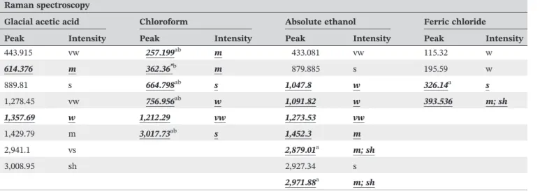
TABLE 3 Characteristic peaks of Carnoy0s solutions components
Raman spectroscopy
Glacial acetic acid Chloroform Absolute ethanol Ferric chloride
Peak Intensity Peak Intensity Peak Intensity Peak Intensity
443.915 vw 257.199ab m 433.081 vw 115.32 w
614.376 m 362.36*b m 879.885 s 195.59 w
889.81 s 664.798ab s 1,047.8 w 326.14a s
1,278.45 vw 756.956ab w 1,091.82 w 393.536 m; sh
1,357.69 w 1,212.29 vw 1,273.53 vw
1,429.79 m 3,017.73ab s 1,452.3 m
2,941.1 vs 2,879.01a m; sh
3,008.95 sh 2,927.34 s
2,971.88a m; sh
Note. vw = very weak; w = weak; m = medium; s = strong; vs = very strong; sh = shoulder.
Underlined items may be identified, but due to overlapping, they do not characterize the“fingerprint”of isolated compounds in Carnoy0s solution.
a
Characterizes the peaks of“fingerprint”identified in Carnoy0s solution.
the solution pH and the recurrence capacity, the authors believe that a more acidic pH can be an advantage for the Carnoy0s solution in a clinical use context.
Concerning the determination of ferric chloride con-centration, the present outcomes showed no difference among solutions during the days of investigation, both intrasolution and intersolution. Indeed, this result dem-onstrates the stability of Carnoy0s solution with ferric
chloride similarly to what was described by Puchtler et al.[29]However, although this relatively adequate stabil-ity, the turbidity of the solution with chloroform from the seventh day after manipulation and the precipitation of ferric crystals in nonfiltered solutions need to be taken into consideration for the use of Carnoy0s solution after
some days after preparation. Indeed, this may alter the properties of solutions when applied in the tissues.
The present study findings reinforce the role of ferric chloride as a coagulative necrosis promoting agent, [35] which from the therapeutic point of view may be strongly associated with low recurrence rates when Carnoy0s solution is used in certain benign odontogenic
neoplasms. [12,24–26] In addition, it showed that the
unfiltered Carnoy0s solutions had a more available ferric
chloride content than the filtered ones, which may be an important information for the orientation of the pharmacist to not filter the solution in order to present
a greater saturation of the ferric chloride, in addition to inform the professional who will use the solution that the turbidity is a normal finding over time in Carnoy0s
solutions with chloroform.
There are no published papers that have indicated that the Carnoy0s solution was handled and used on the same
day or if it was stored and used after 7 days or more. According to our practice and what we observe, it is underlined that what happens is the first option. This may be justified by the fact that there are no studies com-paring the effect of Carnoy0s solution with different
stor-age periods prior to its use.
Raman spectroscopy was used herein to survey the chemical composition of samples, as many materials pos-sess characteristic Raman spectra, proving to be a reliable analytical method for several areas of science. Raman spec-troscopy associated with microscopy is widely applied in the biological field, once it may provide important informa-tion regarding chemical composiinforma-tion with nondestructive sample preparation and without the water interference. Furthermore, this analyseis does not require vast prepara-tion to obtain biochemical and structural informaprepara-tion,[41]
thereby identifying possible degradation of Carnoy0s
solutions over time without affecting the samples.
Carnoy0s solution as a tissue fixative agent was firstly
analyzed by Meade et al.,[42]in which the authors verified TABLE 4 Multifactorial analysis of time, chloroform, and filtration factors in the relative percentage of height, width, area, and area/width ratio of the relative percentage of the peaks in relation to the total of peaks found
Filtration factor Chloroform factor Time factor
Height Width Area Area/width Height Width Area Area/width Height Width Area Area/width
Chloroform
257 0.364 0.516 0.069 0.753 ‐ ‐ ‐ ‐ 0.404 0.881 0.474 0.592
362 0.862 0.621 0.376 0.555 ‐ ‐ ‐ ‐ 0.606 0.992 0.914 0.980
664 0.309 0.896 0.922 0.281 ‐ ‐ ‐ ‐ 0.594 0.765 0.075 0.591
756 0.966 0.152 0.674 0.503 ‐ ‐ ‐ ‐ *.016 0.083 *.021 0.173
Ferric chloride
326 0.451 0.686 0.304 0.435 *.027 *<.001 *<.001 0.473 0.077 0.792 *.025 0.201
Glacial acetic acid
614 0.163 0.219 0.148 0.505 0.551 *<.001 0.154 0.313 *.004 0.686 *.025 0.258
Absolute ethanol
433 0.663 0.642 0.459 0.383 *.007 *<.001 *<.001 0.502 0.382 0.132 *.005 0.307
879 0.172 0.097 *.033 0.351 *.001 *<.001 *<.001 0.610 0.659 0.287 0.479 0.576
1,047 0.283 0.542 0.145 0.409 *.002 *<.001 *<.001 0.534 0.563 0.673 0.705 0.533
1,091 0.381 0.968 0.860 0.845 0.054 *.002 *<.001 *.017 0.373 0.390 0.076 0.148
1,273 0.348 0.511 0.089 0.581 0.093 *<.001 *<.001 0.393 0.364 0.814 0.068 0.481
1,452 0.287 0.520 0.433 0.269 *.004 *.003 *<.001 0.524 0.760 0.309 0.689 0.449
the effects of chemical fixatives in human cells through Raman spectroscopy.[42] However, there are no reports about the characterization of Carnoy0s solution
incorpo-rated with ferric chloride (with and without chloroform), applied as cauterizing agent for oral and maxillofacial sur-gery, surveyed by Raman spectroscopy.
In the present investigation, the laser with 638‐nm wavelength was undertaken, although an infrared laser with 785‐nm wavelength was used by Meade et al.[42] Although this study has not used the same laser of the pre-viously cited authors, the data obtained by Meade et al.[42] (666, 757 [chloroform], 881, 1,050, 1,093, 1,274, and 1,452 cm−1 [absolute etanol]) were similar to the ones
observed in Figure 2. This can be explained by the fact that both lasers in red/infrared region may justify the similar results obtained. In addition, this investigation detected new peaks that had not been previously reported; specifi-cally, the peaks at 257, 362 (chloroform), and 326 cm−1
(ferric chloride) were also detected in the present spectra. These peaks contrast the modified Carnoy0s solution from
traditional Carnoy0s solution and identify ferric chloride
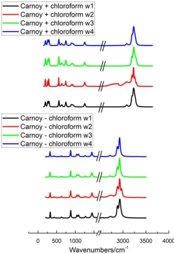
in the solutions. Spectroscopic alterations on the behavior of peaks 257, 362 (chloroform), and 326 cm−1(ferric
chlo-ride) were found, suggesting an instability of the solution in the second week after preparation and posterior return to the stability condition in the third week (Figure 3). This may likely explain the turbidity of the solution with chloro-form after 7 days. Such behavior did not occur intensely in the solution without chloroform (Figure 3). Moreover, the pattern observed in filtered and nonfiltered solutions were contrasting regardless the period of evaluation (Figure 4). Indeed, such event might be explained by high saturation of ferric chloride in nonfiltered solution, as, in tempera-tures lower than 125 °C and acidic media, the formation of iron oxide or iron oxide‐hydroxide may occur precipitat-ing in less than 6 hr.[43,44]
Statistical analysis of Raman spectra (considering the assessment of height, width, area, and area/width ratio of isolated peaks) of solutions0 components have evidenced
significant alterations on filtration, chloroform, and time factors. Regarding filtration, only absolute ethanol peak (879 cm−1) presented statistical difference (p < .05;
Table 4). Stability of further peaks (p> .05) suggests that fil-tration factor seems not affecting the stability of solutions. Concerning chloroform factor, peaks of ferric chloride (326 cm−1), acetic acid (614 cm−1), and absolute ethanol
(433, 879, 1,047, 1,091, 1,273, and 1,452 cm−1) suffered
sig-nificant alteration (p< .05). Such modifications suggest that the presence of chloroform attains higher instability for the solution, as observed by chloroform‐containing solutions, which depicted turbidity after long periods.
Regarding the time factor, chloroform peak (756 cm−1), ferric chloride (326 cm−1), acetic acid
(614 cm−1), and absolute ethanol (433 cm−1) showed
sig-nificant differences (p< .05; Table 4). Peaks from chloro-form and ethanol were modified in only one peak from each component, which likely shows no noteworthy alter-ation for these components in solutions over time. Con-versely, ferric chloride (326 cm−1) and acetic acid
(614 cm−1) attained more alterations over time. This
may have a potential correlation with iron precipitation and volatilization of components over time. Clinical trials employing Carnoy0s solution in oral and maxillofacial
sur-gery only cited that the solution was prepared for that spe-cific moment, but there is no information if the solution should be discarded right after using, or rationale based on alterations on its properties and local effects. The pres-ent investigation is the first to assay the “aging” of Carnoy0s solutions in normal storage conditions.
This study showed the long‐term stability of Carnoy0s
solution. Such information is important for the clinician because the practitioner can use the solution after storage. Indeed, there will likely have no significant changes on the clinical effect of the solution when used as an FIGURE 3 Spectra of Carnoy0s solution with chloroform in
adjuvant therapy on different days up to 6 days after preparation.
5
|
C O N C L U S I O N S
The absence of chloroform in solution may affect the per-formance of the solution due to the reduction of viscosity and ethanol penetration in tissues. Overall, Carnoy0s
solu-tions are acidic, stable in the initial 7 days, and prone to suffer alterations in the formulation with chloroform after the seventh day (turbidity) as well as iron precipitation in the container when used without filtering.
C O N F L I C T O F I N T E R E S T
The authors declared that they have no conflict of interest.
O R C I D
Francisco Samuel Rodrigues Carvalho http://orcid.org/ 0000-0002-3142-1268
R E F E R E N C E S
[1] F. W. G. Costa, E. C. S. Soares, S. H. B. Batista,RSBO.2010,
7(2), 208.
[2] F. W. G. Costa, G. A. C. Brito, R. M. A. Pessoa, E. C. S. Soares,J. Appl. Oral. Sci.2011,19(6), 604.
[3] E. A. Al‐Moraissi, A. A. Dahan, M. S. Alwadeai, F. O. Oginni, J. M. Al‐jazmali, A. S. Alkhutari, N. H. Al‐Tairi, A. A. Almaweri, J. S. Al‐Sanabania, J. Craniomaxillofac. Surg.
2016; Oct 31. pii: S1010‐5182(16)30258‐X. https://doi.org/
10.1016/j.jcms.2016.10.013. [Epub ahead of print].
[4] R. A. Voorsmit, P. J. Stoelinga, U. J. van Haelst,J. Maxillofac. Surg.1981,9(4), 228.
[5] P. J. Stoelinga, F. B. Bronkhorst,J. Craniomaxillofac. Surg.1988,
16(4), 184.
[6] R. Dammer, H. Niederdellmann, P. Dammer, M. Nuebler‐ Moritz,Br. J. Oral. Maxillofac. Surg.1997,35(1), 46.
[7] I. Nish, G. Sandor, S. Weinberg,Plast. Surg.1997,5(3), 161.
[8] H. T. Chow,Oral. Surg. Oral. Med. Oral. Pathol. Oral. Radiol. Endod.1998,86(5), 573.
[9] M. Chiapasco, A. Rossi, J. J. Motta, M. Crescentini, J. Oral. Maxillofac. Surg.2000,58(9), 942; discussion 9.
[10] P. J. Stoelinga,Int. J. Oral. Maxillofac. Surg.2001,30(1), 14.
[11] Y. F. Zhao, J. X. Wei, S. P. Wang,Oral. Surg. Oral. Med. Oral. Pathol. Oral. Radiol. Endod.2002,94(2), 151.
[12] T. A. Morgan, C. C. Burton, F. Qian,J. Oral. Maxillofac. Surg.
2005,63(5), 635.
[13] D. Chirapathomsakul, P. Sastravaha, P. Jansisyanont, Oral. Surg. Oral. Med. Oral. Pathol. Oral. Radiol. Endod. 2006,
101(1), 5; discussion 10.
[14] M. Gosau, F. G. Draenert, S. Muller, B. Frerich, R. Burgers, T. E. Reichert, O. Driemel,Clin. Oral. Investig.2010,14(1), 27.
[15] J. L. Schussel, R. T. Stramandinoli, J. L. Dissenha, L. F. Avila, L. M. Sassi,J Contemp Dent Pract.2011,12(2), 100.
[16] O. Ribeiro Junior, A. M. Borba, C. A. Alves, M. M. de Gouveia, F. L. Coracin, J. Guimaraes Junior,Oral. Dis.2012,
18(6), 548.
[17] J. Dashow, J. I. Helman, S. P. Edwards, J. McHugh, B. B. Ward,
J. Oral. Maxillofac. Surg.2013,71(9), e4.
[18] N. Wuscher,Rev. Fr. Histotechnol.1999,12(1), 9.
[19] J. R. Baker. Principles of biological microtechnique; a study of fixation and dyeing.London: Methuen;1958.
[20] R. Zollinger, A. R. Moritz,J. Nerv. Ment. Dis.1933,78(1), 86.
[21] E. C. Cutler, R. Zollinger,Am. J. Surg.1933,19(3), 411.
[22] R. A. Voorsmit,Ned. Tijdschr. Tandheelkd.2010,117(5), 278.
[23] J. Ecker, R. T. Horst, D. Koslovsky,J. Oral. Maxillofac. Surg.
2016,74(2), 278.
FIGURE 4 Spectra of Carnoy0s solutions with and without
[24] J. E. Dashow, J. B. McHugh, T. M. Braun, S. P. Edwards, J. I. Helman, B. B. Ward,J. Oral. Maxillofac. Surg.2015,73(11), 2132.
[25] E. A. Al‐Moraissi, M. A. Pogrel, E. Ellis 3rd.,J. Oral. Maxillofac. Surg.2016,74(10), 1974.
[26] Y. Y. Leung, S. L. Lau, K. Y. Tsoi, H. L. Ma, C. L. Ng,Int. J. Oral. Maxillofac. Surg.2016,45(9), 1154.
[27] US Food and Drug Administration, Section 460.200, in FDA Compliance Policy Guides, Food and Drug Administration, Washington, DC1992, 219.
[28] J. Hellstein, T. Hopkins, T. Morgan,Oral Surg Oral Med Oral Pathol Oral Radiol Endod.2007,103(4), 24.
[29] H. Puchtler, F. Sweat Waldrop, H. M. Conner, M. S. Terry,
Histochemie.1968,16(4), 361.
[30] H. Puchtler, F. S. Waldrop, S. N. Meloan, M. S. Terry, H. M. Conner,Histochemie.1970,21(2), 97.
[31] D. A. Luz, U. Ribeiro Jr., C. Chassot, E. S. F. S. Collet, I. Cecconello, C. E. Corbett,Histopathol.2008,53(6), 740.
[32] A. R. Dias, M. A. Pereira, E. S. Mello, B. Zilberstein, I. Cecconello, U. Ribeiro Junior,Gastric Cancer.2016,19(1), 136.
[33] C‐J. Tsai. Comparing dna damage caused by formaldehyde, glutaraldehye, Carnoy0s and methacarn in cancer tissue
fixa-tions. Thesis of Bowling Green State University;2006.
[34] J. B. Carnoy. La biologie cellulaire; étude comparée de la cellule dans les deux règnes. Lierre; Paris: J. Van In & Cie; O. Doin;1884.
[35] S. Nouri, M. R. Sharif, S. Sahba,Trauma Mon.2015,20(1), e18042.
[36] B. Kann, H. L. Offerhaus, M. Windbergs, C. Otto,Adv. Drug Delivery Rev.2015,89, 71.
[37] K. Kong, C. Kendall, N. Stone, I. Notingher,Adv. Drug Delivery
Rev.2015,89, 121.
[38] A. N. D. V. Sanitária,Farmacopeia Brasileira, Anvisa, Brasília 2010, 546.
[39] R. P. Buck, S. Singhadeja, L. B. Rogers,Analytic Chem. 1954,
26(7), 1240.
[40] J. K. Kauppinen, D. J. Moffatt, H. H. Mantsch, D. G. Cameron,
Appl. Spectrosc.1981,35(3), 271.
[41] H. J. Butler, L. Ashton, B. Bird, G. Cinque, K. Curtis, J. Dorney, K. Esmonde‐White, N. J. Fullwood, B. Gardner, P. L. Martin‐ Hirsch, M. J. Walsh, M. R. McAinsh, N. Stone, F. L. Martin,
Nat. Protoc.2016,11(4), 664.
[42] A. D. Meade, C. Clarke, F. Draux, G. D. Sockalingum, M. Manfait, F. M. Lyng, H. J. Byrne,Anal. Bioanal. Chem.2010,
396(5), 1781.
[43] P. A. Riveros, J. E. Dutrizac,Hidrometallurgy.1997,46, 85.
[44] J. E. Dutrizac, P. A. Riveros,Mettalurgical and Materials Trans-actions B.1999;(B), 993.
How to cite this article: Carvalho FSR, Feitosa VP, da Cruz Fonseca SG, et al. Physicochemical and rheological characterization of different Carnoy0s
![TABLE 2 Concentration of [Fe 3+ ] by UV/Vis calculated by the line equation in calibration curve (Abs = 0.1521.[Fe 3+ ] + 0.0077)](https://thumb-eu.123doks.com/thumbv2/123dok_br/15294018.545925/4.892.73.435.735.856/table-concentration-vis-calculated-line-equation-calibration-curve.webp)
![FIGURE 4 Spectra of Carnoy 0 s solutions with and without chloroform filtered and nonfiltered [Colour figure can be viewed at wileyonlinelibrary.com]](https://thumb-eu.123doks.com/thumbv2/123dok_br/15294018.545925/9.892.68.426.66.664/figure-spectra-carnoy-solutions-chloroform-filtered-nonfiltered-wileyonlinelibrary.webp)
