UNIVERSIDADE FEDERAL DO CEARÁ
FACULDADE DE FARMÁCIA, ODONTOLOGIA E ENFERMAGEM
DEPARTAMENTO DE CLÍNICA ODONTOLÓGICA
PROGRAMA DE PÓS-GRADUAÇÃO EM ODONTOLOGIA
VANARA FLORÊNCIO PASSOS
INFLUÊNCIA DE AGENTES PROTETORES NA PREVENÇÃO DA EROSÃO
EXTRÍNSECA OU INTRÍNSECA: ESTUDOS IN VITRO E IN SITU
FORTALEZA
VANARA FLORÊNCIO PASSOS
INFLUÊNCIA DE AGENTES PROTETORES NA PREVENÇÃO DA EROSÃO
EXTRÍNSECA OU INTRÍNSECA: ESTUDOS IN VITRO E IN SITU
Tese apresentada ao Programa de Pós-Graduação em Odontologia da Faculdade de Farmácia, Odontologia e Enfermagem da Universidade Federal do Ceará, como um dos requisitos para a obtenção do título de Doutor em Odontologia.
Área de Concentração: Clínica Odontológica.
Orientador: Prof. Dr. Sérgio Lima Santiago
FORTALEZA
Dados Internacionais de Catalogação na Publicação Universidade Federal do Ceará
Biblioteca de Ciências da Saúde
P324i Passos, Vanara Florêncio.
Influência de agentes protetores na prevenção da erosão extrínseca ou intrínseca: estudos in vitro e in situ. / Vanara Florêncio Passos. – 2012.
85 f. : il. color., enc. ; 30 cm.
Tese (doutorado) – Universidade Federal do Ceará; Centro de Ciências da Saúde; Faculdade de Farmácia, Odontologia e Enfermagem; Departamento de Odontologia; Programa de Pós-Graduação em Odontologia; Doutorado em Odontologia, Fortaleza, 2012.
Área de Concentração: Clínica Odontológica. Orientação: Prof. Dr. Sérgio Lima Santiago.
1. Erosão Dentária. 2. Flúor. 3. Hidróxido de Magnésio. 4. Dentina. 5. Abrasão. 6. Esmalte. I. Título.
VANARA FLORÊNCIO PASSOS
INFLUÊNCIA DE AGENTES PROTETORES NA PREVENÇÃO DA EROSÃO
EXTRÍNSECA OU INTRÍNSECA: ESTUDOS IN VITRO E IN SITU
Tese apresentada ao Programa de Pós-Graduação em Odontologia da Faculdade de Farmácia, Odontologia e Enfermagem da Universidade Federal do Ceará, como um dos requisitos para a obtenção do título de Doutor em Odontologia.
Aprovada em: ___/___/___
BANCA EXAMINADORA
________________________________________
Prof. Dr. Sérgio Lima Santiago (Orientador)
Universidade Federal do Ceará – UFC
________________________________________
Prof. Dr. Jaime Aparecido Cury
Universidade Estadual de Campinas - UNICAMP
________________________________________
Profa. Dra. Cecília Pedroso Turssi
Instituto e Centro de Pesquisas Odontológicas São Leopoldo Mandic
________________________________________
Profa. Dra. Monica Yamauti
Universidade Federal do Ceará – UFC
________________________________________
Prof. Dr. Juliano Sartori Mendonça
2
A Deus e Nossa Senhora,
Obrigada, por me fazerem chegar até aqui, por
me guiarem e por me ajudarem em todos os
AGRADECIMENTOS
Aos meus pais, Hermes Florêncio da Costa e Lindamina Passos Araújo da Costa, pelo apoio, paciência e incentivo aos meus estudos, ao meu irmão, Ícaro Florêncio Passos, e ao meu namorado, Fernando Vasconcelos Gentil Farias, pela participação como voluntários
e pela ajuda efetiva para a concretização da pesquisa realizada, além de paciência e
companheirismo.
Ao Prof. Jaime Aparecido Cury, assim como todos integrantes da FOP, pelo apoio,
ajuda, ideias, acolhimento e incentivo.
Ao Prof. Sérgio Lima Santiago, pela confiança, oportunidade de orientação e
incentivo a perpetuação de futuras pesquisas. Aprendi e cresci muito sob a sua orientação.
Seus ensinamentos foram e serão fundamentais para minha formação e amadurecimento
profissional. Muito obrigada pela contribuição para meu crescimento pessoal e profissional.
A todos os voluntários que participaram efetivamente da fase in situ, pois sem vocês,
não seria possível a realização do estudo.
Às amigas e companheiras de trabalho, Juliana Paiva e Mary Anne Melo, pela grande
ajuda, companheirismo e incentivo durante todo o curso.
Aos meus colegas de turma de Doutorado e professores, pela amizade, sugestões e
reflexões.
Aos alunos de graduação e de iniciação científica, Weslanny Morais, Bruna Melo, Camille, Cícero Leonardo, Sarah Guedes e Felipe Carvalho que sempre se mostraram
disponíveis, ajudando a qualquer momento.
À Coordenação de Aperfeiçoamento de Pessoal de Nível Superior (CAPES), pela
4
Ao CNPq (Conselho Nacional de Desenvolvimento Científico e Tecnológico) pelo
auxílio financeiro por meio do processo 620107/2008-1, e pela concessão de bolsa de auxílio
financeiro nos últimos dois anos.
À Universidade Federal do Ceará, pela realização do Curso de Pós-Graduação.
A todas as pessoas que, de algum modo, contribuíram para a concretização deste
RESUMO
Considerando o declínio da prevalência da doença cárie e periodontal na
sociedade, observa-se uma maior longevidade dentária. Assim, lesões cervicais não-cariosas
têm sido observadas com maior freqüência. Portanto, produtos que possibilitem a redução do
desgaste dentário, sendo de fácil acesso e de uso diário, são alternativas ideais para a redução
da perda de tecido mineralizado. Dessa forma, esta tese é constituída por três artigos que
objetivaram, respectivamente: (1) verificar o efeito preventivo de três pastas comerciais
contendo AmF, AmF/SnF2 or SnF2 através de um modelo erosivo/abrasivo em dentina
humana; (2) investigar o efeito do Mg(OH)2 presente em dentifrícios comercializados na
prevenção do processo erosivo no esmalte por ácidos extrínseco ou intrínseco, bem como, a
influência do número de dias experimentais na progressão da erosão; (3) avaliar in situ a ação do Mg(OH)2 e NaF na prevenção da erosão por ácidos de origem intrínseca em esmalte
humano. Nos artigos 1 e 2, foram realizados modelos in vitro, cíclico, randomizado, cego, no qual os espécimes foram submetidos a processo de des- e remineralização. No artigo 1, foi
simulado processo erosivo por ácido de origem intrínseca, enquanto no artigo 2, foi avaliada
erosão por ácido de origem intrínseca e extrínseca. No artigo 1, foi acrescentado o processo
abrasivo através da escovação. Para ambos os estudos, o ciclo foi repetido três vezes ao dia
durante cinco dias. No artigo 3, foi realizado um estudo in situ, randomizado, duplo-cego, cruzado, em três fases de 5 dias cada, com a participação de 18 voluntários, que utilizaram
dispositivos palatinos, contendo 2 blocos de esmalte dentário humano tratados com diferentes
dentifrícios: controle (0 ppm F), Mg(OH)2 (2%) e NaF (1450 ppm F). Os espécimes foram
submetidos à erosão por imersão do dispositivo em um copo contendo HCl 0,01 M (pH=2)
por 60 segundos, 4 vezes ao dia, em horários pré-determinados. Em seguida, os voluntários
escovaram seus dentes por 25 segundos e, com o dispositivo na boca, bochecharam a
suspensão dentifrício/saliva formada por 60 segundos. As alterações ocasionadas nos
espécimes de todos os artigos foram avaliadas por testes de dureza e/ou perfilometria
mecânica. Os dados obtidos foram testados usando ANOVA (p< 0,05). O teste de Tukey foi
aplicado, quando necessário, em casos no qual ANOVA revelou diferença estatística. Os
resultados do artigo 1 mostraram que dentifrícios contendo AmF/SnF2 ou SnF2 reduziram
significativamente (p<0,05) a perda de superfície dentinária após o processo erosivo/abrasivo.
No artigo 2, dentifrícios contendo Mg(OH)2 ou NaF foram efetivos em reduzir a
desmineralização ocasionada por ácido cítrico 0,05 M (p<0,001) comparados ao grupo
7
para ambos os produtos. Os resultados do artigo 3 demonstraram que houve efeito dos
dentifrícios testados na redução da perda de superfície de esmalte (p = 0,021). Entretanto, os
mesmos não evidenciaram efeitos na redução da desmineralização (p = 0,349). Dessa forma,
conclui-se que dentifrícios contendo fluoreto estanhoso ou hidróxido de magnésio são
efetivos em realizar algum efeito protetor no substrato dentário após ação de ácidos de origem
exógena ou endógena.
ABSTRACT
Considering the decline in the prevalence of caries and periodontal diseases in society,
there is greater tooth longevity. Thus, non-carious cervical lesions have been observed with
greater frequency. Therefore, products that enable the reduction of tooth wear, with easy
access and daily use, are ideal alternatives for reducing the loss of mineralized tissue. This
thesis consists of three papers that aim, respectively: (1) verify the preventive effect of three
commercial AmF, AmF/SnF2 or SnF2-containing dentifrices through an erosive/abrasive
model on human dentin; (2) investigate the effect of Mg(OH)2 present in commercial
dentifrices in preventing the erosion of enamel by extrinsic and intrinsic acids, as well as, the
influence of the number of experimental days in erosion progression; (3) evaluate in situ the action of Mg(OH)2 and NaF in prevention of intrinsic erosion on human enamel. In Articles 1
and 2, were conducted in vitro models, randomized, blind, cyclic, in which the specimens were subjected to the process of de- and remineralization. In Article 1, the erosion was
simulated by intrinsic acid, while in Article 2, was intrinsic and extrinsic acids. In Article 1,
was added the process of abrasion for toothbrushing. For both studies, the cycle was repeated
three times daily for 5 days. In Article 3, the study was in situ, randomized, double-blind, crossover in three phases of five days each, with the participation of 18 volunteers who wore
palatal appliances containing 2 blocks of human enamel treated with different toothpastes:
control (0 ppm F), Mg(OH)2 (2%) and NaF (1450 ppm F). The specimens were subjected to
erosion by immersion them in a cup containing 0.01 M hydrochloric acid (pH = 2) for 60
seconds, 4 times a day, in predetermined times. Then, the volunteers brushed their teeth for 25
seconds with the device in the mouth, and the dilution dentifrice/saliva was rinsed mouthwash
for 60 seconds. The alterations caused in specimens of all articles were evaluated by hardness
and/or stylus profilometry. Data were tested using ANOVA (p <0.05). The Tukey test was
used when required, in cases in which ANOVA revealed difference statistically significant.
The results showed that in the article 1, AmF/SnF2 or SnF2 significantly reduced (p <0.05) the
loss of dentin surface after erosion/abrasion. In Article 2, Mg(OH)2 or NaF-containing
dentifrices were effective in reducing demineralisation caused by citric acid 0.05 M (p
<0.001) compared to control. However, none prevention was observed for simulation of
intrinsic acid, for both products. The results of the article 3 showed that tested dentifrices
reduced the loss of surface enamel (p = 0.021). However, these products not present
9
fluoride or magnesium hydroxide are effective in performing some protective effect on the
dental substrate after action of acids exogenous or endogenous.
SUMÁRIO
1 INTRODUÇÃO... 11
2 PROPOSIÇÃO... 15
3 CAPÍTULOS... 17
3.1 CAPÍTULO 1... 19
3.2 CAPÍTULO 2... 36
3.3 CAPÍTULO 3... 53
4 CONCLUSÃO GERAL... 70
REFERÊNCIAS………... 72
APÊNDICES……… 79
1 INTRODUÇÃO
O declínio da prevalência da doença cárie e periodontal, devido a estratégias de
promoção de saúde, tem permitido que outras patologias orais como o desgaste dentário sejam
observadas com maior frequência, como evidenciado por estudos epidemiológicos presentes
na literatura (LARSEN; POULSEN; HANSEN, 2005; JAEGGI; LUSSI, 2006). Na última
década, muitas pesquisas têm sido realizadas a fim de obter melhor compreensão sobre o
processo de desenvolvimento do desgaste dentário (HUYSMANS; CHEW; ELLWOOD,
2011). Esse processo caracteriza-se pela perda não-cariosa de tecido dentário, apresentando
uma etiologia multifatorial com o envolvimento de processos interrelacionados incluindo
abrasão durante a mastigação e a escovação, a atrição entre dentes antagonistas, dissolução
química ocasionada pela erosão e a abfração (IMFELD, 1996).
O processo de erosão dentária ocorre como resultado da dissolução química do
dente por ácidos não-bacterianos, no qual íons hidrogênio dos ácidos reagem com a
hidroxiapatita [Ca10(PO4)6(OH)2], resultando na liberação de íons minerais (Ca2+,OH- e PO43-)
(BARTLETT 2005; LUSSI et al., 2011). Os ácidos responsáveis por esse processo podem ser de origem intrínseca ou extrínseca. A dieta é a causa extrínseca mais comum de erosão
dentária. Entretanto, medicamentos, como a vitamina C, e, mais raramente, a exposição aos
ácidos no trabalho ou em ambientes de lazer também podem causar a desmineralização da
superfície dentária (GANDARA; TRUELOVE, 1999; LUSSI; JAEGGI, ZERO, 2004;
AMAECHI; HIGHAM, 2005; LUSSI; JAEGGI, 2008; LUSSI et al., 2011; JOHANSSON et al., 2012). O principal ácido envolvido neste processo é o ácido cítrico, um constituinte de muitos sucos de frutas e bebidas ácidas, com um potencial erosivo que pode induzir a
desmineralização do tecido dentário. Adicionalmente, o ácido cítrico age como um quelante
tendo seu efeito exacerbado pela sua união com o cálcio (SAURO et al., 2008; SERRA; MESSIAS; TURSSI, 2009).
Os fatores responsáveis por erosão de origem intrínseca que ocasionam elevada
acidez na cavidade bucal incluem vômitos, desordens alimentares, doença do refluxo
gastroesofágico (DRGE), regurgitação, e ruminação (SERRA; MESSIAS; TURSSI, 2009;
LAZARCHIL; FRAZIER, 2009; LUSSI et al., 2011). Atualmente, a DRGE tem sido descrita como um importante fator etiológico da erosão dentária, sendo definido como um fluxo
retrógrado de conteúdo gástrico para o esôfago que ocorre principalmente após as refeições
(RANJITKAR; KAIDONIS; SMALES, 2012). Essa condição apresenta importante interesse
_______________________________________________________________________Introdução13
de aproximadamente 1,2 (RANJITKAR; KAIDONIS; SMALES, 2012), capaz de dissolver os
cristais de hidroxiapatita do esmalte. Pace et al. (2008) mostraram, através de uma revisão sistemática, uma forte associação entre DRGE e erosão dentária, na qual observaram uma
prevalência média de 24% de erosão em pacientes com o referido distúrbio. Devido aos
maiores cuidados com a higiene bucal, alguns pesquisadores têm avaliado a associação dos
processos erosivos e a escovação dentária (MAGALHÃES et al., 2007; SALES-PERES; PESSAN; BUZALAF, 2007; HUGLES et al., 2008, MAGALHÃES et al., 2008; MORETTO et al., 2010; AUSTIN et al., 2011; GANSS et al., 2011; ROCHEL et al., 2011). Diversos estudos comprovam a ocorrência de maior perda de estrutura dentária por meio da ação
sinérgica dos processos erosivos e abrasivos, uma vez que, a superfície dentária submetida à
erosão encontra-se amolecida sendo mais suscetível ao desgaste pelo processo abrasivo
(HARA et al., 2008; MAGALHÃES et al., 2008; WIEGAND; EGERT; ATTIN, 2008; MORETTO et al., 2010; ROCHEL et al., 2011).
A avaliação da perda de estrutura dentária por erosão e/ou abrasão pode ser
realizada por diversos métodos de análise. Dessa forma, a perfilometria é o método
quantitativo mais comumente aplicado para determinar efeitos erosivos em esmalte ou
dentina, seguida pela análise de dureza de superfície quando o substrato for o esmalte dentário
e a micro-radiografia quando empregada a dentina (SCHLUETER et al., 2011). A dureza de superfície é uma técnica mais sensível para estágios iniciais da erosão, apresentando
limitações em lesões mais avançadas, a passo que a perfilometria permite uma melhor
observação em estágios avançados de erosão, verificando a perda de tecido dentário por
erosão, abrasão ou sua combinação, mas não é sensível para detectar alterações iniciais de
superfície (HARA; ZERO 2008; SCHLUETER et al., 2011).
Assim, estratégias preventivas à perda de tecido dentário têm sido avaliadas. Um
método rotineiramente usado para higienização e de fácil acesso é o dentifrício, que
apresenta-se como um veículo de agentes fluoretados ou produtos neutralizantes (HARA et al., 2009; HUYSMANS et al., 2011; GANSS et al., 2011; MESSIAS et al., 2011; GHASSEMI et al., 2012). Desse modo, o uso de dentifrícios fluoretados pode ser uma estratégia preventiva para a erosão, reduzindo a desmineralização ou perda de estrutura
O efeito protetor do flúor frente à perda de substância dentária por erosão é devido
à formação de fluoreto de cálcio (CaF2) na superfície do esmalte (CHRISTOFFERSEN et al., 1988; LAGERWEIJ et al., 2006; GANSS et al., 2007). O uso de fluoreto estanhoso tem apresentado bons resultados no controle de lesões de erosão, provavelmente pela formação de
uma camada protetora que contém produtos da reação do SnF2 com a hidroxiapatita, como
sais de Sn2OHPO4, Sn3F3PO4, Ca(SnF3)2 ou CaF2 (ADDY; MOSTAFA, 1988).
Do mesmo modo, substâncias neutralizantes também têm sido sugeridas para
ajudar no controle da erosão por ácido hidroclorídrico e para minimizar os efeitos destrutivos
em tecidos dentários. A literatura relata que o emprego de bochechos de bicarbonato de sódio
ou suspensões neutralizantes como de hidróxido de magnésio ou hidróxido de alumínio pode
reduzir a perda de esmalte dentário (MESSIAS et al., 2010; LINDQUIST et al., 2011; TURSSI et al., 2012). Esses compostos agem por neutralização dos ácidos da cavidade bucal e inibindo a redução do pH (MEURMAN et al., 1988). Contudo, poucos trabalhos avaliam o efeito de agentes neutralizantes contidos em cremes dentais na prevenção da erosão
(MESSIAS; SERRA; TURSSI, 2008).
Outro fator que pode influenciar o processo de desgaste dentário é a saliva, que
tem sido considerada o fator biológico mais importante na prevenção da erosão devido à
habilidade de agir diretamente no agente erosivo por diluição, limpeza, neutralização e
tamponamento de ácidos. A saliva permite a formação de uma membrana protetora, a película
adquirida salivar, que age como uma barreira de difusão ou membrana de permeabilidade
seletiva prevenindo o contato direto entre os ácidos e a superfície dentária. A saliva também
reduz a taxa de desmineralização e age na remineralização por possuir cálcio, fosfato e flúor
disponível para o esmalte e a dentina erosionadas (HARA; LUSSI; ZERO, 2006). De acordo
com Hara et al. (2006), a película adquirida formada por duas horas sobre o esmalte apresenta potencial protetor frente à desmineralização.
A busca constante por produtos que possam prevenir ou até mesmo evitar a perda
de estrutura dentária, que sejam de fácil disponibilidade, baixo custo e elevada eficácia,
enfatiza a significância clínica dos estudos que compõe esta tese que objetivaram avaliar
através de metodologias in vitro e in situ o efeito de diferentes dentifrícios fluoretados e de agentes neutralizantes disponíveis no comércio na prevenção da erosão associada ou não a
2 PROPOSIÇÃO
Os objetivos do presente estudo foram:
CAPÍTULO 1 - Avaliar in vitro o efeito do tipo de flúor presente nos dentifrícios – fluoreto de amina ou fluoreto estanhoso - na prevenção da perda de superfície após erosão de origem
intrínseca e abrasão em dentina;
CAPÍTULO 2 - Avaliar in vitro o efeito do hidróxido de magnésio presente em dentifrícios na prevenção da desmineralização e perda de superfície de esmalte humano submetidos à erosão
de origem extrínseca ou intrínseca, bem como avaliar o efeito do número de dias
experimentais na progressão da erosão;
CAPÍTULO 3 - Avaliar in situ o efeito do fluoreto de sódio e do hidróxido de magnésio presentes em dentifrícios na redução da desmineralização e perda de superfície do esmalte
humano submetidos à erosão por ácido hidroclorídrico.
3 CAPÍTULOS
Esta tese esta baseada no Artigo 46 do Regimento Interno do Programa de
pós-graduação em Odontologia da Universidade Federal do Ceará que regulamenta o formato
alternativo para dissertações de Mestrado e teses de Doutorado e permite a inserção de artigos
científicos de autoria ou co-autoria do candidato. Por se tratarem de pesquisas envolvendo
seres humanos, ou partes deles, o projeto de pesquisa deste trabalho foi submetido à
apreciação do Comitê de Ética em Pesquisa da Universidade Federal do Ceará, tendo sido
aprovado (Anexo A e B). Assim sendo, esta tese é composta três capítulos, sendo dois estudos
in vitro e um in situ, conforme descrito abaixo:
3 Capítulo 1
“EFFECT OF COMMERCIAL FLUORIDE DENTIFRICES AGAINST SIMULATED GASTRIC ACID IN AN EROSION-ABRASION MODEL”
Passos VF, Vasconcelos AA, Pequeno JHP, Rodrigues LKA, Santiago SL. Este artigo será
submetido à publicação no periódico “Clinical Oral Investigations”.
3 Capítulo 2
“MAGNESIUM HYDROXIDE-CONTAINING DENTIFRICE ON EXTRINSIC AND INTRINSIC ENAMEL EROSION MODEL”
Passos VF, Rodrigues LKA, Santiago SL. Este artigo será submetido à publicação no
periódico “Journal of Dentistry”.
3 Capítulo 3
“MAGNESIUM HYDROXIDE-BASED DENTIFRICE AS AN ANTI-EROSIVE AGENT
IN AN IN SITU INTRINSIC EROSION MODEL”
Passos VF, Rodrigues LKA, Santiago SL. Este artigo será submetido à publicação no
CAPÍTULO 1
3.1 Capítulo 1
EFFECT OF COMMERCIAL FLUORIDE DENTIFRICES AGAINST SIMULATED GASTRIC ACID IN AN EROSION-ABRASION MODEL
Vanara Florêncio Passos, DDS, MSc, PhD 1
Faculty of Pharmacy, Dentistry and Nursing, Federal University of Ceará
2
University of Fortaleza
Fortaleza-CE-Brazil
Andréa Araújo de Vasconcellos, DDS, MSc. 3
Piracicaba Dental School, University of Campinas
Campinas-SP-Brazil
José Heriberto Pinheiro Pequeno, DDS 1
Faculty of Pharmacy, Dentistry and Nursing, Federal University of Ceará
Fortaleza-CE-Brazil
Jaime Aparecido Cury, DDS, MSc, PhD 3
Piracicaba Dental School, University of Campinas
Campinas-SP-Brazil
Lidiany Karla Azevedo Rodrigues, DDS, MSc, PhD 1
Faculty of Pharmacy, Dentistry and Nursing, Federal University of Ceará
Fortaleza-CE-Brazil
Sérgio Lima Santiago, DDS, MSc, PhD 1
Faculty of Pharmacy, Dentistry and Nursing, Federal University of Ceará
Fortaleza-CE-Brazil
Running title: Dentinal erosive-abrasive inhibition by dentifrices
_______________________________________________________________________Capítulo 1 21
Full address of the author to whom correspondence should be sent:
Dr. Sérgio Lima Santiago Rua Monsenhor Furtado s/no. CEP 60430-355 Fortaleza, CE Brazil
Phone- +55-85-88242704 Fax- +55-85-33668232
Effect of commercial fluoride dentifrices against simulated gastric acid in an erosion-abrasion model
Abstract
Objectives: This study assessed the effect of three commercial dentifrices with different
fluoride-containing compounds using an erosion-abrasion in vitro model. Materials and
Methods: Dentin specimens were randomized into four groups (n=10): Control (no F); Elmex
(1,400 ppm AmF); Meridol (1,400 ppm AmF/SnF2) and Crest Pro-Health (1,100 ppm SnF2).
Dentin specimens were submitted to cycles of demineralization (HCl 0.01 M for 60s),
remineralization (artificial saliva for 60 min), and immersion in 1:3 w/w of dentifrice/artificial
saliva, followed by toothbrushing (150 brushing strokes). The cycle was repeated three times
daily for five days. Surface loss was quantified by stylus profilometry. Data were submitted
to one-way ANOVA and Tukey tests (p<0.05). Results: Wear (µm±SD) was: Control:
4.1±1.2; Elmex: 3.7±1.5; Meridol: 1.3±0.4; Crest Pro-Health: 2.1±0.7. So, all products
(except Elmex) reduced mineral loss significantly (p < 0.05) when compared to the control.
Conclusion: None of dentifrices avoid the erosive-abrasive process; however, dentifrices
SnF2-containing were effective in to reduce significantly dentin loss.
Clinical Relevance: Scientific literature shows evidence that fluoride can strengthen dental
tissue against erosive acid damage. However, the beneficial effect of different fluorides
presents in commercial dentifrices is questionable. So, the determination of an effective
fluoride dentifrice may be beneficial in the reduction of erosive process in patients with
gastric disorders.
_______________________________________________________________________Capítulo 1 23
Introduction
Tooth erosion is a loss of dental hard tissues due to the chemical dissolution of teeth
by non-bacterial acids, and is caused by both intrinsic and extrinsic factors. The intrinsic
source of erosive acids is hydrochloric acid, which is produced by the stomach. The oral
presence of this acid is frequently observed in patients who suffer from eating disorders, such
as recurrent vomiting, regurgitation or gastro-esophageal reflux [1,2]. Moreover, the extrinsic
sources responsible for erosion are acidic drinks and food [2-5]. According Packer [6], the
changes in lifestyle have increased consumption of acid-containing products.
The excessive contact with dietary acids can be avoided through of oral health
education. In the meantime, individuals presenting chronic reflux or gastrointestinal disorders
that cause the secretion of the hydrochloric acid present in the gastric juice into the oral cavity
require a prolonged medical treatment until stop to expose your teeth to acid contact [7].
According Ranjitkar, Kaidonis and Smales [7], the success of medical intervention is variable
and the adequate treatment if difficult to manage. So, additionally to eliminate the cause, the
use of products that reduce the effect of endogenous acid in dental surface should be
beneficial.
Application of fluoride at high concentrations or metal ion fluoride may lead to the
formation of mainly CaF2- like products or may form a physical barrier, protecting it against
acid attack [8-10]. In recent years, however, researchers have evaluated various regimens
regarding fluoride levels existing in toothpastes, solutions, gels and varnishes and their effects
on dental erosion and inconsistent results have been found [11-14].
An ideal preventive measure would be the use of fluoridated dentifrices, which are
excellent vehicles for fluoride delivery [15] given their global use. Nevertheless, some degree
shown a minor mineral loss when brushing with fluoride as opposed to non fluoride
dentifrices [17-19]. Therefore, new toothpastes with different composition have been
developed for protection against caries and erosion. However, only little is known about
preventive measures against tissue loss caused by combined erosive-abrasive impacts, in
particular if the complex histological structure of eroded dentin is considered [20].
Consequently, this in vitro study aimed to investigate the effects of three commercial
dentifrices (Elmex, Crest Pro-health and Meridol) containing respectively AmF, SnF2 or
AmF/SnF2 compared with a non-fluoride dentifrice in the reduction of dentin loss after
erosion/abrasion caused by intrinsic acid and toothbrushing. The null hypothesis tested was
that there is no difference among the preventive effects of tested dentifrices on dentinal
erosion/abrasion.
2. Materials and Methods
2.1 Ethical Aspects:
The protocol (# 131/2009) was approved by the local Ethics Committee. The extracted
human teeth were collected after the patient’s informed consent. The collected third molars
were stored in 0.01% (w/v) thymol solution at 4°C and used within one month after
extraction.
2.2. Dentin Sample Preparation
Root dentin slabs (4 x 4 x 2 mm) were obtained from freshly extracted and caries-free
third molars. The dentin slabs were obtained using a water-cooled low-speed diamond disc
mounted in a sectioning machine (IsoMetTM Low Speed Saw Buehler, Lake Bluff, IL, USA).
Subsequently, the slabs were embedded in self-polymerized acrylic resin cylinders (Arotec
_______________________________________________________________________Capítulo 1 25
facilitate handling. The slabs were ground flat with wet 320-, 600- and 1,200-grit silicon
carbide paper (Extec, Enfiel, CT, USA) and polished with a diamond spray of 1 μm (Extec,
Enfiel, CT, USA). After each flattening and polishing procedure, the specimens were
sonicated for 2 min (Ultra Cleaner 1400, UNIQUE, Indaiatuba, SP, Brazil) in deionized
distilled water, and polished surfaces free of visual defects or cracks were selected for this
study.
An initial surface hardness (SH) test was performed with five indentations in the
center of the slab with a Knoop diamond under a 25 g load for 5 s (FM7 AMRS; Future Tech,
Tokyo, Japan) to select 50 slabs. The slabs, which presented microhardness values ranging
from 52-64 Kg/mm2, were randomly assigned into five groups (Table 1) according to a
computer generated randomization list (n=10). The pH-values were obtained of slurry
containing 1:3 w/w of dentifrice/artificial saliva and all the toothpastes present silica as
abrasive.
In order to maintain reference surfaces for lesion depth determination, a layer of an
acid-resistant varnish with a dark color (Colorama, CEIL Coml Exp Ind Ltd., São Paulo,
Brazil) was applied on one-half of the surface of each slab.
2.3. Erosion-abrasion Cycling Model
Demineralization was performed by the immersion of each slab in 3 mL of
hydrochloric acid (HCl 0.01 M, pH 1.96) for 60 s. Subsequently, each sample was
remineralized in 5 mL of artificial saliva (1.45 mM Ca, 5.4 mMPO4, 0.1 M Tris buffer, 2.2
g/L porcine gastric mucin, pH 7.0) under constant agitation for 60 min. The slabs were then
immersed in 3 mL of 1:3 w/w of dentifrice/artificial saliva for 60 s. Next, the specimens were
positioned in a brushing machine, (MSEt – 1500W – Marcelo Nucci ME – São Carlos, SP –
were plated for suspensions of dentifrice and artificial saliva (1:3; w/w), according to each
group. After each cycle, which was composed of demineralization, remineralization and
toothbrushing, the specimens were again rinsed in deionized distilled water. All solutions
were used at room temperature. The described cycle was repeated three times daily for five
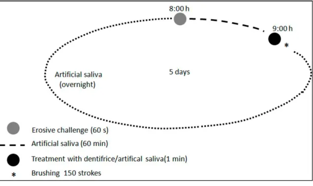
days at room temperature, as shown in Figure 1. After the daily cycles, the samples were
stored overnight in artificial saliva at 37ºC.
2.4. Tissue Loss Measurements
After the experimental period, the nail varnish layer over the surfaces was carefully
removed using acetone and a scalpel blade without touching the dentinal surfaces. The
specimens were allowed to dry for 10 min before analysis in order to reduce the possible
interference caused by the shrinkage of dentin organic content. Dentinal wear was determined
in relation to the reference surfaces, using a profilometer (Hommel Tester T1000,
Hommelwerke GmbH, Germany). Five readings were performed on each slab, at intervals of 100 µm. These profilometric traces were taken by moving the stylus from the reference to the
exposed surfaces. For each sample, mean was calculated from the values obtained from the
five traces.
2.6. Statistical Analysis
Mean and standard deviation (SD) values of wear per group were calculated.
Statistical procedures were performed with the Statistical Package for Social Sciences (SPSS
17.0) for Windows. A Kolmogorov-Smirnov test was applied to all groups to test for the
normal distribution of errors. Because the values were normally distributed across all of the
groups, ANOVA and Tukey’s post hoc tests were used for comparative purposes. The level of
_______________________________________________________________________Capítulo 1 27
3. Results
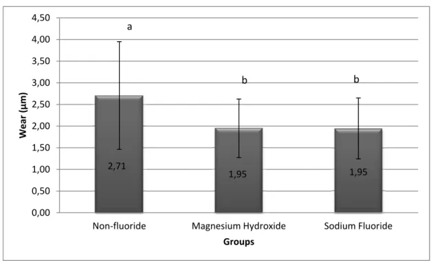
Mean dentinal loss of all experimental groups is presented in Figure 2. After five days
of erosion and abrasion cycles, tissue loss was highest in the control group, and all products
(except Elmex) reduced mineral loss significantly in the order of 37 – 67% (p < 0.05 when
compared to the control). However, Meridol, Crest Pro-health and Sensodyne Pronamel were
statistically similar to each other. There were no no significant difference between control
group and Elmex (p = 0.948).
4. Discussion
This study evaluated the effect of three commercial dentifrices containing AmF, SnF2
or AmF/SnF2 on reduction of dentin erosion using an erosive-abrasive model. The aim of
erosion-abrasion model is to reduce the mineral loss by improving the characteristics of
resistance of tooth surfaces. So, a preventive method that can be used daily and that acts
against caries and erosion would be an excellent option.
Fluoride delivery by dentifrice has been evaluated as an alternative to reduce erosion
[21] considering that dentifrices are used daily and can be easily accessed by the general
population. Some authors have showed that fluoride dentifrices can enhance the resistance of
the eroded dental substrate [17, 19, 22, 23]. However, the level of wear can be modulated not
only by fluoride, but also by abrasiveness of dentifrice [17]. In this study, dentifrices tested
presented values of Relative Dentin Abrasion raging from 70 to 144, below the limit of 250
recommend by ISO standard 11609 and ADA. This way, all dentifrices tested display a
From the present results, the hypothesis that there are no differences in the effect
between dentifrices tested was rejected, because dentifrices containing fluoride in the forms of
SnF2 (Crest Pro-Health Enamel Shield) and AmF/SnF2 (Meridol) reduced the tooth wear
caused by hydrochloric acid on dentin in vitro. Thus, the results reinforce the relevance of
dentifrices containing fluoride in the reduction of dental erosion, similar to other in vitro [21]
and in situ studies [19, 23, 25].
To our knowledge, few studies have evaluated the erosion/abrasive effects of
AmF-containing dentifrices on dentin. Application of AmF fluoride may lead the formation of CaF2
products that can protect against acid attack. However, the formation of CaF2- like products
does not sufficiently resistant to avoid loss of dental structure after a strong erosive process
occasioned by a simulated gastric acid and a brushing with dentifrice of medium abrasivity.
Hara et al. [17] verified the interaction of fluoride and abrasivity of dentifrices obtained that
in root dentin the abrasivity had a higher impact than fluoride in the reduction of tissue loss.
In respect of AmF, the results of the present study are corroborated with previous
studies [13, 26], which have used different fluoridated solutions or dentifrices to evaluate the
mineral loss in enamel and found no differences between AmF and the absence of any
fluoride in inhibiting tooth wear. Otherwise, different results were demonstrated by Wiegand
et al. [27] and Wegehaupt et al. [28], which showed a statistically significant capacity of AmF
in preventing dental erosion. Probably, the result observed by Wiegand et al. [27] may be
attributed to use of a weak acid (Sprite Zero) to perform the erosive challenge, which might
have promoted a lower wear rate. Besides, the authors used a more acidic AmF solution (pH =
4.5) that may have increase the fluoride action, since the lower the pH of fluoride greater the
formation of calcium fluoride [29]. Wegehaupt et al. [28], also, observed a beneficial effect,
_______________________________________________________________________Capítulo 1 29
The best effects were found when Sn-containing dentifrices were used, corroborating
with other studies that assessed solutions or dentifrices containing same fluoride types [11,
14, 30-32]. Probably, this result can be attributed to the fact that when Sn-products are used,
the dentinal surface is covered by a layer containing the reaction products of SnF2 and
hydroxyapatite, such as Sn2OHPO4, Sn3F3PO4, Ca(SnF3)2 or CaF2 salts, which obliterated
patent dentinal tubules [10]. According to other studies, this distinct coating is capable of
survival even after a 2-min immersion in citric acid solution [13]or a 50% decrease in the
etching depths after acid exposure for 6 min on enamel [12]. In addition, the simple presence
of the cited salts may be relevant for protection against erosion. Recently, Ganss et al. [33],
showed that tin is retained in the demineralized organic matrix, beyond what is diffused
through the dentin structure and is accumulated in the underlying mineralized tissues. These
authors suggest that the main mechanism of action of SnF2 is the uptake of tin in the
underlying mineralized tissues, rather than surface precipitation.
Nevertheless, when Sn fluoride was used, the wear was modulated by the fluoride
effect, and both Meridol and Crest Pro-health present similar wear rate. So, the kind of
fluoride may affect the ability of dentifrices to remineralize eroded substrate, that was no
observed when Am-F dentifrices were used. The results of study show that the protective
effect of SnF2 is effective even using strong acid challenge. These SnF2 products can improve
the fluoride action and minimize the effect of abrasivity, without harming the dental cleaning.
The in vitro experimental model used suggests that the kind of fluoride influences in
the reduction of erosive process by simulated gastric acid, presenting a effective impact on the
wear. So, considering the limitations of the study, it can be concluded that Meridol and Crest
Pro-Health, both containing SnF2, are effective in reducing mineral loss after erosion by
6. References
1. Imfeld T (1996) Dental erosion: Definition, classification and links. Eur J Oral Sci 104:151-155.
2. Lussi A, Schlueter N, Rakhmatullina E, Ganss C (2011) Dental erosion – an overview with
emphasis on chemical and histopathological aspects. Caries Res 45(Suppl 1):2-12. doi:
10.1159/000325915.
3. Amaechi BT, Higham SM (2005) Dental erosion: possible approaches to prevention and
control. J. Dent 33:243-252.
4. Lussi A, Jaeggi T (2008) Erosion-diagnosis and risk factors. Clin Oral Invest 12(Suppl
1):5-13. doi: 10.1007/s00784-007-0179-z.
5. Johansson AK, Omar R, Carlsson GE, Johansson A (2012) Dental erosion and its growing
importance in clinical practice: from past to present. Int J Dent. doi: 10.1155/2012/632907
6. Packer CD (2009) Cola-induced hypokalaemia: a super-sized problem. Int J Clin Pract
63:833-835. doi: 10.1111/j.1742-1241.2009.02066.x.
7. Ranjitkar S, Kaidonis JA, Smales RJ (2012) Gastroesophageal reflux disease and tooth
erosion. Int J Dent. doi: 10.1155/2012/479850.
8. Christoffersen J, Christoffersen MR, Kibalczyc W, Perdok WG (1988) Kinetics of
dissolution and growth of calcium fluoride and effects of phosphate. Acta Odontol Scand
46:325-36.
9. Messias DC, Maeda FA, Turssi CP, Serra MC (2011) Effect of dentifrices against
hydrochloric acid-induced erosion. Oral Health Prev Dent 9:269-273.
10. Addy M, Mostafa P (1988) Dentine hypersensitivity. I. Effects produced by the uptake in
vitro of metal ions, fluoride and formaldehyde onto dentine. J Oral Rehabil 15:575-585.
11. Willumsen T, Ogaard B, Hansen Bf, Rølla G (2004) Effects from pretreatment of
stannous fluoride versus sodium fluoride on enamel exposed to 0.1 M or 0.01 M hydrochloric
_______________________________________________________________________Capítulo 1 31
12. Hove L, Holme B, Øgaard B, Willumsen T, Tveit AB (2006) The protective effect of
TiF4, SnF2, and NaF on erosion of enamel by hydrochloric acid in vitro measured by white
light interferometry. Caries Res 40:440-443.
13. Ganss C, Schlueter N, Hardt M, Schattenberg P, Klimek J (2008) Effect of fluoride
compounds on enamel erosion in vitro: a comparison of amine, sodium and stannous fluoride.
Caries Res 42:2-7.
14. Schlueter N, Klimek J, Ganss C (2011) Efficacy of tin-containing solutions on erosive
mineral loss in enamel and dentine in situ. Clin Oral Investig 15:361-367. doi:
10.1007/s00784-010-0386-x.
15. Hara AT, Kelly SA, González-Cabezas C, Eckert GJ, Barlow AP, Mason SC, Zero DT
(2009) Influence of fluoride availability of dentifrices on eroded enamel remineralization in
situ. Caries Res 43:57-63. doi: 10.1159/000201591.
16. Forward GC (1991) Role of toothpastes in the cleaning of teeth. Int Dent J 41: 164–170.
17. Hara AT, Gonza´ lez-Cabezas C, Creeth J , Parmar M, Eckert GJ, Zero DT (2009)
Interplay between fluoride and abrasivity of dentifrices on dental erosion–abrasion. Journal of
dentistry 37:781-785. doi: 10.1016/j.jdent.2009.06.006.
18. Attin T, Deifuss H, Hellwig E (1999) Influence of acidified fluoride gel on abrasion
resistance of eroded enamel. Caries Research 33:135–139.
19. Passos VF, Santiago SL, Tenuta LMA, Cury JA (2010) Protective effect of
NaF/triclosan/copolymer and MFP dentifrice on enamel erosion. Am J Dent 23:193-195.
20. Ganss C, Hardt M, Blazek D, Klimek J, Schlueter N (2009) Effects of toothbrushing force
on the mineral content and demineralized organic matrix of eroded dentine. Eur J Oral Sci
117:255-260. doi: 10.1111/j.1600-0722.2009.00617.x.
21. Lussi A, Megert B, Eggenberger D, Jaeggi T (2008) Impact of different toothpastes on the
prevention of erosion. Caries Res 42:62-67.
22. Zero DT, Lussi A (2006) Behavioral factors. Monogr Oral Sci 20:100-5.
23. Zero DT, Hara AT, Kelly SA, González-Cabezas C (2006) Evaluation of a desensitizing
24. Giles A, Claydon NCA, Addy M, Hughes N, Sufi F, West NX (2009) Clinical in situ
study investigating abrasive effects of two commercially available toothpastes. J Oral Rehabil
36:498-507.
25. Ganss C, Schlueter N, Friedrich D, Klimek J (2007) Efficacy of waiting periods and
topical fluoride treatment on toothbrush abrasion of eroded enamel in situ. Caries Res
41:146-151.
26. Lagerweij MD, Buchalla W, Kohnke S, Becker K, Lennon AM, Attin T (2006) Prevention
of erosion and abrasion by a high fluoride concentration gel applied at high frequencies.
Caries Res 40:148-153.
27. Wiegand A, Magalhães AC, Navarro RS, Schmidin PR, Rios D, Buzalaf MAR, et al
(2010) Effect of titanium tetrafluoride and amine fluoride treatment combined with carbon
dioxide laser irradiation on enamel and dentin erosion. Photomed Laser Surg 28:219-226. doi:
10.1089/pho.2009.2551.
28. Wegehaupt FJ, Sener B, Attin T, Schmidlin PR (2011) Anti-erosive potential of amine
fluoride, cerium chloride and laser irradiation application on dentine. Arch Oral Biol
56:1541-1547. doi: 10.1016/j.archoralbio.2011.06.010.
29. Saxegaard E, Rölla G (1988) Fluoride acquisition on and in human enamel during topical
application in vitro. Scand J Dent Res 96:523–535.
30. Wiegand A, Bichsel D, Magalhães AC, Becker K, Attin T (2009) Effect of sodium, amine
and stannous fluoride at the same concentration and different pH on in vitro erosion. J Dent
37:591-595. doi: 10.1016/j.jdent.2009.03.020.
31. Ganss C, Lussi A, Sommer N, Klimek J, Schlueter N (2010). Efficacy of fluoride
compounds and stannous chloride as erosion inhibitors in dentine. Caries Res 44:248-252.
doi: 10.1159/000314671.
32. Ganss C, Neutard L, Von Hinckeldey J, Klimek J, Schlueter N (2010) Efficacy of a
Tin/fluoride rinse: a randomized in situ trial on erosion. J Dent Res 89:1214-1218. doi:
10.1177/0022034510375291
33. Ganss C, Hardt M, Lussi A, Cocks A, Klimek J, Schlueter N (2010) Mechanism of action
_______________________________________________________________________Capítulo 1 33
tissue loss, and scanning electron microscopy study. Eur J Oral Sci 118:376-384. doi:
Table
Table 1. Dentifrice product information and pH-values obtained of slurry (dentifrice/artificial
saliva)
TRADENAME FLUORIDE PH RDA MANUFACTURER
Control No F 7.33 70 Manufactured
Elmex 1,400 AmF 5.86 100 Gaba international,
Münchenstein, Switzerland
Meridol 1,400
AmF/SnF2
6.00 100 Gaba international,
Münchenstein, Switzerland
Crest
Pro-Health Enamel
Shield
1,100 SnF2 5.64 144 Crest, Procter and Gamble,
_______________________________________________________________________Capítulo 1 35
Figures
Figure 1. 24-hour cycle followed during experiment. This sequence was repeated during 5
days.
_______________________________________________________________________Capítulo 2 37
3.2 Capítulo 2
Magnesium hydroxide-containing dentifrice on extrinsic and intrinsic enamel erosion model
Vanara Florêncio Passos, DDS, MSc, PhD 1
Faculty of Pharmacy, Dentistry and Nursing, Federal University of Ceará
2
University of Fortaleza
Fortaleza-CE-Brazil
Lidiany Karla Azevedo Rodrigues, DDS, MSc, PhD 1
Faculty of Pharmacy, Dentistry and Nursing, Federal University of Ceará
Fortaleza-CE-Brazil
Sérgio Lima Santiago, DDS, MSc, PhD 1
Faculty of Pharmacy, Dentistry and Nursing, Federal University of Ceará
Fortaleza-CE-Brazil
Running title: Erosive inhibition by fluoride or magnesium hydroxide-dentifrices
Keywords: Magnesium hydroxide; Dentifrice; Hydrochloric Acid; Sodium Fluoride; Citric
acid.
Full address of the author to whom correspondence should be sent:
Dr. Sérgio Lima Santiago Rua Monsenhor Furtado s/no. CEP 60430-355 Fortaleza, CE Brazil
Abstract
Objective: To evaluate, in vitro, the effect of MgOH2 dentifrice as well as the influence of the
number of experimental days on extrinsic (citric acid - C6H8O7) and intrinsic (hydrochloric
acid - HCl) enamel erosion models. Methods: Human enamel slabs were selected by surface
hardness and randomly assigned to 3 groups (n=9) as follows: non-fluoridated (control), NaF
(1,450 ppm F) and Mg(OH)2 (2%) dentifrices. In thisstudy, the slabs were submitted daily to
a previous 2 hour-acquired pellicle formation and, during 5 days, submitted to cycles (3x/day)
of demineralization (C6H8O7 0.05 M, pH 3.75 or HCl 0.01 M, pH 2 for 30 s), treatment (1 min
- 1:3 w/w of dentifrice/distilled water) and remineralization (artificial saliva for 120 min).
Enamel changes were determined by surface hardness loss percentage (SHL) at each day and
mechanical profilometry analysis. Data were analyzed by two-way ANOVA followed by
Tukey's test to %SHL and one-way ANOVA to profilometry (p<0.05). Results: The number
of experimental days influenced the erosion process in the two types of erosion (p<0.001).
NaF and Mg(OH)2-containing dentifrices were effective in reducing enamel extrinsic acid
erosion for %SHL (p<0.001) compared to the control group, however, they were not effective
for the intrinsic one (p = 0.295). With regard to surface wear, no statistically significant
difference was found among treated and control groups for citric acid (p=0.225) and
hydrochloric acid (p=0.526). Conclusion: The findings suggest that NaF and MgOH2
dentifrices might protect enamel against slight erosion, but protection was not effective in the
strong acid erosion.
Keywords: Magnesium hydroxide; Dentifrice; Hydrochloric Acid; Sodium Fluoride; Citric
_______________________________________________________________________Capítulo 2 39
Introduction
The frequent ingestion of citrus fruits, pure acidic juices or carbonated sports drinks,
as well as gastrointestinal disorders that cause the secretion of gastric acid into the oral cavity
may lead to demineralization and loss of dental hard tissues.1,2 According to Duffley et al.3, adolescents consume an average of 360 ml of acid drinks per day, which shows a high
ingestion of acid-containing products. A systematic review showed a median prevalence of
24% for tooth erosion in adult patients with gastro esophageal reflux disease,4 proving that
gastric acid is also an important etiological factor to dental erosion.
The avoidance of lifelong contact of erosive acidic contents with dental surfaces is an
impossible task. Therefore, the development of early diagnostic methods and adequate
preventive approaches should be searched.1 Most preventive measures are based on the
release of compounds by oral solutions or dentifrices due to daily use and easy access to over
the counter merchandise. These products can introduce chemical protective substances into
the oral cavity as buffer agents.5 Sodium bicarbonate and magnesium hydroxide are the most
commonly found buffer agents6,7 available as solution or incorporated into dentifrices.
Different neutralizing products have been assessed to reduce intrinsic erosion.5,8-10 In
this regard, the protective effect of antacid products on dental erosion has been shown in some
studies.8-10 According to Lindquist et al.9, these products increase the intra-oral pH after the erosive process, presenting a buffering effect.6 Furthermore, this effect may relate to the fact
that these products could react with the acid, forming a salt.10 However, there is no published
data on the effect of dentifrices containing magnesium hydroxide on enamel surfaces exposed
to a simulated exogenous or endogenous erosive model.
The aim of the present study was to assess the effect of magnesium hydroxide,
fluoridated and non-fluoridated based dentifrices in terms of reducing the progression of
enamel surface erosion originated by extrinsic acid (experiment 1) or intrinsic acid
(experiment 2). In addition, the influence of the number of experimental days in enamel
surface softening. The null hypothesis tested was that there is no difference among the tested
Material and methods
Preparation of enamel samples
The study protocol was reviewed and approved in the local Research and Ethics
Committee (protocol #75/12). Enamel slabs were obtained from caries free human third molar
that had been stored in 0.01% (w/v) thymol solution at 4°C.11 Enamel slabs (4 x 4 x 2 mm)
were cut from the middle third of the coronal surface. Each slab was ground flat with the
dentin and enamel surfaces planar parallel, and polished. The enamel surface was sequentially
ground in a water-cooled mechanical grinder (Ecomet/Automet 250 Grinder-Polisher;
Buehler, Lake Bluff, IL, USA) with 400-, 600-, and 1,200-grit Al2O3 papers and polished on
cloths with a 1 µm diamond suspension (Alpha Micropolish; Buehler).
A total of fifty-four enamel slabs were randomly divided into experimental groups
based upon their baseline surface hardness values (SHbas), using a computer generated list
(Microsoft Excel 2007). The SHbas values were determined by placing five indentations, 100
µm apart from each other at the center of the specimens using Knoop indenter with a load of
50 g and a dwell time of 5 seconds (FM100, Future Tech, Tokyo, Japan). Enamel specimens
presenting a mean hardness of 328.1 ± 13.1 Kg/mm² were selected and allocated to three
groups to experiment 1 and to experiment 2 (n=9) generating balanced groups.
Subsequently, two parts of each specimen were covered with a dark-colored
acid-resistant varnish (Jordana Cosmetics Corp., Los Angeles, CA, USA) to serve as the reference
area for profilometry analysis. The exposed area of 2 x 4 mm in their central area was
subjected to treatments. In experiment 1, the acid challenge was performed using 0.05 M
citric acid (citric acid dehydrated, pH 3.75; Dinâmica®, Diadema, SP, Brazil), while in
experiment 2, 0.01 M hydrochloric acid (pH 2.0; Merck, Darmstadt, Germany) was used. The
experimental groups were: non-fluoridated (control, 0 ppm F; pH = 6.86), NaF (1450 ppm F;
pH = 7.36) and Mg(OH)2 (0 ppm F; 2%; pH = 9.96) dentifrices.
_______________________________________________________________________Capítulo 2 41
In each experimental day, fresh saliva samples were collected from groups of 15-20
volunteers without active carious lesions, erosions, or salivary dysfunction. The subjects did
not eat or smoke during the 8-hour period before sampling. Saliva was stimulated with
paraffin wax for 5 min. Saliva from the first minute of chewing was swallowed, and the rest
was collected and deposited into a 50-ml centrifuge tubule. The saliva samples were
centrifuged for 10 min at 2000 rpm in a pre-cooled centrifuge (4°C) (5415R, Eppendorf, Brazil). The clear fluid above the sediments was pooled and used for pellicle formation.12
Each group of enamel slabs was independently immersed in clarified saliva and incubated
during 2 hours before each experimental day, prior to erosive challenges, under agitation at
100 rpm (5 ml per slab) at 37°C to simulated oral cavity temperature.
Experimental procedure
The study consisted of 2 separate experiments. Both experiments consisted of cyclic
procedures repeated over a five–day period, including pellicle formation, erosion, treatments
with test dentifrices and remineralization with artificial saliva (1.5 mM Ca; 0.9 mM PO4; 150
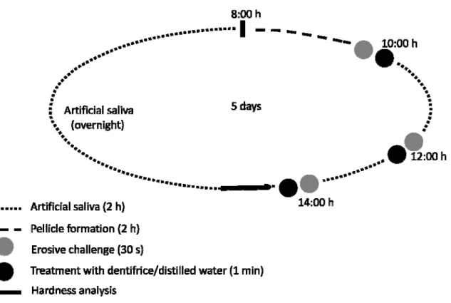
mM KCl and 0.1 M Tris buffer, pH 7.0)13 (Figure 1).
In each experimental day, all procedures were performed under agitation at 100 rpm,
at 37°C. All specimens were immersed in clarified saliva during 2 hours to allow formation of
the acquired pellicle. Subsequently, each slab was submitted to citric acid or hydrochloric acid
solution during 30 seconds. After, the specimens were also treated with fresh dentifrice slurry
(5 ml for specimen) during 1 minute prepared from non-fluoridated, magnesium hydroxide or
sodium fluoride dentifrices (1 part of toothpaste to 3 parts of distilled water solution, by
weight). The slurries were freshly prepared at the beginning of each experimental day. Next,
each slab was rinsed with distilled water and immersed in artificial saliva during 2 hours. This
cycle was repeated three times a day during 5 days, in the end of each experimental day, the
slabs were evaluated by surface hardness, as shown in Figure 1.
Measurements were performed with a stylus profilometer Hommel Tester T1000
(Hommelwerke GmbH, Germany) after the last experimental day. The measurement of
interest was the difference between the heights of the surfaces of the reference and the treated
areas. Before the analysis, the nail varnish was carefully removed exposing the untreated
references areas. On each sample, at intervals of 100 µm, five profile traces (1.5 mm in
length) were recorded, being the levels of enamel wear determined in relation to the reference
surfaces. For each sample, the mean values obtained from the five traces were calculated.
Percentage of surface hardness loss assessment
Immediately after completion of each experimental day, slabs were placed in the
hardness machine and five new indentations (SHafter), similarly described for the baseline
determinations, spaced 100 µm apart from the previous measurements (SHbas) were made
with a Knoop diamond under a 50 g load for 5 s. The percentage of SH loss (% SHL) was
then calculated for each day according to the equation:
%SHL = [(SHbas. – SHafter) x 100/ SHbas.]
Statistical analysis
Mean values of wear and %SHL were calculated. A Kolmogorov-Smirnov test was
applied to all groups to test for the normal distribution of errors. Because the values were
normally distributed across all of the groups, two-way ANOVA was carried out to analyze
percentage of SH loss to evaluate the influence of treatment and of number of experimental
days. One-way ANOVA was used to evaluate profilometry among the groups. Tukey’s post
hoc test was applied, when necessary, in cases where ANOVA revealed significant
differences. Statistical analyses were performed with the Statistical Package for Social
Sciences (SPSS 17.0) for Windows. The level of significance was set at 5%.
Results
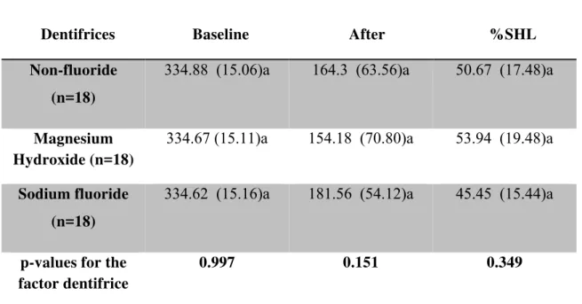
In experiment 1, two-way ANOVA revealed a significant difference among the
_______________________________________________________________________Capítulo 2 43
by number of experimental days (p<0.001; F=68.0). However, the interaction between the
factors was not significant (p=0.982; F=0.24). It was observed that with an increase in the
number of experimental days, all specimens displayed a statistically significant surface
softening from day 1 to day 3, which stabilizes at days 4 and 5. The results showed that
magnesium hydroxide reduced significantly the %SHL compared to non-fluoridated
(p=0.0001) and fluoridated dentifrices (p=0.001). Sodium fluoride reduced the percentage of
surface loss compared to non-fluoridated dentifrice (p=0.001) [Figure 2]. The dentifrices
tested showed no significant effect on wear compared to non-fluoride dentifrice (Table 1).
In experiment 2, two-way ANOVA did not show a significant difference among the
tested dentifrices (p=0.295; F=1.23). However, the %SHL to the number of experimental days
differed significantly (p<0.001; F=48.43). The interaction between factors (tested dentifrices
and number of experimental days) was not significant (p=0.326; F=1.16). Demineralization
influenced by number of experimental days was similar between experiment 1 (citric acid)
and 2 (hydrochloric acid) (Figures 2 and 3). Dentifrices tested showed no significant
preventive effect against wear compared to non-fluoride dentifrice (Table 1).
Discussion
Our in vitro de-remineralization cycling model investigated two products with respect to their capacity to protect enamel from intrinsic or dietary acids. This study confirmed the
expected surface softening and enamel tissue loss due to action of citric acid or hydrochloric
acid, even after the pellicle formation for 2 h before each experimental day. The use of an in vitro multiple-exposure acid model allows a better understanding of the erosive challenges faced by the dentition, and performed a controlled investigation, reducing experimental time
and cost.14
The null hypothesis that there is no difference in the preventive effect among the
tested dentifrices on extrinsic enamel erosion caused by citric acid was partially rejected, due
to an observed difference in the percentage of surface hardness analysis. Magnesium
hydroxide and sodium fluoride reduced the surface softening compared with a non-fluoridated
dentifrice.
The results of this extrinsic erosion model reinforce the relevance of magnesium
caused by dietary acids. However, magnesium hydroxide was more effective than sodium
fluoride in protecting the human enamel against citric acid erosion. This result may be
explained through the acid buffering that occurs immediately after the contact of Mg(OH)2
and C6H8O7, that produce a salt and H2O. The use of magnesium hydroxide-dentifrice may
also help saliva to neutralize and clean erosive products from the oral cavity.8 Lindquist et al.9 showed that antacid products can increase the intra-oral pH after an erosive challenge, which
is desirable to obtain acid neutralization. Similarly, Messias et al.8 using sodium bicarbonate solution observed a preventive effect even in intrinsic erosion in situ.
Furthermore, the relevance of sodium fluoride-containing dentifrices in the reduction
of enamel erosion caused by dietary acids was described by other in vitro15,16 and in situ studies.17,18 White et al.19 verified that low concentrations (10 ug/g F-) of sodium fluoride solutions are already capable of reducing hydroxyapatite dissolution in citric acid in vitro. Therefore, 1450 ppm F present in the NaF dentifrice even after dilution in saliva can be
effective. The preventive effect of sodium fluoride dentifrice in erosive lesions is mainly
based on the deposition of high amounts of CaF2-like products on enamel surface, which can
protect it against acid attack.20,21 This layer is formed on enamel surfaces even within a short
exposure time of 20 s - 2 min,22 reducing initial erosion. However, the protective action will
be reduced after repeated and excessive acid contact. Another point to consider is the pH of
fluoride, which presents an important role in the efficacy of the preventive agent, since acidic
fluoride increases the formation of CaF2-like deposit. In the present study, sodium fluoride
dentifrice may be less effective than magnesium hydroxide in the protection of enamel
erosion, because the NaF dentifrice presented a neutral pH.
Meanwhile, sodium fluoride and magnesium hydroxide dentifrices were not effective
to reduce the enamel loss. In fact, surface profilometry analysis is more indicated to evaluate
advanced erosion,23 which did not occur in experiment 1. Based on the present results, enamel
surface loss was only of approximately 0.2 µm after five experimental days for extrinsic acid
exposure, whereas intrinsic acid exposure produced a surface loss sevenfold more severe,
proving a moderate action of acidic beverages. However, profilometric analysis is widely
accepted as a technique to evaluate erosive challenge.24 Despite the outcome of the
profilometric analysis not properly assess the prejudicial effect of erosion in the case of
experiment 1, this analysis serves as a complementary assessment to prove the loss of tooth
_______________________________________________________________________Capítulo 2 45
In both analyses of experiment 2, the tested dentifrices failed to reject the null
hypothesis. A possible explanation for the lack of benefit of sodium fluoride and magnesium
hydroxide dentifrice used in this in vitro report may be the severe action of hydrochloric acid, which may have masked the preventive effect of these products. Despite the methodological
differences, studies using hydrochloric acid to simulate intrinsic erosion and using fluoride or
antacid dentifrices agree with our findings.5,21 Moreover, in the case of NaF dentifrice, the
time of application (1 min) using a neutral sodium fluoride may be not sufficient to form a
resistant CaF2-layer able to prevent simulated intrinsic erosion. Besides, the use of diluted
dentifrices (1:3) may have also reduced the formation of CaF2, damaging the protective effect
of sodium fluoride to strong acids.
Dental biofilm may serve as a reservoir of magnesium hydroxide. However, in the
present in vitro report this complementary action is not observed, probably reducing the protecting effect exerted by Mg(OH)2. Thus, further studies mimicking in vivo conditions and reproducing intra-oral influences as a constant action of saliva may be needed to verify the
real effect of this agent. Turssi et al.10 shows that magnesium hydroxide suspension can provide a significant reduction on surface enamel loss. Nevertheless, this effective result may
be based on high concentration of magnesium hydroxide in the suspension used by the
authors (80 mg ml-¹), while the concentration used in the present study was approximately 6
mg/ml. Besides, the erosive cycling model used was less aggressive (5 cycles) than the one
used in our study (15 cycles).
In both experiments, the surface hardness and tissue loss results (Figure 2 and 3; Table
1) showed that all dentifrices were unable to avoid the simulated extrinsic or intrinsic erosive
challenges on human enamel. Moreover, an increase in the number of experimental days
generated a longer exposure time to acids, leading to greater softening of surface enamel,
confirmed the progressive destruction of the enamel structures. According to Amaechi et al.25 and West et al.26, the frequent contact of the teeth with acidic products leads to the loss of a protective layer on the enamel surface, exposing a more vulnerable surface to future acid
exposure. In addition, Creanor et al.27, evaluating continuous or intermittent orange juice tooth exposure, observed that intermittent erosive protocol performed considerably greater
lesion depths, being more realistic. Intermittent erosive challenge does not allow mineral ions
dissolved from the tooth to influence the undersaturated condition generated by acid
beverages. Hence, new acid solution constantly remove more calcium and phosphate from the
Conclusion
In conclusion, within the limitations of this in vitro study, the use of fluoride or hydroxide magnesium-containing dentifrices can present a protective role in extrinsic enamel
erosion. However, the intrinsic erosive lesions caused by hydrochloric acid were not
prevented by these therapeutic products. Furthermore, frequent and chronic contact of acids
with the dentition progressively increases erosion, even in the presence of protective products.
Acknowledgments
This research was supported by National Council for Scientific and Technological
Development (Process # 620107/2008-1). The first author thanks National Council for
Scientific and Technological Development for concession of scholarships during this study
_______________________________________________________________________Capítulo 2 47
References
1. Lussi A, Schlueter N, Rakhmatullina E, Ganss C. Dental erosion--an overview with
emphasis on chemical and histopathological aspects. Caries Research 2011; 45 Suppl 1: 2-12.
2. Johansson AK, Omar R, Carlsson GE, Johansson A. Dental erosion and its growing
importance in clinical practice: from past to present. International Journal of Dentistry 2012; Article ID 632907, 17 pages.
3. Duffey KJ, Huybrechts I, Mouratidou T, Libuda L, Kersting M, De Vriendt T, et al.
Beverage consumption among European adolescents in the HELENA study. European Journal of Clinical Nutrition 2012; 66: 244-52.
4. Pace F, Pallotta S, Tonini M, Vakil N, Bianchi Porro G. Systematic review:
gastro-oesophageal reflux disease and dental lesions. Alimentary Pharmacology & Therapeutics 2008; 27: 1179-86.
5. Messias DC, Serra MC, Turssi CP. Potential effect of sodium bicarbonate-containing
dentifrice in controlling enamel erosion in situ. American Journal of Dentistry 2008; 21: 300-2.
6. Meurman JH, Kuittinen T, Kangas M, Tuisku T. Buffering effect of antacids in the
mouth--a new tremouth--atment of dentmouth--al erosion? Scandinavian Journal of Dental Research 1988; 96: 412-7.
7. Serra MC, Messias DC, Turssi CP. Control of erosive tooth wear: possibilities and
rationale. Brazilian Oral Research 2009; 23 Suppl 1: 49-55.
8. Messias DC, Turssi CP, Hara AT, Serra MC. Sodium bicarbonate solution as an
anti-erosive agent against simulated endogenous erosion. European Journal of Oral Sciences 2010; 118: 385-8.
9. Lindquist B, Lingström P, Fändriks L, Birkhed D. Influence of five neutralizing products
on intra-oral pH after rinsing with simulated gastric acid. European Journal of Oral Sciences 2011; 119: 301-4.
10. Turssi CP, Vianna LM, Hara AT, do Amaral FL, França FM, Basting RT. Counteractive