Submitted: May 06, 2016
Accepted for publication: Mar 27, 2017 Last revision: Apr 20, 2017
A randomized clinical trial on the
sealing of occlusal carious lesions:
3–4-year results
Abstract: This randomized clinical trial aimed to assess the eficacy of sealing occlusal carious lesions in permanent teeth. The sample consisted of 54 occlusal carious lesions in permanent molars and premolars of 49 patients aged 8–43 years (median: 19 years). The inclusion criteria comprised the presence of a cavity with no access allowing bioilm control. The maximum depth of the lesion was the middle third of the dentin thickness, as assessed by bitewing radiography. The teeth were randomly assigned to sealant treatment (n = 28) or restorative treatment (n = 26). Clinical and radiographic examinations were performed after 1 year and after 3–4 years. The outcomes depended on the clinical performance of the sealant/restoration and the control of caries progression observed radiographically. Survival analysis was performed to assess success rates. Over the 3-4 years of monitoring, 2 sealants were totally lost, 1 needed repair, and 1 showed caries progression, totaling 4 failures in the sealant group. In the restoration group, 1 failure was observed (in need of repair). The success rates were 76% and 94% in the sealant and the restoration groups, respectively (p > 0.05). The sealing of occlusal carious lesions in permanent teeth succeeded in controlling caries over a 3–4-year period. However, sealed carious lesions require patient compliance in attending regular follow-ups to control the occurrence of clinical failures of the sealants.
Keywords: Dental Caries; Pit and Fissure Sealants; Dentition, Permanent; Longitudinal Studies.
Introduction
The traditional treatment for cavitated carious lesions consists of the removal of caries tissue prior to the placement of a restoration. Since cavity preparation usually involves some loss of healthy dental tissue, alternative approaches have been proposed to preserve tooth structure, such as the sealing of carious tissue beneath sealants or restorations. These approaches may even reduce or completely eliminate the population of viable microorganisms,1,2,3,4,5,6,7,8 thus controlling caries progression.
Only one randomized clinical trial has evaluated the effect of sealing decayed tissue without the previous excavation of frank cavitated lesions in permanent teeth.9 After 10 years, this study showed that a resin
composite restoration placed over carious tissue performed similarly to Luana Severo ALVES(a)
Fernanda Cristina Mendes de Santa GIONGO(b)
Bruna MUA(b)
Vanessa Balbé MARTINS(b)
Berenice BARBACHAN E SILVA(b)
Vibeke QVIST(c)
Marisa MALTZ(b)
(a) Universidade Federal de
Santa Maria – UFSM, School of Dentistry, Department of Restorative Dentistry, Santa Maria, RS, Brazil.
(b) Universidade Federal do Rio Grande
do Sul – UFRS, Faculty of Odontology, Department of Social and Preventive Dentistry, Porto Alegre, RS, Brazil.
(c) University of Copenhagen, School of
Dentistry, Department of Cariology and Endodontics, Copenhagen, Denmark.
Declaration of Interest: The authors certify that they have no commercial or associative interest that represents a conflict of interest in connection with the manuscript.
Corresponding Author:
Luana Severo Alves
E-mail: [email protected]
a conventional amalgam restoration. More recently, 49 sealants placed directly over occlusal carious lesions were compared with 12 conventional restorations.10 The authors showed that the majority of the sealed lesions were arrested successfully over 2–3 years of monitoring. In primary molars, one study comparing sealing versus partial caries removal showed that both strategies had similar eficacy in arresting the caries progression of cavitated occlusal lesions.11 However, the authors found a higher clinical survival rate at 18 months for restorations, with a higher frequency of retreatments in the sealant group.
More randomized controlled clinical trials are needed to investigate whether the placement of a sealant may be seen as a clinical alternative to control caries progression in lesions with shallow-to-moderate depth. Therefore, the aim of this randomized controlled clinical trial was to assess the eficacy of sealing occlusal carious lesions in permanent teeth over a 3–4-year period.
Methodology
Forty-nine patients aged 8–43 years (median 19 years), under dental treatment at the Faculty of Odontology, Federal University of Rio Grande do Sul, Brazil, were included in the study. The recruitment phase lasted 12 months. The study protocol was approved by the Ethics Committee of the Federal University of Rio Grande do Sul, Brazil (Protocol no. 01/08). A written informed consent was obtained from all the patients or their parents/legal guardians.
Sample
The sample size was calculated based on the results by Bakhshandeh et al.,10 and used the following parameters: success rate of sealants of 70.4% (annual failure rate of 7.4%), success rate of restorations of 100%, α = 5% and 1-β = 80%. Thus, 21 teeth per group were required, considering a dropout rate of 25%, to result in a sample of 27 treatments per group. Sample size calculation did not account for data clustering.
The sample consisted of 54 carious permanent posterior teeth (3 premolars and 51 molars). Overall, 45 patients had 1 lesion; 3 patients had 2 lesions; and 1 patient had 3 lesions. Occlusal lesions had to present
a carious cavity with no access allowing biofilm control to be included in the study. Radiographically, lesions could not exceed the middle third of the dentin thickness, as assessed by bitewing radiography. Teeth presenting any sign/symptom of pulp involvement were excluded from the study. All the lesions were selected by one author (FCMSG).
Interventions
Treatments were performed by 20 dental students from the Federal University of Rio Grande do Sul (n = 34 teeth) and by one researcher (FCMSG) (n = 20 teeth), who supervised all the procedures at chairside. This researcher accompanied each clinical step, to ensure the standardization of the clinical procedures, including clinical and radiographic diagnosis, rubber dam installation, caries removal, and application of the restoration/sealing technique. The occlusal surface was cleaned with a pumice/water slurry and Robinson bristle brushes. Local anesthesia was performed prior to the installation of the rubber dam. Then, the teeth were randomly assigned to 2 groups:
a. test group, sealant placed directly over the carious lesion; and
b. control group, conventional restorative treatment. The teeth allocated to the test group (n = 28) were submitted to the following protocol: acid etching with 37% phosphoric acid gel for 30s; cavity washing and drying; application of the sealant material (Fluroshield, Caulk/Dentsply®, Rio de Janeiro, Brazil)
on the occlusal lesion, with an explorer; light-curing for 20s; removal of the rubber dam; and occlusal adjustments when necessary. The teeth allocated to the control group (n = 26) received the following treatment: removal of all carious dentin using a slowly rotating, sterile, round steel bur, according to the clinical hardness criteria; acid etching with 37% phosphoric acid gel for 30 s in enamel and for 15s in dentin; cavity washing and drying; application of the bonding agent (Excite Adhesive, Ivoclar Vivadent®,
São Paulo, Brazil) on the enamel and dentin cavity walls; light-curing for 20s; restoration with a resin composite (Tetric Ceram Ivoclar-VivaDent®, São Paulo,
Randomization
The randomization unit was the tooth. The sequence of treatments to be executed was deined using a table of random numbers. Sealed dark envelopes were numbered consecutively and kept in this sequence. After rubber dam installation, the next envelope was taken and the indicated treatment was performed accordingly.
Follow-up assessments
The teeth were assessed clinically and radiographically after 1 year (FCMSG and BM) and after 3–4 years (VBM). At the follow-up assessments, the teeth were cleaned and clinically assessed for retention/integrity of sealants and restorations, and for occurrence of secondary caries, as previously described.10 In brief, the sealants were classiied as complete retention, partial retention or lost retention, whereas restorations were classiied as optimal, acceptable or unacceptable. Lost retention of sealants and unacceptable restorations, as well as the occurrence of secondary caries, were all regarded as failures. Bitewing radiographs were taken to perform the radiographic analysis described below. The occurrence of pain/ sensitivity was also investigated.
Radiographic analysis
Standardized bitewing radiographs were taken using a ilm holder (Jon®, São Paulo, Brazil). Digital
radiographs (VistaScan Perio®; Bietigheim-Bissingen,
Germany) were taken using phosphor storage plates with an exposure of 0.6 s. The image plates were read using the VistaScan system (Dürr Dental®,
Bietigheim-Bissingen, Germany) immediately after exposure. The images were exported using dbsWin®4
software, saved, and displayed on the monitor screen for visual evaluation.
Baseline and 3–4-year follow-up radiographs were analyzed qualitatively with the images displayed side-by-side on the monitor screen. Lesion depth was classiied as “progression” or “arrest.” Tertiary dentin deposition was classiied as “present” or “absent.”
The teeth pertaining to the test group were also submitted to digital subtraction radiography, to assess changes in the mineral content at the radiolucent zone beneath the sealants, over the 3–4 years. A methodology
similar to that previously described by Alves et al.12 was used for geometric alignment of the images with Regeemy software (Image Registration and Mosaicking, v0.2.43: Instituto National de Pesquisas Espaciais, São José dos Campos, Brazil), and subtraction with Adobe Photoshop CS2 (v. 9.0, Adobe Systems Inc, San Jose, USA). By deinition, unchanged areas are displayed in a neutral gray shade in the subtraction image, whereas regions that have changed between the 2 radiographic examinations are displayed in darker or lighter shades of gray, representing areas of mineral loss or mineral gain, respectively.13 Based on this distinction, the examiner qualitatively evaluated the presence of dark or light areas beneath the sealant, after performing the subtraction.
All radiographic analyses were performed by a single calibrated examiner (VBM). The radiographs were evaluated twice within a time interval of at least one week. Cohen’s kappa score was 0.78 for lesion depth and 0.84 for tertiary dentin deposition.
Outcome
Success was defined as a combination of clinical and radiographic parameters: integrity of sealant/restoration, lack of caries progression and absence of pain/sensitivity. Accordingly, the partial/total loss of sealant, the need for repair/replacement of restoration, and/or caries progression were regarded as failures. The occurrence of secondary caries in the sealant/restoration margins was assessed as a secondary outcome.
Statistical analysis
The Mann-Whitney and the Pearson Chi-Square tests were used to compare the sealant and the conventional restoration groups, in regard to baseline characteristics of the subjects (age, gender, DMF-T14 and Gingival Bleeding Index15). Fischer’s exact test was used to compare the deposition of tertiary dentin between the groups.
model with individual level frailty was used to assess the association between treatment and failure. The time to the event was counted and analyzed in days. Sealants replaced by restorations (n=3) were considered as lost cases in the main analysis. A secondary analysis was performed, designating these cases as failures.
A p-value <0.05 was considered statistically signiicant. The unit of analysis was the tooth. The data were analyzed using STATA software, version 12.0.
Results
Table 1 shows the sample distribution according to clinical and radiographic assessments at baseline. The majority of the lesions (61%) were radiographically located in ≥ outer third of the dentin, and about a quarter were located in the enamel-dentin junction.
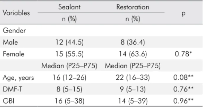
Table 2 compares the baseline characteristics of the subjects according to the intervention group. There was no signiicant difference between the subjects who received sealants versus restorations, in regard to baseline characteristics.
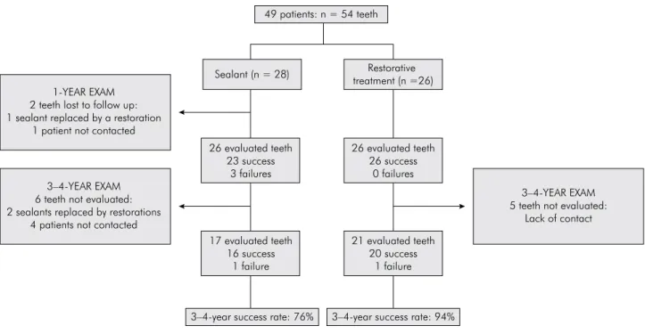
Figure 1 illustrates the lowchart of the study. Two cases were lost to follow-up in the sealant group, over the study period: 1 sealant was replaced by a restoration for unknown reasons, before the 1-year recall, and 1 patient could not be contacted. At the 1-year recall, 3 clinical failures were detected in the test group: 2 sealants were totally lost and 1 was partially lost (in need of repair). No lesion progression was observed in these teeth. In one case of total loss, which presented a cavity in the dentin, the remaining tissue was hard and dark brown. At the 3–4-year follow-up, 11 teeth could not be evaluated: 2 sealants were replaced by restorations between the two follow-ups, and 9 patients could not be contacted. These cases were included in the survival analysis containing the 1-year data. One clinical failure was detected in the control group at the 3–4-year recall: 1 restoration repair was necessary. No case of secondary caries was detected.
Table 3 shows the radiographic analysis of lesion depth, comparing the baseline with the 3–4-year images. Of the 17 teeth evaluated in the sealant group, 1 tooth showed caries progression. The tooth with caries progression presented the sealant as clinically acceptable in the clinical analysis. No case of caries progression was detected in the restoration group. Digital subtraction radiography performed in the sealant group showed 1 case with mineral loss (the same case detected in the side-by-side analysis), 14 cases that remained unchanged, and 2 cases with mineral gain, over the study period.
Combining the results of the clinical and the radiographic analyses, a total of 4 failures were observed in the sealant group, and 1 failure in the restoration group. Survival analysis showed success rates of 76% and 94% for the sealant and the restoration groups, respectively (Figure 2). No statistically significant difference was observed between the success rates of the restorations and the sealants (Weibull regression analysis, p = 0.16). When sealants replaced by restorations were considered as failures, their success rate decreased to 63%, and a borderline p-value was observed in the Weibull regression (p = 0.048), with an increased risk for sealant failure.
Regarding the deposition of tertiary dentin, one tooth could not be evaluated in the sealant group, Table 1. Sample distribution according to clinical and
radiographic assessments at baseline.
Radiographic assessment
Clinical assessment
CE CE +
Shadow CD Total
Absent 4 2 2 8
Caries at EDJ 9 2 2 13
Caries ≤ 1/3 D 13 5 6 24
Caries 1/3–2/3 D 0 3 6 9
Total 26 12 16 54
CE: cavity in enamel; CD: cavity in dentin; EDJ: enamel-dentin junction; D: dentin.
Table 2. Baseline characteristics of the subjects according to group.
Variables Sealant Restoration p
n (%) n (%)
Gender
Male 12 (44.5) 8 (36.4)
Female 15 (55.5) 14 (63.6) 0.78*
Median (P25–P75) Median (P25–P75)
Age, years 16 (12–26) 22 (16–33) 0.08**
DMF-T 8 (5–15) 9 (5–13) 0.76**
GBI 16 (5–38) 14 (5–39) 0.96**
Table 4. Radiographic analysis of tertiary dentin deposition, comparing the baseline with 3-4-year images. n (%)
Tertiary dentin Sealant* Restoration p**
Present 5 (31) 1 (5) 0.07
Absent 11 (69) 20 (95)
Total 16 (100) 21 (100)
*Figure in the sealant group totals 16, because the pulp chamber was not visible in the radiographic image of one tooth; **Fischer’s exact test.
because its pulp chamber was not visible in the bitewing radiograph. Sealed teeth showed a numerically higher proportion of cases of tertiary dentin deposition than restored teeth, but this difference was not statistically signiicant (p = 0.07) (Table 4).
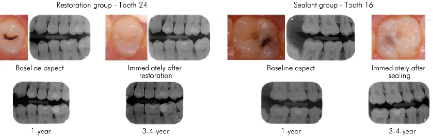
Figure 3 shows examples of teeth allocated to the test and the control groups (clinical and radiographic images at baseline and at 3–4-year follow-up). Figure 4 shows the radiographic images of sealed teeth (baseline and 3–4-year follow-up).
49 patients: n = 54 teeth
Sealant (n = 28)
26 evaluated teeth 23 success
3 failures
26 evaluated teeth 26 success
0 failures
17 evaluated teeth 16 success
1 failure
21 evaluated teeth 20 success
1 failure 1-YEAR EXAM
2 teeth lost to follow up: 1 sealant replaced by a restoration
1 patient not contacted
3–4-YEAR EXAM 6 teeth not evaluated: 2 sealants replaced by restorations
4 patients not contacted
3–4-YEAR EXAM 5 teeth not evaluated:
Lack of contact Restorative
treatment (n =26)
3–4-year success rate: 76% 3–4-year success rate: 94%
Figure 1. Flowchart of the study
Survival
analysis time Weibull regression
R
S 1
.8 .9
.7
.5 .6
.2 .3 .4
.1
0 500 1000 1500
Figure 2. Survival rates of sealants (S) and restorations (R) over the study period
Table 3. Radiographic analysis of lesion depth, comparing the baseline with 3-4-year images. n (%)
Lesion depth Sealant Restoration
Side-by-side analysis
Progression 1 (6) 0
Arrest 16 (94) 21 (100)
Regression 0 0
Digital subtraction analysis
Progression 1 (6)
-Arrest 14 (82)
-Regression 2 (12)
Discussion
The present study investigated the clinical and radiographic performance of sealants and conventional restorative treatments in occlusal carious lesions. Our results suggest that sealing initial cavitated lesions may arrest caries progression, as seen over a period of 3–4 years of monitoring.
In the present study, the majority of failures observed in the sealant group (3/4) were detected in the clinical examination. This inding is consistent with the results by Bakhshandeh et al.,10 who evaluated the sealing of occlusal carious lesions, achieving ≤ 2/3 of dentin in
permanent teeth. After 2–3 years, the authors observed that 9 out of a total of 10 failures were related to the clinical performance of the sealants: 7 cases showed loss of retention and 2 cases showed partial retention (annual failure rate of resin sealants of 7.4%). Similarly, Hesse et al.11 showed that all the failures detected in sealants placed in primary molars were clinical failures, resulting in a higher frequency of retreatments in this group. Collectively, these indings indicate that the use of dental sealants to control caries lesions requires patient compliance in attending regular follow-ups to assess the need for retreatment. As stated in a recent meta-analysis, although invasive techniques could require fewer retreatments, they may also trigger an earlier cycle of restorations; furthermore, more extensive/expensive interventions could also be required earlier.16 In this sense, sealants may avoid or, at least, postpone having to resort to restorative treatment.
Of the 20 sealed teeth followed for 3–4 years, only one case showed caries progression. The lack of caries progression in the vast majority of the sealed teeth conirms the knowledge that sealants a. form a physical barrier, b. isolate the caries lesion from the oral environment, c. restrict the access of nutrients to cariogenic bacteria, and 4. control caries progression. The tooth with total loss of sealant in our study clearly demonstrated this effect: the remaining dentin was hard and dark brown, highly indicative of lesion arrestment. Digital subtraction radiography was the method of choice to detect slight radiographic alterations, because it is a more sensitive technique. Even so, only one case was found to be associated with lesion progression. 1-year
Baseline aspect
Restoration group - Tooth 24 Sealant group - Tooth 16
Immediately after
restoration Baseline aspect Immediately after sealing
3-4-year 1-year 3-4-year
Figure 3. Clinical and radiographic images of teeth allocated to the test and the control groups (baseline and 3–4-year follow-up).
Tooth 16
Baseline 3-4-year
Tooth 16
Tooth 38
Moreover, this technique was able to show 2 cases with mineral gain in the carious lesion beneath the sealant. Interference with the cariogenic environment provides favorable conditions and enhances defensive responses of the pulp-dentin complex by promoting hard tissue formation,17 such as the focal deposition of tertiary dentin. In this study, tertiary dentin was observed more often in the sealant group, in agreement with Bakhshandeh et al.10. This inding evidences the reactionary capability of the pulp-dentin complex of sealed teeth. A 10-year study assessing the radiographic outcomes of sealing deep carious lesions has shown that the deposition of tertiary dentin is a slow and chronic process that may take many years to be radiographically evident12. Based on this inding, we could speculate that further reassessments of these patients may show a higher proportion of teeth presenting tertiary dentin deposition.
In the present study, 3 sealants were replaced by restorations, performed by another dental professional for unknown reasons. One could argue that these cases should automatically be computed as sealant failures. However, it is important to point out that the lesions included in our sample needed restorative treatment, according to the standard criteria for indicating restorations. This considered, the fact that the sealed lesions were restored does not entail lesion progression; rather, it probably means that the patient went for a dental visit and the professional performed a conventional treatment (removal of all carious dentin and restoration). Although we could judge that such cases would be better dealt with using the 1-year data, when the sealants were still available for evaluation, we performed a secondary analysis, and considered them as failures.
The comparison between the sealant and the restoration groups, regarding the subjects’ baseline characteristics, showed no significant differences between the groups. It is important to highlight that these comparisons are usually underpowered, and it is likely that statistically signiicant differences would be detected if we had a larger sample size (mainly in regard to age). Among the four subjects with more than one tooth included in the study, only one received both treatments. In this case, the baseline characteristics of this participant were included in both the sealant and the restorative treatment groups for this analysis (Table 2).
The fact that the treatments were performed by dental students could be seen as a limitation of this study; on the other hand, this increases the external validity and the generalizing scope of our results.
One could argue that the number of dropouts in this study may have had some negative impact on our indings. In fact, we are aware that if our inal sample size had been larger, we could have detected a statistically signiicant difference between the clinical performance of the sealants and the restorations. Previous studies have already shown better longevity for resin restoration than for sealants placed over initial active lesions. Bakhshandeh et al.10 found an annual failure rate for a sealant placed on initial lesions, i.e. 7.4% compared with 2% for posterior
resin restorations.18 However, the authors recognize that their main inding was that sealants were able to arrest caries progression over a 3–4-year period. The placement of a restoration causes the removal of a substantial amount of dental tissue, including healthy tissue. In this sense, the use of sealants to manage cavitated occlusal caries may avoid, or, at least, postpone having to resort to restorative treatment, thus improving oral health and tooth longevity. Although sealed carious dentin becomes more mineralized over time,12 this greater hardness may result in reduced tissue removal, in the event of a possible restoration placed at a future time.
In conclusion, this randomized controlled clinical trial demonstrated that sealing occlusal carious lesions in permanent teeth was able to control caries progression over a 3–4-year period. However, sealed carious lesions require patient compliance to attend regular follow-ups in order to control the occurrence of the clinical failures of sealants. Even if the tooth must be restored at a future time, the use of a sealant will postpone the restorative treatment, mineralize the carious tissue beneath the sealant, reduce tissue removal, and ultimately improve tooth prognosis.
Acknowledgements
1. Besic FC. The fate of bacteria sealed in dental cavities. J Dent Res. 1943;22(5):349-54.
https://doi.org/10.1177/00220345430220050101 2. Orhan AI, Oz FT, Ozcelik B, Orhan K. A clinical and
microbiological comparative study of deep carious lesion treatment in deciduous and young permanent molars. Clin Oral Investig. 2008;12(4):369-78. https://doi.org/10.1007/s00784-008-0208-6
3. Mertz-Fairhurst EJ, Schuster GS, Williams JE, Fairhurst CW. Clinical progress of sealed and unsealed caries. Part I: depth changes and bacterial counts. J Prosthet Dent. 1979;42(5):521-6. https://doi.org/10.1016/0022-3913(79)90245-2
4. Going RE, Loesche WJ, Grainger DA, Syed SA. The viability of microorganisms in carious lesions five years after covering with a fissure sealant. J Am Dent Assoc. 1978;97(3):455-62. https://doi.org/10.14219/jada.archive.1978.0327
5. Jensen ØE, Handelman SL. Effect of an autopolymerizing sealant on viability of microflora in occlusal dental caries. Scand J Dent Res. 1980;88(5):382-8. https://doi.org/10.1111/j.1600-0722.1980.tb01243.x 6. Maltz M, Oliveira EF, Fontanella V, Bianchi R. A clinical,
microbiologic, and radiographic study of deep caries lesions after incomplete caries removal. Quintessence Int. 2002;33(2):151-9. 7. Weerheijm KL, Kreulen CM, de Soet JJ, Groen HJ,
Amerongen WE. Bacterial counts in carious dentine under restorations: 2-year in vivo effects. Caries Res. 1999;33(2):130-4. https://doi.org/10.1159/000016506 8. Pinto AS, Araújo FB, Franzon R, Figueiredo MC, Henz S, García-Godoy F et al. Clinical and microbiological effect of calcium hydroxide protection in indirect pulp capping in primary teeth. Am J Dent. 2006;19(6):382-6.
9. Mertz-Fairhurst EJ, Curtis JW Jr., Ergle JW, Rueggeberg FA, Adair SM. Ultraconservative and cariostatic sealed restorations: results at year 10. J Am Dent Assoc. 1998;129(1):55-66. https://doi.org/10.14219/jada.archive.1998.0022
10. Bakhshandeh A, Qvist V, Ekstrand KR. Sealing occlusal caries lesions in adults referred for restorative treatment: 2-3 years of follow-up. Clin Oral Investig. 2012;16(2):521-9. https://doi.org/10.1007/s00784-011-0549-4
11. Hesse D, Bonifácio CC, Mendes FM, Braga MM, Imparato JC, Raggio DP. Sealing versus partial caries removal in primary molars: a randomized clinical trial. BMC Oral Health. 2014;14(1):58. https://doi.org/10.1186/1472-6831-14-58 12. Alves LS, Fontanella V, Damo AC, Oliveira EF, Maltz M.
Qualitative and quantitative radiographic assessment of sealed carious dentin: a 10-year prospective study. Oral Surg Oral Med Oral Pathol Oral Radiol Endod. 2010;109(1):135-41. https://doi.org/10.1016/j.tripleo.2009.08.021
13. Christgau M, Hiller KA, Schmalz G, Kolbeck C, Wenzel A. Quantitative digital subtraction radiography for the determination of small changes in bone thickness: an in vitro study. Oral Surg Oral Med Oral Pathol Oral Radiol Endod. 1998;85(4):462-72. https://doi.org/10.1016/S1079-2104(98)90076-2
14. Silva BB, Maltz M. [Prevalence of dental caries, gingivitis and fluorosis in 12-year-old schoolchildren from Porto Alegre - RS, Brazil, 1998/1999]. Pesqui Odontol Bras. 2001;15(3):208-14. http://dx.doi.org/10.1590/S1517-74912001000300006 15. Ainamo J, Bay I. Problems and proposals for recording
gingivitis and plaque. Int Dent J. 1975;25(4):229-35. 16. Schwendicke F, Jäger AM, Paris S, Hsu LY, Tu YK.
Treating pit-and-fissure caries: a systematic review and network meta-analysis. J Dent Res. 2015;94(4):522-33. https://doi.org/10.1177/0022034515571184
17. Bjørndal L, Mjör IA. Pulp-dentin biology in restorative dentistry. Part 4: Dental caries: characteristics of lesions and pulpal reactions. Quintessence Int. 2001;32(9):717-36. 18. Qvist V. Longevity of restorations: “The death spiral”.
In: Fejerskov O, Kidd E, editors. Dental caries: the disease and its clinical management. Oxford: Blackwell; 2008. p. 443-56.