405
Rev Bras Cir Cardiovasc | Braz J Cardiovasc Surg
Rev Bras Cir Cardiovasc 2013;28(3):405-7 Soubihe Junior NV, et al. - Presentation of a needle for direct or percutaneous
myocardium stem cells injection
RBCCV 44205-1488 DOI: 10.5935/1678-9741.20130062
Presentation of a needle for direct or percutaneous
myocardium stem cells injection
Apresentação de uma agulha para injeção direta ou percutânea de células tronco no miocárdio
Nathan Valle Soubihe Junior
1, MD; Andre Schmidt
1, MD, PhD; Agnes Afrodite Sumarelli
Albuquerque
1, BsC; Paulo Roberto Barbosa Evora
1,
MD, PhD;
1 Faculty of Medicine of Ribeirão Preto, University of São Paulo
(FMRP--USP), Ribeirão Preto, SP, Brazil.
Work carried out at Faculty of Medicine of Ribeirão Preto – USP (FM-RP-USP), Ribeirão Preto, SP, Brazil.
Work funded by CNPq, FAPESP, FAEPA and USP
Correspondence address: Paulo Roberto Barbosa Evora
Department of Surgery and Anatomy, Faculty of Medicine of Ribeirão Preto, University of São Paulo
Avenida Bandeirantes, 3900 – Monte Alegre – Ribeirão Preto, SP, Brazil – Zip Code: 14048-900
E-mail: [email protected]
Article received on April 7th, 2013
Article accepted on June 25th, 2013
SHORT COMMUNICATION
Abstract
The instrument has a locking mechanism and is composed of an external needle with blunt tip, with multiple 0.5 mm diam-eter holes. Internally it is itted with a mandrill needle, which can be mobilized inside occluding or releasing the lateral holes. The procedure for producing micro lesions is done by exchang-ing the blunt mandrill for a brush-mandrill, provided with mi-cro bristles that are structurally designed to ill the holes with small exteriorization of bristles. As an option to brush mandrill there is a second self-expandable feather shaped mandrill.
Descriptors: Stem cells. Myocardial infarction. Biopsy. Myo-cardium.
Resumo
O instrumento tem um mecanismo de bloqueio e é composto por uma agulha externa de ponta romba com vários furos de diâmetro de 0,5 mm. Internamente é equipada com uma agulha de mandril, que pode ser mobilizada ocluindo ou liberando os orifícios laterais. O procedimento para a produção de microlesões é feito por meio da troca do mandril de ponta romba por um mandril escova provido de microcerdas metálicas estruturalmente concebidas para preencher os orifícios com a exteriorização das pequenas cerdas. Como alternativa para mandril escova há um segundo mandril autoexpansível com o formato de uma pequena pena.
Descritores: Células-tronco. Infarto do miocárdio. Biópsia. Miocárdio.
INTRODUCTION
Sutton et al. [1] wrote, in 1964, a brief historical review reporting that percutaneous introduction of a needle into the cardiac ventricular cavities of human beings was performed many years ago for therapeutic reasons. Summaries of indications and technics were published by Henschen [2] and Lauter [3]. The feasibility of obtaining ventricular myocardial specimens from human beings either by open or closed
percutaneous routes was demonstrated in 1956 [4]. These old experiences proved that the heart tolerated these procedures very well, the myocardium quickly contracting around the small effect so that bleeding spontaneously ceased.
406
Rev Bras Cir Cardiovasc | Braz J Cardiovasc Surg
Rev Bras Cir Cardiovasc 2013;28(3):405-7 Soubihe Junior NV, et al. - Presentation of a needle for direct or percutaneous
myocardium stem cells injection
There is a clear superiority of results when direct myocardial injections are compared with intracoronary injections. Alternative methods of application of cell therapy products in myocardial infarction, acute or subacute, were tested. Direct injection as adjunctive therapy to revascularization surgery is a viable proposition, but it caters to a select group of patients referred for surgical revascularization immediately after an acute ischemic event [5-8].
The aim of this presentation is to describe a method of percutaneous puncture and injection of stem cells in the myocardium. This device is an adaptation Soubihe needle that was used in the ifties and sixties for myocardial biopsies [9].
METHODS
The instrument is equipped with a locking mechanism, which allows its perfect mobilization as one single unit for micro lesions, and it can be used only as a needle, so it becomes a biological injection instrument. The instrument for myocardium puncturing and injection of biological material is composed of an external needle, which contains at its end a blunt tip and multiple 0.5 mm diameter holes. Internally it is itted with a blunt mandrill, which when introduced into the external needle, can be mobilized inside to ill the lateral holes occluding or releasing them. This instrument is harmless to myocardial ibers and coronaries as it had been proved when its irst designed shape was used to perform heart biopsies, in the past (Figures 1 and 2).
Fig. 1 – Schematic presentation. The instrument has a locking mechanism (1,2,3) and is composed of an external needle with blunt
tip, with multiple 0.5 mm diameter holes (4,5). Internally it is itted
with a mandrill needle, which can be mobilized inside occluding or releasing the lateral holes. The procedure for producing micro lesions is done by exchanging the blunt mandrill for a brush-mandrill,
provided with micro bristles that are structurally designed to ill the
holes with small exteriorization of bristles (6). As an option to brush mandrill there is a second self-expandable feather shaped mandrill (7)
The main objective of the puncturing procedure is to release steam cells into the myocardium. For that, micro lesions should be performed to “receive” the stem cells. The procedure for producing micro lesions is done by exchanging the blunt mandrill by a brush-mandrill, provided with micro bristles that are structurally designed to ill the holes with small exteriorization of bristles. As an option to brush mandrill there is a second self-expandable feather shaped mandrill.
DISCUSSION
A very important characteristic of this instrument is safety. The blunt end of the needle guaranties that during the punching process, in case of unexpected touch of a coronary artery, the vessel will not be hurt or cut, but the needle will slide sideways from it. Therefore, steam cells are injected into the heart while it is perfectly pumping, with the chest unopened. A real possibility is the direct intramyocardial injection during thoracotomy through speciic or during the surgical treatment of coronary artery disease [10,11].
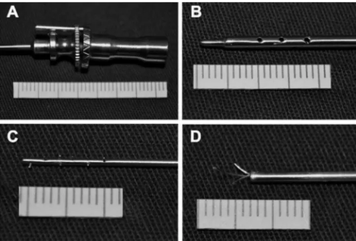
Fig. 2 – Photo presentation. A: Locking mechanism; B: External needle with blunt tip, with multiple 0.5 mm diameter holes. C: Brush-mandrill; D: Self-expandable feather shaped mandrill
Authors’ roles & responsibilities
NVSJ Original idea and design of the project AS Project planning
AASA Technical support
407
Rev Bras Cir Cardiovasc | Braz J Cardiovasc Surg
Rev Bras Cir Cardiovasc 2013;28(3):405-7 Soubihe Junior NV, et al. - Presentation of a needle for direct or percutaneous
myocardium stem cells injection
REFERENCES
1. Sutton GC, Driscoll JF, Gunnar RM, Tobin JR Jr. Exploratory mediastinotomy in primary myocardial disease. Progr Cardiovasc Dis. 1964;7:83-97.
2. Henschen K. Die wiederbelebung des herzens dureh peri- und intrakardiale injektion dureh herzaderlass und herzinfusion. Schweiz Med Wchnschr. 1920;1:261.
3. Lauter S. Kreislaufprobleme. Miinchener Med Wclmschr. 1930;77:526.
4. Kent G, Sutton DC, Sutton GC. Needle biopsy of the human ventricular myocardium. Q Bull Northwest Univ Med Sch. 1956;30(3):213-4.
5. Strauer BE, Kornowski R. Stem cell therapy in perspective. Circulation. 2003;107(7):929-34.
6. Perin EC, Geng YJ, Willerson JT. Adult stem cell therapy in perspective. Circulation. 2003;107(7):935-8.
7. Ladage D, Ishikawa K, Tilemann L, Müller-Ehmsen J, Kawase Y. Percutaneous methods of vector delivery in preclinical models. Gene Ther. 2012;19(6):637-41.
8. Krause K, Jaquet K, Schneider C, Haupt S, Lioznov MV, Otte KM, et al. Percutaneous intramyocardial stem cell injection in
patients with acute myocardial infarction: irst-in-man study.
Heart. 2009;95(14):1145-52.
9. Soubihe NV. Herzbiopsie. Thoraxchirurgie. 1961;9:31.
10. Galantier M, Moreira GB, Bub RF, Galantier J, Buffolo E, Carvalho AC, et al. Revascularização transmiocárdica a laser. Rev Bras Cir Cardiovasc. 1996;11(2):67-74.
11. Dallan LA, Gowdak LH, Lisboa LA, Schettert I, Krieger JE, Cesar LA, et al. Cell therapy plus transmyocardial laser revascularization: a proposed alternative procedure for refractory angina. Rev Bras Cir Cardiovasc. 2008;23(1):46-52.
ACKNOWLEDGEMENTS