Epidemiology
Raquel Gonçalves Vieira- Andrade(a)
Clarissa Lopes Drumond(b) Laura Pereira Azevedo Alves(b) Leandro Silva Marques(c) Maria Letícia Ramos-Jorge(b)
(a)Department of Orthodontics and Pediatric Dentistry, School of Dentistry, Federal University of Minas Gerais - UFMG, Belo Horizonte, MG, Brazil.
(b)Department of Pediatric Dentistry, School of Dentistry, Federal University of the Jequitinhonha and Mucuri Valleys, Diamantina, MG, Brazil.
(c)Department of Orthodontics, School of Dentistry, University of Rio Verde Valley - UNINCOR, Três Corações, MG, Brazil.
Corresponding Author: Raquel Gonçalves Vieira-Andrade E-mail: raquelvieira.andrade@gmail.com
Received for publication on Feb 09, 2012 Accepted for publication on May 10, 2012
Inflammatory root resorption in
primary molars: prevalence and
associated factors
Abstract: This study aimed at determining the prevalence of inlamma-tory root resorption and associated factors in 1068 primary mandibular molars in 453 children 3 to 12 years of age. Age, dental history and med-ical history were recorded using a questionnaire administered to the chil-dren’s parents/caregivers. Previously trained and calibrated examiners as-sessed radiographic images of the primary molars by direct observation, with the aid of a viewing box. Root resorption (physiological or inlam-matory), dental crown status (healthy, carious with no pulp involvement, carious with pulp involvement and evidence of restoration), and pulpot-omy or pulpectpulpot-omy were determined. Data analysis involved descriptive statistics, the chi-square test and a multiple logistic regression (p < 0.05). The prevalence of inlammatory root resorption was 16.2% (n = 173). The male gender (OR: 1.4; 95% CI), the 3-to-7-years age bracket (OR: 1.5; 95% CI), an unhealthy dental crown (OR: 8.7; 95% CI), caries with pulp involvement (OR: 7.4; 95% CI), pulpotomy (OR: 3.1; 95% CI), and pulpectomy (OR: 5.4; 95% CI) were risk factors for the occurrence of in-lammatory root resorption in primary molars. In conclusion, the preva-lence of inlammatory root resorption in the present sample was 16.2%. Gender, age, an unhealthy tooth, caries with pulp involvement, pulpot-omy, pulpectpulpot-omy, and the absence of a restoration were associated with a higher occurrence of inlammatory root resorption in primary molars.
Descriptors: Root Resorption; Prevalence; Epidemiology.
Introduction
Inlammatory root resorption in primary teeth is a frequent inding in the clinical practice of pediatric dentists.1 Radiographically, inlammato-ry root resorption is characterized by the gradual loss of tooth substance associated with persistent, progressive radiolucency of the adjacent alveo-lar bone.2,3 This leads to an imbalance in the stomatognathic system and problems such as unerupted permanent successors and the early loss of primary teeth.1,3-10
Inlammatory root resorption is generally diagnosed by clinical and radiological examinations, especially periapical radiographs.11,12 This type of resorption is a consequence and/or complication of different clini-cal situations, such as pulp, periodontal and/or periradicular infectious processes, dental injury, orthodontic forces,or excessive occlusal forces.1,6
Anatomical aspects peculiar to the primary teeth, such as reduced Declaration of Interests: The authors
enamel and dentin thickness, greater permeability and lower hardness and strength, further contribute to the more rapid spread of infectious processes in the pulp tissue, which can trigger an inlammatory process and root resorption.13,14 Treatment consists of removing the triggering factors, whose determi-nation is critical to choosing the most appropriate protocol.9
Although studies in the literature report the prevalence of and factors associated with the devel-opment of inlammatory resorption in permanent teeth, little is known about how these factors inlu-ence the occurrinlu-ence of inlammatory resorption in primary teeth.3,15 Since the occurrence of this type of resorption is highly frequent in the clinical prac-tice of pediatric dentists, the study of this phenom-enon is of considerable importance to the diagnosis and planning of dental care for children.
This study was conducted to determine the prev-alence of inlammatory pathological root resorp-tion, and associated factors, in the primary mandib-ular molars of children 3 to 12 years of age, based on radiographic images and clinical records.
Methodology
A retrospective cross-sectional study was car-ried out on a sample of 1068 primary mandibular molars assessed on 671 periapical radiographs re-trieved from the radiographic iles of the pediatric clinic at the Federal University of the Jequitinhonha and Mucuri Valleys (Brazil). The periapical radio-graphs were taken with four identical x-ray ma-chines (Dabi Atlante, Ribeirão Preto, Brazil) and processed manually. Only high-quality periapical radiographs (with adequate details, good sharpness, density and contrast, and no distortions) exhibiting the root apex of primary mandibular molars and the tooth bud of the permanent successor were used. The radiographs were obtained from 453 male and female children 3 to 12 years of age, from the city of Diamantina, state of Minas Gerais, Brazil.
The research team was composed of four exam-iners who underwent a training program and cali-bration process involving 80 randomly selected ra-diographic images of primary mandibular molars, either involved in an inlammatory process or not.
These images were not part of the main study. Seven days following the initial evaluation, the same 80 radiographs were re-evaluated by the examiners to determine intraexaminer agreement (kappa index). The examiners’ results were compared to those of a doctoral student with expertise on the subject, considered the gold standard. Kappa values ranged from 0.82 to 0.91. Diagnostic differences were dis-cussed and resolved by consensus.
A questionnaire was previously administered to the children’s parents/caregivers to acquire baseline data on gender, age, dental history and medical his-tory. Medical history involved the presence/absence of general health problems, such as systemic and chronic diseases. Dental history was taken to gain information on treatments performed on the teeth under study.
The periapical radiographs were analyzed by the researchers by direct observation, with the aid of a viewing box (CARP, Ribeirão Preto, Brazil). This assessment was divided into two phases. The researchers irst determined the type of root resorp-tion (physiological or inlammatory). Inlammatory root resorption was deined as the presence of a periapical lesion radiographically characterized by radiolucency of the adjacent alveolar bone, associat-ed with the loss of tooth substance. The researchers then determined the dental crown status (healthy, carious with no pulp involvement or carious with pulp involvement). The researchers also checked whether the primary molar showed any evidence of restoration, pulpotomy or pulpectomy, which was conirmed by examining the patient’s records.
Statistical analysis
was set at 5% (p < 0.05). All independent variables with a p-value ≤ 0.20 were included in a multivari-ate logistic regression model.
The present study received the approval of the Institutional Review Board of the Federal University of the Jequitinhonha and Mucuri Valleys (Brazil) under protocol number 135/10.
Results
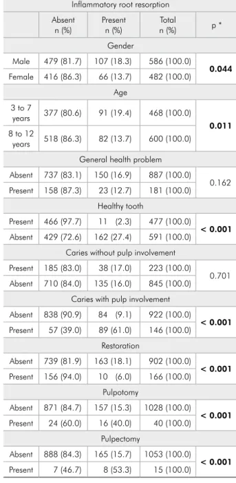
A total of 1068 primary mandibular molars were assessed from 453 children (mean age: 7.9 years; standard deviation: 2.12 years), 54.4% (n = 252) of whom were male and 45.6% (n = 211) were female. The prevalence of inlammatory root resorption was 16.2% (n = 173). Statistically signiicant associations were found between inlammatory root resorption and gender (p < 0.05), age (p < 0.05), an unhealthy tooth (p < 0.001), caries with pulp involvement (p < 0.001), a restored tooth (p < 0.001), pulpotomy (p < 0.001) and pulpectomy (p < 0.001) (Table 1). No statistically signiicant association was found between inlammatory root resorption and general health problems or caries with no pulp involvement.
Table 2 shows the results of the multivariate lo-gistic regression analysis. The following variables were risk factors for inlammatory root resorption in primary mandibular molars: male gender (OR: 1.4; 95% CI), 3-to-7-years age bracket (OR: 1.5; 95% CI), an unhealthy tooth (OR: 8.7; 95% CI), caries with pulp involvement (OR: 7.4; 95% CI), ab-sence of a restoration (OR: 3.1; 95% CI), pulpotomy (OR: 3.1; 95% CI) and pulpectomy (OR: 5.4; 95% CI).
Discussion
In the present study, inlammatory root resorp-tion was associated with gender, age, an unhealthy tooth, caries with pulp involvement, pulpotomy, pulpectomy and absence of a restoration. This diver-sity of clinical situations corroborates the indings of a previous study, deining inlammatory resorp-tion as a complicaresorp-tion and/or consequence of dif-ferent clinical situations, such as pulp, periodontal and/or periradicular infectious processes.1
A statistically signiicant association was found between inlammatory root resorption and age
Table 1 - Associations between independent variables and inflammatory root resorption.
Inflammatory root resorption Absent
n (%) Presentn (%) Totaln (%) p * Gender
Male 479 (81.7) 107 (18.3) 586 (100.0)
0.044 Female 416 (86.3) 66 (13.7) 482 (100.0)
Age 3 to 7
years 377 (80.6) 91 (19.4) 468 (100.0)
0.011 8 to 12
years 518 (86.3) 82 (13.7) 600 (100.0) General health problem Absent 737 (83.1) 150 (16.9) 887 (100.0)
0.162 Present 158 (87.3) 23 (12.7) 181 (100.0)
Healthy tooth
Present 466 (97.7) 11 (2.3) 477 (100.0)
< 0.001 Absent 429 (72.6) 162 (27.4) 591 (100.0)
Caries without pulp involvement Present 185 (83.0) 38 (17.0) 223 (100.0)
0.701 Absent 710 (84.0) 135 (16.0) 845 (100.0)
Caries with pulp involvement Absent 838 (90.9) 84 (9.1) 922 (100.0)
< 0.001 Present 57 (39.0) 89 (61.0) 146 (100.0)
Restoration
Absent 739 (81.9) 163 (18.1) 902 (100.0)
< 0.001 Present 156 (94.0) 10 (6.0) 166 (100.0)
Pulpotomy
Absent 871 (84.7) 157 (15.3) 1028 (100.0)
< 0.001 Present 24 (60.0) 16 (40.0) 40 (100.0)
Pulpectomy
Absent 888 (84.3) 165 (15.7) 1053 (100.0)
< 0.001 Present 7 (46.7) 8 (53.3) 15 (100.0)
* chi-square test.
Pathological processes involving the hard tissues of the root (cementum and dentin) and adjacent tis-sues (periodontal ligament and alveolar bone) may occasionally be associated with inlammatory re-sorption.9,18 Dental caries was the most prevalent pathological process in the present study. Caries with pulp involvement represented an
approximate-ly sevenfold greater chance of having inlammatory root resorption. Dental pulp attacked by microor-ganisms that are part of the cariogenic process suf-fers primary inlammation/infection.19 In primary molars, this infection spreads more quickly to the inter-radicular region, owing to the lesser thickness of the pulp chamber loor and the presence of fo-ramina in the furcation areas. As a result, bone loss occurs in the inter-radicular area, which is one of the irst signs of a compromised pulp.10,20
Restorative procedures, pulpotomy and pulp-ectomy are among the various treatment options available for tooth decay.21-23 In the present study, the absence of a restoration and pulpotomy or pulp-ectomy were related to a greater occurrence of in-lammatory root resorption. Pulpectomy was related to the greatest risk, with an approximately ivefold greater chance of being related to this type of root resorption. Pulpectomy is indicated for cases of ad-vanced degenerative pulp alterations or pulp necro-sis.24 According to some studies, this procedure may accelerate the process of inlammatory root resorp-tion.19 Pulpotomy is a more conservative treatment option commonly used on teeth having deep carious lesions with no istula or swelling, and with no peri-apical or furcation lesions.20,25 In the present study, however, the teeth submitted to pulpotomy were ap-proximately threefold more likely to have a periapi-cal lesion, a inding which agrees with that reported by Aminabadi et al.,26 who found that root resorp-tion was the second most common inding among teeth submitted to pulpotomy. Based on the above-mentioned information, the occurrence of inlam-matory root resorption in primary molars treated by pulpotomy or pulpectomy in the present study may be attributed to diagnostic errors made while assess-ing pulp condition, or to technical failure while per-forming the treatment chosen.
As mentioned earlier, primary molars with no restorations had an approximately threefold greater chance of displaying inlammatory root resorption. This association may be explained by the fact that some teeth had untreated caries. However, although restorative procedures are among the various treat-ment options available for tooth decay,21-23 the in-creased risk of an inlammatory resorption
occur-Table 2 - Results of the multivariate logistic regression for inflammatory root resorption.
Independent variables
Unadjusted OR
(95% CI) p
* Adjusted OR
(95% CI) p Gender
Female 1.00 1.00
Male (1.0–1.9)1.40 0.044 (0.8–1.8)1.24 0.295
Age 8 to 12
years 1.00 1.00
3 to 7
years (1.1–2.1)1.52 0.011* (0.7–1.7)1.13 0.548 Health problem
Absent 1.00 1.00
Present (0.4–1.1)0.71 0.163 (0.5–1.6)0.89 0.705
Healthy tooth
Present 1.00 1.00
Absent (8.5–29.8)15.99 < 0.001 (4.3–17.4)8.70 < 0.001
Caries with pulp involvement
Absent 1.00 1.00
Present (10.4–23.2)15.57 < 0.001 (4.6–11.9)7.40 < 0.001
Restoration
Present 1.00 1.00
Absent (1.7–6.6)3.44 < 0.001 (1.5–6.6)3.15 0.002
Pulpotomy
Absent 1.00 1.00
Present (1.9–7.1)3.69 < 0.001 (1.5–6.5)3.12 0.002
Pulpectomy
Absent 1.00 1.00
Present (2.2–17.1)6.15 0.001 (1.8–15.8)5.40 0.002
ring in primary molars with no restorations should be considered with caution, since this group also in-cluded healthy teeth.
Periapical radiographs were used to assess the type of resorption in the present study. The course of a root resorption is monitored by radiographic examination only in the more advanced stages of this process, and periapical radiographs are the most commonly used diagnostic tools for this pur-pose.11,12 However, today there are more accurate di-agnostic methods to evaluate resorption processes, including cone-beam computed tomography.27 This method is thus suggested for future investigations with the same objective as that of the present study.
One of the limitations of this study was the fact that the evaluation of radiographic images does not allow the determination of the level of caries activity or of the exact degree of pulp involvement. A study carried out by Di Nicolo et al.28 found that normal or transitioning pulp (in the treatable category) was diagnosed in 50% of teeth with inactive le-sions and 11.1% of teeth with active carious lele-sions. Therefore, conducting a combined clinical and ra-diographic examination is essential to obtaining a proper diagnosis and to choosing the most appro-priate treatment for each clinical situation. Another limitation of this study was that it did not allow de-termining the time elapsing between the completion of pulpotomy or pulpectomy and the occurrence of
inlammatory root resorption in each patient, since both events—pulp intervention and root resorption —were identiied on the same radiograph at a single moment in time. Because some information was ob-tained through a questionnaire administered to the children’s parents/caregivers, one must consider the possibility of memory bias, especially with regard to the children’s medical history. Moreover, some in-formation may have been recorded inadequately or even omitted from the clinical charts.
Because the cross-sectional design does not in-clude temporality (exposure and outcome data are collected on a single occasion), it was not possible to determine a possible cause-effect relationship be-tween the results. Further studies with a longitudi-nal design should thus be conducted to gain a better understanding of the relationship between inlam-matory root resorption and the possible risk factors addressed herein.
Conclusion
The prevalence of inlammatory root resorption in the primary mandibular molars investigated in the present study was 16.2%. Gender, age, an un-healthy tooth, caries with pulp involvement, pulp-otomy, pulpectomy and the absence of a restoration were related to a higher occurrence of inlammatory root resorption in primary molars.
References
1. Santos BZ, Bosco VL, Silva JYB, Lamb MMR. [Physiological and pathological factors and mechanisms in the process of root resorption of deciduous teeth]. RSBO (Online). 2010;7(3):332-9. Portuguese.
2. Kinirons MJ, Boyd DH, Gregg TA. Inflammatory and replace-ment resorption in reimplanted permanent incisor teeth: a study of characteristics of 84 teeth. Endod Dent Traumatol. 1999 Dec;15(6):269-72.
3. Cardoso M, Rocha MJ. Identification of factors associated with pathological root resorption in traumatized primary teeth. Dent Traumatol. 2008 Jun;24(6):343-9.
4. Tronstad L. Root resorption – etiology, terminology and clinical manifestations. Endod Dent Traumatol. 1988 Dec;4(6):241-52.
5. Camm JH, Schuler JL. Premature eruption of the premolars. ASDC J Dent Child. 1990 Mar-Apr;57(2):128-33.
6. Gunraj MN. Dental root resorption. Oral Surg Oral Med Oral Pathol Oral Radiol Endod. 1999 Dec;88(6):647-53.
7. Sahara N. Cellular events at the onset of physiological root resorption in rabbit deciduous teeth. Anat Rec. 2001 Dec;264(4):387-96.
8. Leroy, RB. Impact of undergraduate students experience in the deciduous molars on the emergence of successors. Eur J Oral Sci 2003;111(2):106-10.
9. Fuss Z, Tsesis I, Lin S. Root resorption: diagnosis, classifica-tion and treatment choices based on simulaclassifica-tion factors. Dent Traumatol. 2003 Aug;19(4):175-82.
11. Andreasen FM, Andreasen JO. Diagnosis of luxation injuries: the importance of standardized clinical, radiographic and photographic techniques in clinical investigations. Endod Dent Traumatol. 1985 Oct;1(5):160-9.
12. Sameshima GT, Asgarifar KO. Assessment of root resorption and root shape: periapical vs panoramic films. Angle Orthod. 2001 Jun;71(3):185-9.
13. Morabito A, Defabianis P. A SEM investigation on pulpal-periodontal connections in primary teeth. ASDC J Dent Child. 1992 Jan-Feb:59(1):53-7.
14. Ne RF, Witherspoon DE, Gutmann JL . Tooth resorption. Quintessence Int. 1999 Jan;30(1):9-25.
15. Borum MK, Andreasen JO. Sequelae of trauma to primary maxillary incisors. I. Complications in the primary dentition. Endod Dent Traumatol. 1998 Feb;14(1):31-44.
16. Ak G, Sepet E, Pinar A, Aren G, Turan N. Reasons for early loss of primary molars. Oral Health Prev Dent. 2005;3(2):113-7.
17. Leroy R, Cecere S, Lesaffre E, Declerck D. Caries experience in primary molars and its impact on the variability in permanent tooth emergence sequences. J Dent. 2009 Nov;37(11):865-71. 18. Hammarstrom L, Lindskog S. General morphological aspects
of resorption of teeth and alveolar bone. Int Endod J. 1985 Apr;18(2):93-108.
19. Nero-Vieira g. La resorción as proceso inflamatorio. Aproxi-mación a La pathogenicity de las resorciones y dentaria perio-dontal. RCOE. 2005;10(5-6):545-56.
20. Kramer PF, Faraco Júnior IM, Meira R. The SEM investiga-tion of accessory foramina in the furcainvestiga-tion areas of primary molars. J Clin Pediatr Dent. 2003 Winter;27(2):157-61. 21. Fuks AB, Eidelman E. Pulp therapy in the primary dentition.
Curr Opin Dent. 1991 Oct;1(5):556-63.
22. JOE Editorial Board . Treatment of the primary tooth: an online study guide. J Endod. 2008 May;34(5 Suppl):e107-10. 23. Blanchard S, Boynton J. Current pulp therapy options for
primary teeth. J Mich Dent Assoc. 2010 Jan;92(1):38,40-1. 24. Mendoza AM, Reina JE, Garcia-Godoy F. Evolution and
prog-nosis of necrotic primary teeth after pulpectomy. Am J Dent. 2010 Oct;23(5):265-8.
25. Souza RA, Gomes SC, Dantas JC, Silva-Sousa YT, Pécora JD. Importance of the diagnosis in the pulpotomy of immature permanent teeth. Braz Dent J. 2007;18(3):244-7.
26. Aminabadi NA, Farahani RM, Gajan EB. A clinical study of formocresol pulpotomy versus root canal therapy of vital pri-mary incisors. J Clin Pediatr Dent. 2008 Spring;32(3):211-4. 27. D’Addazio PS, Campos CN, Özcan M, Teixeira HG, Passoni RM, Carvalho AC. A comparative study between cone-beam computed tomography and periapical radiographs in the di-agnosis of simulated endodontic complications. Int Endod J. 2011 Mar;44(3):218-24.
