Forensic Dentistry
Sudhanshu Saxena(a)
(a) Department of Public Health Dentistry, People’s College of Dental Sciences and Research Centre, People’s group, Bhanpur, Bhopal, Madhya Pradesh, India.
Correponding author: Sudhanshu Saxena
Department of Public Health Dentistry, People’s College of Dental Sciences and Research Centre, People’s group, Bhanpur Karond bypass road
Bhopal - Madhya Pradesh - India ZIP: 462037
E-mail: [email protected]
Received for publication on Oct 11, 2010 Accepted for publication on Dec 15, 2010
Age estimation of indian adults from
orthopantomographs
Abstract: The aim of this study was to develop a method for estimating the chronological age of Indian adults based on the relationship between age and various morphological variables of canine teeth, obtained us-ing orthopantomographs. Orthopantomographs of 120 selected patients were digitized, and radiographic images of the right maxillary canine in each case were processed using a computer aided drafting program. Pulp/ tooth area ratio, pulp/root length ratio, pulp/tooth length ratio, pulp/root width ratio at the cemento-enamel junction level, pulp/root width ratio at midroot level, and pulp/root width ratio at the midpoint between the cemento-enamel junction and the midroot of the canine were calculat-ed by measuring various features on the images. Pearson’s correlation, multiple linear regression, one way analysis of variance (ANOVA), and Student’s t-test were used for statistical analysis. Regression equations were developed to estimate age from morphological variables. The ob-served minus the estimated age in the total study sample ranged from -2.2 to +1.5 years, in males from -0.9 to +0.8 years, while in females it was from -1 to +0.8 years. Differences between observed and estimated ages of subjects were not statistically signiicant. In conclusion there is a linear relationship of pulp/root width ratio at mid-root level and pulp/ tooth area ratio of the right maxillary canine with chronological age in the Indian population. Age of subjects can therefore be estimated with a good degree of accuracy using regression equations.
Descriptors: Forensic Dentistry; Age Determination by Teeth; Adult.
Introduction
It is often necessary to estimate an individual’s age due to certain questions related to legal requirements in paleodemographic research or in a forensic context.
Although several parts of the body can be used for age estimation, the poor condition of the remains in particularly severe crashes or ires in cases of those recently dead; or of moisture and burial conditions in the case of historic subjects, make many parts of the body unusable. For these reasons, the teeth are the part of the body most frequently used for identiication and age estimation.1
In children, age determination from teeth is a relatively simple, ac-curate procedure and is based on the stages of development and eruption of teeth. However, in adults it is a challenge to medico-legal science.2
including methods which analyze the various forms of tooth modiication such as wear, dentin transpar-ency, tooth cementum annulations, racemization of aspartic acid and apposition of secondary dentin. Some of the methods are very complex and destruc-tive and are therefore not normally used; wear and the apposition of secondary dentin are the currently available nondestructive methods. Tooth wear is inluenced by various external factors (masticato-ry function, type of food, timing and sequence of tooth eruption), tooth form, position of teeth, thick-ness and hardthick-ness of enamel and predisposition to enamel hypoplasia. However, apposition of second-ary dentin is a continuing, regular process which is only modiied by caries or particular abrasion. Sec-ondary dentin has been studied by sectioning and radiography.1
The study of tooth radiographs is a nondestruc-tive and simple process which can be applied to both living and deceased persons, in contrast to other time consuming, expensive, less reliable and de-structive methods which may not be acceptable for ethical, religious, cultural or scientiic reasons.3
Fur-ther, procedures such as digitization of panoramic radiographs and computer assisted image analysis avoid the bias inherent in observer subjectivity and improve reliability, accuracy and precision.4
Any tooth can be used to assess age. Canine teeth are a good candidate for age estimation be-cause they are often present in old age. They are less likely than other anterior teeth to suffer wear as a result of particular work and are the single-rooted teeth with the largest pulp area and thus are the eas-iest to analyze.1
Based on the above background, this study was conducted to develop a method for estimating the chronological age of Indian adults based on the re-lationship between age and various morphological variables of canine teeth, obtained using orthopan-tomographs.
Materials and Methods
Based on the results of a pilot study, to achieve a 5% level of signiicance and 80% power, a study with a sample size of 120 participants was required, calculated using Power and Precision v. 4 (Biostat
Inc., Englewood, USA). One hundred and twenty orthopantomographs (Planmeca Oy, Helsinki, Fin-land) from patients of both sexes seen at the People’s College of Dental Sciences and Research Centre, Bhopal, Madhya Pradesh state, India, were collected based on the following criteria:
a. Patients aged between 21 to 60 years b. Fully erupted right maxillary canine
c. Fully formed root of right maxillary canine d. Right maxillary canine free from any pathology
such as caries, periodontitis, abrasion, erosion, fracture or impaction
e. Absence of restoration and endodontic illing of the right maxillary canine
f. Absence of malaligned or rotated right maxillary canine
Orthopantomographs were taken as part of rou-tine treatment that was being rendered to the pa-tient. Participation in the study was voluntary. Rel-evant data including date of birth and gender were entered into the proforma after obtaining informed consent. Ethical clearance was obtained from the ethical committee of the People’s College of Dental Sciences and Research Centre.
Methods
Orthopantomographs of selected patients were obtained and digitized using an ASTRA 4000U scanner (UMAX Technologies Inc., Dallas, USA). Radiographic images of canines (RIC) were pro-cessed using the computer-aided drafting program, Auto CAD 2005 (Autodesk Inc., San Rafael, USA).
Twenty points around the edge of the tooth out-line and ten points around the pulp outout-line of the right maxillary canine were identiied. Measure-ments of the canine tooth area and the pulp area from the radiographic images were then evaluated.
RIC were re-examined after an interval of one week. The following morphological variables were cal-culated from the above measurements:
• AR = pulp/tooth area ratio • p = pulp/root length ratio • r = pulp/tooth length ratio
• a = pulp/root width ratio at CEJ level • c = pulp/root width ratio at midroot level
• b = pulp/root width ratio at midpoint level be-tween CEJ level and midroot level
The observed age was calculated by subtracting the date of birth from the date of the radiograph.
Statistical analysis was performed using SPSS v. 17 (SPSS Inc., Chicago, USA). Pearson’s correla-tion coeficients were calculated for the relacorrela-tionship between observed age and predictive variables. A multiple linear regression model for age estimation was developed by selecting those variables which contributed signiicantly to age estimation. One way analysis of variance (ANOVA) was used to compare differences between observed and estimated age in different age groups. Student’s t-test was used to compare the morphological variables of males with those of females and to compare observed age with estimated age. A p value < 0.05 was considered sta-tistically signiicant.
Results
Kappa statistics showed that the intra-examiner agreement score (k = 0.94) was almost perfect.
In the present study, 60 males and 60 females (to-tal 120 subjects) were equally divided into four age groups, i.e. 21-30 years, 31-40 years, 41-50 years, and 51-60 years.
The morphological variables did not show any signiicant difference between the sexes (p > 0.05). Pearson’s correlation coeficients between observed age and predictive morphological variables in males, females and in the total sample population were highly signiicant and inversely correlated for AR (pulp/tooth area ratio) and c (pulp/root width ratio at midroot level) (p < 0.001).
The subjects’ ages were modeled as a linear func-tion of the morphological variables (predictors). A multiple linear regression procedure was therefore
applied to optimize this model.
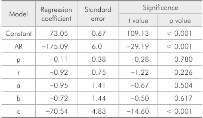
Table 1 shows the regression analysis of the total sample, while Table 2 shows the results in males and Table 3 in females. Among all the morphological variables tested, only AR (pulp/tooth area ratio) and c (pulp/root width ratio at midroot level) showed signiicant results. Hence, a regression model uti-lizing AR and c was used (Table 4) which yields the following linear regression formula to estimate chronological age:
• For subjects of unknown gender: Age = 72.48 - 203.74(AR) - 51.69(c) • For male subjects:
Age = 73.33 - 209.97(AR) - 54.06(c) • For female subjects:
Age = 72.12 - 179.12(AR) - 68.66(c)
Table 1 - Regression analysis with all morphological vari-ables (predictors) and age as the dependent variable in the total study sample (n = 120).
Model Regression coefficient
Standard error
Significance
t value p value
Constant 71.65 0.60 120.30 < 0.001
AR -204.52 3.78 -54.16 < 0.001
p 0.09 0.44 0.21 0.833
r 0.59 0.70 0.84 0.402
a 0.63 0.59 1.07 0.285
b 1.48 1.02 1.46 0.148
c -52.21 3.66 -14.24 < 0.001
Dependent variable: Age
Table 2 - Regression analysis with all morphological vari-ables (predictors) and age as the dependent variable in males (n = 60).
Model Regression coefficient
Standard error
Significance
t value p value
Constant 73.05 0.67 109.13 < 0.001
AR -175.09 6.0 -29.19 < 0.001
p -0.11 0.38 -0.28 0.780
r -0.92 0.75 -1.22 0.226
a -0.95 1.41 -0.67 0.504
b -0.72 1.44 -0.50 0.617
c -70.54 4.83 -14.60 < 0.001
The observed minus the estimated age in the total sample population ranged from -2.2 to +1.5 years. There was no signiicant difference between observed and estimated age for any of the age groups (Table 5). In addition, the differences of observed and estimated age between age groups were also not statistically signiicant (ANOVA value = 2.259, p = 0.085).
The observed minus the estimated age in males ranged from -0.9 to +0.8 years, while in females it ranged from -1 to +0.8 years. There was no signii-cant difference between observed and estimated age for either males or females (Table 6).
Discussion
The results of the present study showed no sig-niicant difference between observed and estimated age. Age was estimated using regression equations based on pulp/tooth area ratio and pulp/root width ratio at mid-root level.
In 1925 Bodecker established that the apposition of secondary dentin correlated with age.5 In 1995,
Kvaal et al. presented a method for age estimation which was based on investigation of periapical ra-diographs,3 while Paewinsky et al. veriied the
ap-plicability of this method on orthopantomograms.6
In 2004 Cameriere et al. found a linear relation-ship between the pulp/root width ratio at mid-root level, the pulp/tooth area ratio, and chronological age.4 This inding was consistent with the indings
in present study. Cameriere et al. stated that the ra-tio between pulp and tooth area correlated best with age.4 With this background they developed
regres-sion equations for age estimation using the pulp/ tooth area ratios of canines from the maxilla and the mandible separately.1
In their study, Babshet et al. found that Cameri-Table 3 - Regression analysis with all morphological
vari-ables (predictors) and age as the dependent variable in fe-males (n = 60).
Model Regression coefficient
Standard error
Significance
t value p value
Constant 72.54 0.59 122.52 < 0.001
AR -208.10 3.11 -66.82 < 0.001
p 1.05 0.57 1.83 0.073
r -0.39 0.73 -0.53 0.597
a 0.49 0.42 1.17 0.247
b 0.23 0.88 -0.26 0.792
c -56.37 3.45 -16.36 < 0.001
Dependent variable: Age
Table 4 - Regression analysis with selected morphological variables (predictors) and age as the dependent variable.
Group Model Regression coefficient
Standard error
Significance
t value p value
To
tal
Constant 72.48 0.19 388.39 < 0.001
AR -203.74 3.71 -54.93 < 0.001
c -51.69 3.63 -14.24 < 0.001
Male
s Constant 73.33 0.21 349.76 < 0.001
AR -209.97 2.99 -70.24 < 0.001
c -54.06 3.27 -16.54 < 0.001
Fe
m
ale
s Constant 72.12 0.16 437.99 < 0.001
AR -179.12 5.00 -35.83 < 0.001
c -68.66 4.53 -15.16 < 0.001
Dependent variable: Age
Table 5 - Comparison of observed and estimated age in subjects grouped according to age.
Age groups
(years)
Age in years (mean ± SD)
t-test
value p value Observed age Estimated age
21-30 24.86 ± 2.98 24.64 ± 3.25 0.267 0.790 (NS)
31-40 35.25 ± 2.73 35.30 ± 2.64 0.061 0.952 (NS)
41-50 44.60 ± 3.02 44.46 ± 3.11 0.171 0.865 (NS)
51-60 55.09 ± 2.55 54.81 ± 2.60 0.432 0.667 (NS)
Over all 39.95 ± 11.58 39.80 ± 11.56 0.100 0.921(NS)
*NS = Not significant
Table 6 - Comparison of observed and estimated age of subjects according to gender.
Gender Age in years (mean ± SD) t-test
value p value Observed age Estimated age
Male 40.20 ± 11.15 40.04 ± 11.13 0.080 0.937 (NS)
Female 39.70 ± 12.08 39.56 ± 12.05 0.062 0.951 (NS)
Over all 39.95 ± 11.58 39.80 ± 11.56 0.100 0.921 (NS)
6. Paewinsky E, Pfeiffer H, Brinkmann B. Quantification of secondary dentine formation from orthopantomograms--a contribution to forensic age estimation methods in adults. Int J Legal Med. 2005 Jan;119(1):27-30.
7. Babshet M, Acharya AB, Naikmasur VG. Age estimation in Indians from pulp/tooth area ratio of mandibular canines. Fo-rensic Sci Int. 2010 Apr 15;197(1-3):125.e1-4. Epub 2010 Jan 27.
8. Singaraju S, Sharada P. Age estimation using pulp/tooth area ratio: a digital image analysis. J Forensic Dent Sci. 2009 May;1(1):37-41.
9. Cameriere R, Cunha E, Sassaroli E, Nuzzolese E, Ferrante L. Age estimation by pulp/tooth area ratio in canines: study of a Portuguese sample to test Cameriere’s method. Forensic Sci Int. 2009 Dec 15;193(1-3):128.e1-6. Epub 2009 Oct 24. ere’s formula, based on the Italian population, is not
as applicable to the Indian population.7 I also found
the same result in my pilot study using this formula. These results indicate that racial and cultural fac-tors might play an important role in age estimation. Therefore I decided to develop regression equations for the Indian population. I used both pulp/root width ratio at mid-root level and pulp/tooth area ra-tio in regression equara-tions to make the equara-tion as close to ideal as possible.
The present study showed no signiicant inlu-ence of gender on age estimation using canine tooth measurements, which was similar to the indings of previous studies.1,4,8 Nevertheless I developed
sepa-rate formulas for males and females to predict age as accurately as achievable.
The present study showed 99.7% variance and 0.60 standard error of the estimate, when selected variables were used. This result was similar to re-sults of the study conducted by Cameriere et al., who reported 97% variance with 2.37 years stan-dard error of the estimate.9
Though my results are promising, I cannot gen-eralize them to other populations. Further research should be aimed at analyzing a larger sample, in-cluding not only age and gender but also racial and cultural parameters. This method of age estimation cannot be applied to multirooted teeth, as accurate measurements are dificult to perform on these. Sim-ilarly, as the curved arch of the jaw is projected on to a lat ilm, there will always be a certain amount of distortion when measuring the image presented there.8 To create a uniform amount of distortion in
all the radiographs used I chose a particular tooth, the right maxillary canine.
Conclusion
Within the limitations of the study it can be con-cluded that there is a linear relationship of pulp/root width ratio at mid-root level, and pulp/tooth area ratio of the right maxillary canine with chronologi-cal age in the Indian population. In unknown sub-jects age can be estimated using regression equa-tions based on the above mentioned variables.
References
1. Cameriere R, Ferrante L, Belcastro MG, Bonfiglioli B, Rastelli E, Cingolani M. Age estimation by pulp/tooth ratio in canines by peri-Apical Xrays. J Forensic Sci. 2007 Jan;52(1):166-70. 2. Jain RK, Rai B. Age estimation from permanent molar’s attri-tion of Haryana populaattri-tion. Indian J Forensic Odontol. 2009 Apr;2(2):59-61.
3. Kvaal SI, Kolltveit KM, Thomsen IO, Solheim T. Age esti-mation of adults from dental radiographs. Forensic Sci Int. 1995 Jul 28;74(3):175-85.
4. Cameriere R, Ferrante L, Cingolani M. Variations in pulp/ tooth area ratio as an indicator of age: a preliminary study. J Forensic Sci. 2004 Mar;49(2):317-9.