Effectiveness of high speed instrument
and air abrasion on different dental
substrates
Abstract: The aim of this study was to compare the effectiveness of high speed (HS) and air abrasion (AA) instruments on groups of teeth (decidu-ous, permanent, bovine), in terms of preparation time, topography and presence of smear layer. Each group consisted of 5 teeth that had their buccal/lingual surfaces prepared by using either HS or AA. All proce-dures were standardized and timed. The teeth were then sectioned and prepared for evaluation of both the topography and the presence of smear layer by scanning electron microscopy. As regards preparation time, HS yielded preparations 1.5 times quicker than AA did on the three types of dental substrates (Wilcoxon test, p < 0.05). In both techniques (Kruskal-Wallis Test, p < 0.05) the preparation time was inluenced by the dental substrate, particularly in deciduous teeth (Mann-Whitney test with Bon-feroni’s correction, p < 0.017), which required a longer preparation time. In the descriptive analysis of the topography, no difference was found between the substrates. Nonetheless, the different instruments used de-termined distinctive topographies. Both techniques produced a smear layer (χ2 McNemar, p > 0.05) in all substrates, but with different forma-tions. In conclusion, the HS instrument was found to be more rapid than the AA. No difference was found between the three dental substrates as regards both the topography and the presence of smear layer. The dif-ferences found in the present study were only in relation to the effects of each instrument used.
Descriptors: Tooth preparation; Air abrasion, dental; Tooth, deciduous;
Microscopy, electron, scanning; In vitro.
Lívia Azeredo Alves Antunes(a)
Rafael Lima Pedro(a)
Áurea Simone Barrôso Vieira(b)
Lucianne Cople Maia(c)
(a) Master of Science Students; (b)Master of
Science; (c)Associate Professor – Department
of Pediatric Dentistry and Orthodontics, School of Dentistry, Federal University of Rio de Janeiro, RJ, Brazil.
Corresponding author:
Lucianne Cople Maia Rua Gastão Gonçalves, 47 apto 501- Santa Rosa
Niterói - Rio de Janeiro - Brazil CEP: 24240-030
E-mail: [email protected]
Introduction
The development of caries removal techniques has become increasingly conservative and biological. This has been made possible by a better understand-ing of the etiology and prevention of dental caries. In addition, an advanced concept of cavity prepara-tion has emerged as a result of the introducprepara-tion of acid-etching techniques, adhesive restorative materi-als, and the development of new cavity preparations systems, such as air abrasion systems.1
According to Eick et al.2 (1972) and Nakabayas-hi et al.3 (1982), the structural integrity and surface characteristics of the tooth after caries excavation may be relevant to the adhesiveness of the restor-ative material to be used. Therefore, it should be borne in mind that even an ideal cutting instru-ment must fulill the requireinstru-ments established for both patient and practitioner, despite the develop-ment of alternative cavity preparation techniques.4 Moreover, speed and effectiveness of carious tissue removal,5 resulting in satisfactory morphology with minimum formation of smear layer, so that the ad-hesive restorative materials can be properly applied, are important features6 for preparing a dental cav-ity without interfering with tooth restoration. Fur-thermore, the increasing use of different dental sub-strates (bovine, deciduous, and permanent teeth) in research emphasizes the importance of cavity prepa-ration techniques when such teeth are involved.
By taking factors such as these into account, the aim of the present study was to compare in vitro the effects of high speed and air abrasion instruments on deciduous, permanent and bovine teeth, in terms of preparation time, topography, and presence of smear layer.
Material and Methods
This study was conducted after being approved by the local Ethics Committee. Altogether, 15 teeth
ing a double-faced diamond disc (KG Sorensen, São Paulo, SP, Brazil) obtaining two surfaces (buccal and lingual) that were ixed on acrylic bases.
Class V cavity preparations (n = 30) were ran-domly performed on the buccal and lingual surfaces by one operator only. The instruments used were a diamond tip (1061, KG Sorensen, São Paulo, SP, Brazil) mounted on a high speed device (605 extra torque, Kavo, Joinville, SC, Brazil) (n = 15) and an air abrasion system (RONDOlex 2013, Kavo, Bib-erach, Riss, Germany) with a 120° tip, 0.64 mm in diameter. Air abrasion was carried out with 50 µm aluminum oxide grains at a pressure of 80 psi and distance of 1 mm from the tooth surface.
As regards the cavity preparations, the dimen-sions were standardized to 2.0 x 2.0 x 2.5 mm by using a millimetric probe (Hu-Friedy, Zweignieder-lassung Deutschland, Leimen, Germany) for mea-suring the depth, and digital caliper (Mitutoyo, To-kyo, Japan) for measuring width and length.
The diamond tips were changed after every 5 cavity preparations and the air abrasion instrument was cleaned after every 2.
The time spent on each cavity preparation was recorded with a chronometer (Technos, Manaus, AM, Brazil) and the data were then tabulated. The Wilcoxon (p < 0.05), Kruskal-Wallis (p < 0.05), and Mann-Whitney tests, as well as Bonferroni’s cor-rection (p < 0.017) for non-paired correlation, were used for statistical analysis of instruments and sub-strates.
Topographic evaluation of the cavity prepara-tions was based on photomicrographs and quantita-tive and qualitaquantita-tive parameters were taken into ac-count.
(2,000 X) of the smear layer (Kp = 0.98), and scores were adopted7 according to McNemar’s chi-square test (p < 0.05).
Results
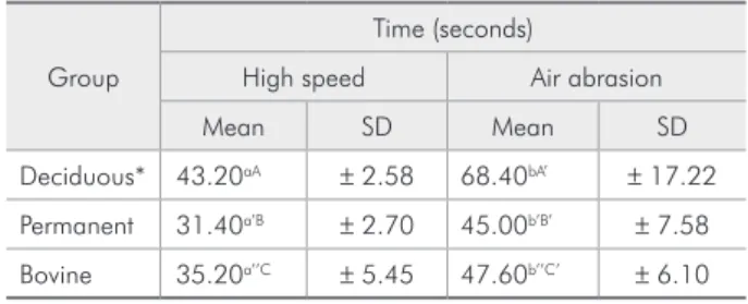
With respect to preparation time (Table 1), it was found that the high speed instruments yielded cavity preparations, on average, 1.5 times more
rap-idly than the air abrasion instruments with regard to the three types of dental substrates. The dental substrates inluenced the preparation time irrespec-tive of the instrument used, and cavities involving permanent teeth were prepared more rapidly than those in bovine and deciduous substrates. Delay in preparing deciduous teeth was found to be statisti-cally signiicant.
With respect to topography, descriptive analysis of Groups D, P, and B showed that the high speed instrument yielded more uniform, U-shaped cavities with deined cavosurface and inner angles, as well as grooves on both enamel and dentine (Figure 1), whereas the air abrasion system yielded more irreg-ular, W- or V-shaped cavities with cavosurface and internal contours forming a margin around the cav-ity preparation. Moreover, enamel and dentine had an irregular aspect with the presence of aluminum oxide (Figure 2).
With respect to the smear layer, both instruments yielded its formation in all teeth (χ2 McNemar,
Table 1 - Cavity preparation time (in seconds) according to instrument used in the three types of dental substrates.
Group
Time (seconds)
High speed Air abrasion
Mean SD Mean SD
Deciduous* 43.20aA ± 2.58 68.40bA’ ± 17.22
Permanent 31.40a’B ± 2.70 45.00b’B’ ± 7.58
Bovine 35.20a’’C ± 5.45 47.60b’’C’ ± 6.10
Different letters indicate statistical significance: row = normal letter (Wil-coxon test); column = capital letter (Kruskal-Wallis test). * Mann-Whitney test with Bonferroni’s correction p < 0.01.
Figure 1 - Substrate topographies resulting from the use of the high speed instrument: (A) deciduous, (B) permanent, and (C) bovine. Note the design of the cavosurface margin (arrows) (Original Magnification 35 X).
A
C
Figure 2 - Substrate topographies resulting from the use of the air abrasion instrument: (A) deciduous, (B) permanent, and (C) bovine. Note the design of the cavosurface margin (arrows) (Original Magnification 35 X).
Figure 3 - Aspect of the smear layer produced by the high speed instrument (Original magnification 2,000 X).
Figure 4 - Aspect of the smear layer produced by the air abrasion instrument impregnated with aluminum oxide par-ticles (Original magnification 2,000 X).
A B
Discussion
Although bovine teeth have commonly been used in research on permeability,8 adhesiveness,9 mor-phology and microleakage10 due to the dificulty in obtaining human permanent and deciduous teeth,9 studies comparing the effects of different prepara-tion systems on bovine teeth are still scarce.
According to Yazici et al.11 (2002), as morphol-ogy and nature of the dentine surface are the main factors in performing adhesive techniques success-fully, it is important to know the type of dentine surface obtained after using cutting instruments, so that alternative treatments, speciically designed for better adaptation and adhesion of restorative ma-terials, can be applied,12 which justiies the present study.
Differently from the new techniques for remov-ing carious tissue, the effectiveness of high speed rotary instruments yielding precise cutting, deined angles, and a rough surface are well established in the literature.13 Moreover, the outcomes from such instruments can be used as analysis parameters since they are widely used and accepted by dental practitioners.
Effective cutting is known to be largely related to the ease with which a given instrument removes tooth tissue, that is, minimum effort in a short time.13 However, it is very dificult to establish the time spent on preparing a cavity with either high speed or air abrasion instruments. In general, stud-ies report that both preparation systems are fast and secure by evaluating the pre-ixation time14,15 or an-alyzing the time/weight relationship.16 In the present study, sample dimensions were standardized, and time measurements were also recorded.
Although there were small differences regard-ing preparation time between the three substrates, the time involved in preparing the deciduous teeth was signiicantly different. This can be explained by the absence of an instrument ixation apparatus. As a result, the operator had to be more careful when preparing the deciduous substrates because of their anatomy, size and smaller thickness. This situation is indeed the clinical reality that dental practitioners face when handling such teeth.
It was observed that the high speed instrument
was 1.5 times quicker than the air abrasion in pre-paring dental cavities, thus corroborating another study.15 However, the results of air abrasion prepa-ration time were not found to be very different, since it tended to become shorter as more preparations were performed (Table 1). Therefore, these results demonstrate that air abrasion can be considered a faster system in comparison with other alternative methods of cavity preparation, such as the ultrason-ic abrasion system. For instance, in two studies per-formed in 200417 and 200518 comparing ultrasonic abrasion with a high speed system, Vieira et al.18 (2005) found that the high speed system was 7.9 and 4.9 times faster, respectively, than the ultrasonic abrasion system, thus evidencing the effectiveness of air abrasion in clinical practice.
According to Yazici et al.11 (2002), SEM obser-vation of the dentine surface treated by different caries removal techniques helps explain the adhe-sive mechanisms involving composite/dentine. Since the behavior of the three types of teeth studied was found to be similar with regard to topography and presence of smear layer, this suggests that different substrates can be compared with each other.
With respect to the instruments used for cavity preparation, it could also be observed that both high speed and air abrasion systems yielded smear layer formation. However, both instruments left morpho-logically differentiated smear layers on the dentine surface, which was also corroborated in other stud-ies.4,6,11
High speed instruments yielded U-shaped cavi-ties with well deined cavosurface and inner angles,19 presence of smear layer,18 grooves on the surface left by the diamond tip, and issures and microfractures on enamel.18,19 Watson, Cook20 (1995) explain that this results from compression and relaxation usu-ally caused by diamond tips and drills. Fissures and microfractures existing in the cavity walls would be the result of changes in the dental surface due to the high impact and cyclic stress caused to the tooth during the use of diamond tips at high speed. These are all disadvantages associated with microleakage and postoperative sensitivity.
abrasion preparation were conservative,21,22 exhibit-ing irregular surfaces due to the impact of alumi-num oxide particles.21 Moreover, a great number of such particles were observed on the dental surfaces, which is corroborated by Yazici et al.11 (2002) but not in another study.19
Nevertheless, the most relevant characteristic re-ported by the previous studies of air abrasion, 19,21-23 and also conirmed by the present study, was a round-shaped margin. This round contour not only favors adhesion and restoration placement, which reduces microleakage,24,25 but is also considered im-portant for the longevity of adhesive restorations26 when associated with typical air abrasion cutting characteristics, namely, rough surfaces and a halo effect.19 This longevity is explained by a reduced in-cidence of fractures in comparison with cavity prep-arations that have deined acute angles. A round contour enables a gradual transition between tooth and restoration, and polymerization shrinkage stress is also diminished.
Restoration material bonding to the dental struc-ture is an important factor in achieving a clinically successful treatment. According to Al-Omari et al.27 (2001), such a bonding depends on several vari-ables, including cavity geometry and type of bond-ing agent. The authors also suggest that dental
to-pography can inluence the quality of the adhesive systems. Therefore, further studies of the behavior of the different adhesive systems (self-etch and to-tal-etch) on dental surfaces treated with either high speed or air abrasion instruments are of crucial im-portance, specially because of the particular char-acteristics resulting from such cavity preparation techniques.
Conclusion
Based on the methodology of the present study, it was observed that the high speed technique was faster than the air abrasion technique for cavity preparation. No difference was found between the three dental substrates with regard to topography and presence of smear layer. The differences found in the present study were only in relation to the ef-fects of the instruments used, since they determined the topography of the surface and type of smear layer.
Acknowledgements
The authors would like to thank the electronic microscopy and microanalysis laboratory (PEMM/ COPPE/ UFRJ) for performing the SEM, and pro-fessor Ronnir Raggio (NESC/ UFRJ) for the statisti-cal analysis support.
References
1. Yip HK, Samaranayake LP. Caries removal techniques and instrumentation: a review. Clin Oral Invest. 1998;2(4):148-54.
2. Eick JD, Johnson LN, Fromer JR, Neumann AW. Surface topography: its influence on wetting and adhesion in a dental adhesive system. J Dent Res. 1972;51(3):780-8.
3. Nakabayashi N, Kojima K, Masuhara E. The promotion of adhesion by the infiltration of monomers into tooth substrates. J Biomed Mater Res. 1982;16(3):265-73.
4. Banerjee A, Watson TF, Kidd EA. Dentine caries
7. Rome WJ, Doran JE, Walker WA 3rd. The efectiveness of
Gly-Oxide and sodium hypochlorite in preventing smear layer formation. J Endod. 1985;2(7):281-8.
8. Tagami J, Tao L, Pashley DH, Horner JA. The permeabil-ity of dentine from bovine incisors in vitro. Arch Oral Biol. 1989;34(10):773-7.
9. Nakamichi I, Iwaku M, Fusayama T. Bovine teeth as possible substitutes in the adhesion test. J Dent Res. 1983;62(10):1076-81.
13. Siegel SS, Fraunhofer JAV. Assessing the cutting efficiency of dental diamond burs. J Am Dent Assoc. 1996;127(6):763-72. 14. Bester SP, de Wet FA, Nel JC, Driessen CH. The effect of air-borne particle abrasion on the dentin smear layer and dentin: an in vitro investigation. Int J Prosthodont. 1995;8(1):46-50. 15. Fernandes MA, Fontana VF. Estudo comparativo in vitro da capacidade de desgaste de estrutura de esmalte dental em dife-rentes tempos, utilizando pontas de diamante e air-touch system. Rev CROMG. 2002;8(1):49-53.
16. Black RB. Air abrasive: some fundamentals. J Am Dent Assoc. 1950;41(6):701-10.
17. Vieira ASB, Alves MPS, Antunes LAA, Primo L, Maia LC. Abra-são ultrasônica versus alta rotação: avaliação do tempo de preparo cavitário e da microinfiltração. In: 21ª Reunião Anual da SBPqO; 2004. Águas de Lindóia. Anais. São Paulo: Sociedade Brasileira de Pesquisa Odontológica; 2004. p. 172.
18. Vieira ASB, Alves MPS, Antunes LAA, Primo L, Maia LC. To-pografia e presença de “smear layer” em dentes decíduos prepa-rados com alta rotação e abrasão ultra-sônica in vitro. In: 22ª Reunião Anual da SBPqO; 2005. Águas de Lindóia. Anais. São Paulo: Sociedade Brasileira de Pesquisa Odontológica; 2005. p. 175.
19. Laurell KA, Hess JA. Scanning electron micrographic effects of air-abrasion cavity preparation on human enamel and den-tin. Quintessence Int. 1995;26(2):139-44.
20. Watson TF, Cook RJ. The influence of bur blade concentricity on high speed tooth cutting interaction: a video-rate confocal microscopic study. J Dent Res. 1995;74(11):1749-55.
21. Antunes LAA, Vieira ASB, Alves MPS, Maia LC. Influência de quatro tipos de pontas na topografia de preparos cavitários ci-néticos em incisivos bovinos. In: 22ª Reunião Anual da SBPqO; 2005. Águas de Lindóia. Anais. São Paulo: Sociedade Brasileira de Pesquisa Odontológica; 2005. p. 220.
22. Peruchi C, Santos-Pinto L, Santos-Pinto A, Barbosa e Silva E. Evaluation of cutting patterns produced in primary teeth by an air-abrasion system. Quintessence Int. 2002;33(4):279-83. 23. Santos-Pinto L, Peruchi C, Cordeiro R, Marker VA.
Evalua-tion of cutting patterns produced with air-abrasion systems using different tip designs. Oper Dent. 2001;26(3):308-12. 24. Hamilton JC, Dennison JB, Stoffers KW, Welch KB. A clinical
evaluation of air-abrasion treatment of questionable carious le-sions. A 12-month report. J Am Dent Assoc. 2001;132(6):762-9.
25. Hicks MJ, Parkins FM, Flaitz CM. Kinetic cavity preparation effects on secondary caries formation around resin restora-tions: a polarized light microscopic in vitro evaluation. ASDC J Dent Child. 2001;68(2):115-21.
26. Nordbo H, Leirskar J, Von Der Fehr FR. Saucer-shaped cav-ity preparations for posterior approximal resin composite restoration: observations up to 10 years. Quintessence Int. 1998;29(1):5-11.