ABSTRACT: This study assessed the surface softening and abrasive wear of eroded bovine enamel with or without the inluence of toothbrushing. Five volunteers took part in this in situ study of 5 days. They wore acrylic palatal appliances containing 6 bovine enamel blocks divided in two rows with 3 blocks, which corresponded to the stud-ied groups: erosion without toothbrushing (GI) and erosion with toothbrushing (GII). The blocks were subjected to erosion by immersion of the appliances in a cola drink for 10 minutes, 4 times a day. After that, no treatment was performed in one row (GI), whereas the other row was brushed (GII). The appliance was then replaced into the mouth. Enamel alterations were determined using proilometry and microhardness tests. Data were tested using paired Student’s t test (p < 0.05). The mean wear values (µm) and percentage of supericial microhardness change (%SMHC) were respectively: GI - 2.77 ± 1.21/91.61 ± 3.68 and GII - 3.80 ± 0.91/58.77 ± 11.47. There was a sig-niicant difference in wear (p = 0.001) and %SMHC (p = 0.001) between the groups. It was concluded that the wear was more pronounced when associated to toothbrushing abrasion. However, toothbrushing promoted less %SMHC due to the removal of the altered supericial enamel layer.
DESCRIPTORS: Tooth erosion; Tooth abrasion; Beverages.
RESUMO: Este estudo avaliou o amolecimento supericial e o desgaste do esmalte bovino erodido com ou sem inlu -ência da escovação. Cinco voluntários izeram parte deste estudo in situ de 5 dias. Eles usaram aparelhos palatinos de acrílico contendo 6 blocos de esmalte bovino divididos em 2 ileiras com 3 blocos, cada ileira correspondeu a um grupo em estudo: erosão sem escovação dentária (GI) e erosão seguida de escovação dentária (GII). Os blocos foram submetidos à erosão por imersão do aparelho em uma bebida à base de cola por 10 minutos, 4 vezes ao dia. Depois disso, nenhum tratamento foi realizado em uma ileira (GI), enquanto a outra ileira foi escovada (GII). Em seguida, o aparelho foi recolocado na boca. Alterações do esmalte foram determinadas por teste de perilometria e microdureza. Os dados foram analisados usando-se teste t de Student (p < 0,05). O valor médio de desgaste (µm) e da porcentagem de perda de microdureza supericial (%PDS) foi respectivamente: GI – 2,77 ± 1,21/91,61 ± 3,68 e GII – 3,80 ± 0,91/58,77 ± 11,47. Houve uma diferença estatisticamente signiicante no desgaste (p = 0,001) e na %PDS (p = 0,001) entre grupos. Conclui-se que o desgaste foi mais pronunciado quando associado à abrasão pela escovação. Entretanto, a escovação promoveu menor %PDS, devido à remoção da camada de esmalte supericial alterada.
DESCRITORES: Erosão de dente; Abrasão dentária; Bebidas.
INTRODUCTION
While the incidence of the major dental dis-ease, caries, has declined in developed countries27,
other dental lesions such as erosion are becoming
increasingly important15. Dental erosion is defined
as the loss of tooth substance by chemical proc-esses not involving bacteria20 caused by a variety
* Assistant Professor, Department of Pediatric Dentistry, School of Dentistry of Maringá, Maringá University Center.
** PhD Student, Department of Pediatric Dentistry, School of Dentistry of Bauru; ****Associate Professor, Department of Biological Science, School of Dentistry of Bauru; ******Associate Professors, Department of Pediatric Dentistry, School of Dentistry of Bauru; *****Associate Professor, Department of Restorative Dentistry, School of Dentistry of Ribeirão Preto – University of São Paulo. *** PhD Student, Department of Pediatric Dentistry, School of Dentistry of Araçatuba, São Paulo State University.
2006;20(2):148-54
Influence of toothbrushing on enamel softening and abrasive
wear of eroded bovine enamel: an
in situ
study
Influência da escovação no amolecimento superficial do esmalte
e desgaste do esmalte bovino erodido: um estudo
in situ
Daniela Rios*
Heitor Marques Honório** Ana Carolina Magalhães*** Marília Afonso Rabelo Buzalaf**** Regina Guenka Palma-Dibb*****
Rios D, Honório HM, Magalhães AC, Buzalaf MAR, Palma-Dibb RG, Machado MAAM, Silva SMB. Inluence of toothbrushing on enamel softening and abrasive wear of eroded bovine enamel: an in situ study. Braz Oral Res 2006;20(2):148-54.
of extrinsic and intrinsic factors9,25.
Intrinsic factors are the result of endogenous acids, generally gastric acids that become in contact with dental surface during nervous anorexia, nerv-ous bulimia and gastroesophagic disturbances28,29.
Extrinsic factors are related to frequent consump-tion of acidic foodstuffs or beverages and exposure to acidic contaminants in the working environ-ment22. In modern societies the extrinsic factors
are becoming more important, due to the increased consumption of acidic drinks as soft drinks, sport drinks, fruit juices and fruit teas22.
The acidic attack leads to irreversible loss of dental hard tissue accompanied by a demineraliza-tion and softening of the surface2,9,17,25. This softened
zone, particularly of enamel, appears to be more susceptible to removal by mechanical forces, like attrition and abrasion, which have little or no effect on the intact tissues1,4,5,8,18. Toothbrushing is a very
important procedure to prevent dental caries. On the other hand it represents a mechanical force that can led to abrasion. Thus, the analysis of alterations that can be promoted by toothbrushing of previ-ously eroded enamel is important and necessary.
The methods used for measurement of the su-perficial alterations of dental enamel can be quan-titative (superficial microhardness, profilometry) and qualitative (scanning electron microscopy)14.
Microhardness testing permits the measurement of the degree of softening of the surface, but does not allow the quantification of the height of surface loss. The profilometric method uses a depth-sens-ing instrument, which gives the height of this sur-face loss. Since the dental enamel can be softened (altering the microhardness) in association with dental wear, a combined study, using microhard-ness and profilometry would be able to quantify the alterations resulted from erosion and abrasion6,14.
Taking into account the association between erosion and abrasion and the importance of com-bined methodologies, the purpose of this study was to assess in situ the surface softening and abrasive wear of eroded bovine enamel with or without the influence of toothbrushing, by using profilometric and microhardness analyses.
MATERIALS AND METHODS
Experimental design
This in situ study was approved by the Re-search and Ethics Committee, School of Dentistry of Bauru, University of São Paulo. Five healthy adult volunteers with a mean age of 23 yr (range
19-28 yr) (Figure 1I), with normal salivary flow (1 ml/min, pH 7.0), and residing in a fluoridated area (0.70 mgF/L) took part in this study, during 5 days11. They wore acrylic palatal appliances, which
contained two rows of 3 bovine dental enamel blocks each. The rows corresponded to one of these conditions: erosion without toothbrushing (GI) and erosion with toothbrushing (GII). The use of two conditions in the same intraoral palatal appliance was supported by the absence of a cross-effect in previous studies5,15,21. Enamel mineral loss was
determined by profilometric and surface micro-hardness tests. Each group comprised 5 experi-mental units (volunteers), which were compared using paired Student’s t test.
Enamel blocks and palatal appliance preparation
Sixty enamel blocks (4 x 4 x 3 mm) were pre-pared from bovine incisors sterilized by storage in a 2% formaldehyde solution (pH 7.0) for 30 days at room temperature11. Using one diamond disk
(Isomet 1000; Buehler, Lake Bluff, IL, USA) the crowns were sectioned from the roots (Figure 1A). Next, using two parallel diamond disks separated by a 4-mm spacer, one slice was cut from the crown of each bovine incisor (Figure 1B). The enamel surface of the blocks (Figure 1C) was ground flat with wa-ter-cooled carborundum discs (320, 600 and 1,200 grades of Al2O3 papers; Buehler, Lake Bluff, IL, USA),
and polished with felt paper wet by diamond spray (1 µm; Buehler) (Figures 1D and 1E) for surface microhardness determination (five indentations in different regions of the blocks, 25 g, 5 s, HMV-2000; Shimadzu Corporation, Tokyo, Japan) (Figure 1F).
Thirty blocks, with a mean (SD) surface micro-hardness of 353 (± 23) KHN, were randomly divided into two groups. In order to maintain reference surfaces for lesion depth determination, two layers of nail varnish were applied on half of the surface of each block (Figure 1G).
On the left and right sides of the intraoral pal-atal appliances, 6 cavities of 5 x 5 x 3 mm were made, and into each of them, one block of enamel was randomly fixed with wax (Figure 1H). The po-sition of two groups (right or left) on the appliance was randomly chosen.
Treatments
During the experimental phase, the volunteers brushed their teeth with fluoride-free dentifrice6.
days16,30. On the first 12 hours, specimens were
not subjected to erosive and abrasive processes, to allow the formation of a salivary pellicle15. On
the following 5 days, erosive and abrasive chal-lenges were made extraorally 4 times a day and at predetermined times (8:00, 12:00, 16:00 and 20:00 h)5,16,30.
In order to submit the enamel blocks to ero-sion, the volunteers were instructed to remove the appliance and immerse it in a cup containing 150 ml of a freshly opened bottle of Coke(pH 2.8, Spal, Porto Real, RJ, Brazil) for 10 minutes (Fig-ure 1J)3,5. After that, no treatment was performed
in one row (GI), whereas the other row (GII) was immediately brushed by the volunteers (Figure 1K). The brushing procedure consisted of 30 extraoral brushing strokes with a soft end-rounded tooth-brush (Bitufo; Sanifil, Jundiaí, SP, Brazil) with a small portion (approximately 0.3 g) of the dentifrice without fluoride (Crest, pH 6.8, silica as abra-sive)7. Volunteers were trained and instructed to
carefully perform this procedure and to avoid a carry-across effect of the treatments. The brushed blocks were washed under deionized water and before the appliance was replaced into the mouth,
the volunteers were instructed to take one sip of the beverage24.
The volunteers received instructions to wear the appliances continuously, including at night, but to remove them during meals (3 times a day), when oral hygiene was performed11,15. Plaque control on
the blocks was achieved by dipping the appliance in 0.2% chlorhexidine gluconate solution (pH 6.8) for 5 minutes at the end of each study day16,30.
Wear assessment
After 5 days, the volunteers stopped wearing the appliances. The enamel blocks were removed from the appliances and the nail varnish over the surfaces was carefully cleaned with acetone-soaked cotton wool4. The blocks were dried and the enamel
Rios D, Honório HM, Magalhães AC, Buzalaf MAR, Palma-Dibb RG, Machado MAAM, Silva SMB. Inluence of toothbrushing on enamel softening and abrasive wear of eroded bovine enamel: an in situ study. Braz Oral Res 2006;20(2):148-54.
formed in micrometers (µm): three blocks x five readings.
Percentage of superficial microhardness change assessment
After the wear assessment, enamel surface microhardness was measured as described ear-lier (Figure 1L) and an average per volunteer was obtained. Ten indentations on each speci-men were made, five on the previously protected enamel surface (SMH) and five on the experimen-tal area (SMH1). Using these measurements, the
percentage of superficial microhardness change (%SMHC) was calculated (%SMHC = [[SMH1
-SMH]/SMH] × 100).
Scanning electron microscopy
Four specimens whose wear and %SMHC val-ues were closest to the mean of each group were selected for scanning electron microscopic observa-tions. The specimens were mounted and covered with palladium-gold by ion sputtering in a Hammer VI cathodic evaporator (Anatech LTD, Alexandria, USA) (Figures 1O and 1P). Then, they were studied and photographed in a JEOL JSM T220A scanning electron microscope (Tokyo, Japan) operating at 15 kV (Figure 1Q).
Statistical analysis
Each group comprised 5 experimental units (volunteers), which were statistically analised. First, the assumptions of equality of variances and
normal distribution of errors were checked for the response variables tested. Since the assumptions were satisfied, the paired Student’s t test was car-ried out for statistical comparisons. The signifi-cance level was set at 5%.
RESULTS
All five volunteers completed the study. Ta-ble 1 shows that Group I (erosion) had a signifi-cantly higher %SMHC than Group II (erosion plus abrasion) (p = 0.001). Nevertheless the wear was less pronounced for Group I when compared to Group II (p = 0.001).
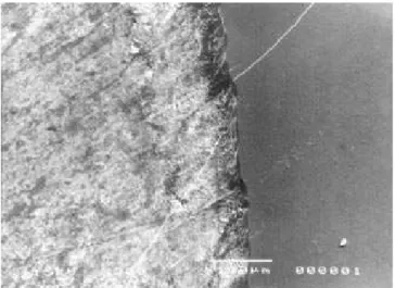
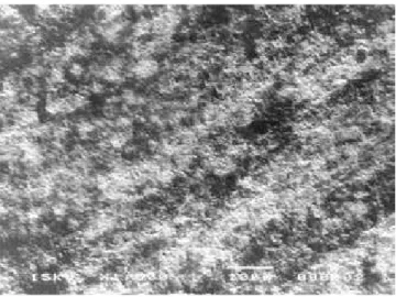
The scanning electron microscopy images permitted the visualization of a distinct line of demarcation at the test-control margin on both groups (Figures 2 and 3). Specimen surfaces that had been covered with nail varnish did not show alterations. At higher magnification of the test area of group I (erosion), prism core dissolution was ob-served (Figure 4). However, the test area of group II (erosion plus toothbrushing) showed a less altered
TABLE 1 - Means and SD of %SMHC and wear (µm) for the experimental groups (n = 5).
Experimental
groups %SMHC Wear
GI-erosion 91.61 ± 3.68a 2.77 ± 1.21a
GII-erosion +
abrasion 58.77 ± 11.47b 3.80 ± 0.91b
Values in the same column followed by distinct lower-case su-perscript letters indicate statistical significance (p < 0.05).
FIGURE 2 - Control-test area of specimen from group I (erosion). The bar represents 100 µm (200 X).
surface, more similar to that of the control area (Figure 5).
DISCUSSION
In order to simulate the everyday situation as closely as possible, an in situ model was chosen in the present study to test erosion plus abrasion on enamel. Bovine enamel was used in agreement with other studies10,11. It was observed that
mi-crohardness of this tissue is similar to that of hu-mans, but bovine enamel is more affected by acid than is human enamel3,10. In addition, removal of
the outermost layer of enamel during specimen preparation was thought to have influenced the se-verity of the erosion process23. Therefore the results
presented may be overestimated when compared to those of human teeth in vivo, but since the study is capable of replicating the biological variations of the oral environment, the results must be an indicator of what would occur in vivo.
The formation of a salivary pellicle during the first 12 h aimed to protect the underlying enamel against erosive destruction. The erosive challenge was performed extraorally because the authors did not want to take the risk of demineralizing the natural teeth of the volunteers by frequent expo-sure to large amounts of acidic solution.
However, it was previously demonstrated that after rinsing with acidic solutions the pH at tooth surfaces drops below the critical pH for enamel dissolution for a period of about 2 min24. Moreover,
rinsing with acidic beverages generates a sharp rise
in parotid salivary flow rate, which returns to rest-ing levels only within 6 min24. Considering these
interactions of acidic substances with saliva, the participants of the present study were instructed to take one sip of the beverage before reinsertion of the intraoral appliance after the extraoral im-mersion of the specimens.
The volunteers were instructed not to touch the enamel blocks with the tongue, in order to avoid the abrasive effect of the tongue13. To
stan-dardize the parameter abrasion as much as pos-sible, the specimens were brushed extraorally with a soft end-tufted toothbrush and the volunteers were trained and instructed to carefully perform this procedure with 30 brushing strokes without applying force15,18. It may be assumed that this
brushing of a single tooth occurs only in the case of performing meticulous tooth cleaning. This fact should be considered when interpreting the total amount of enamel abraded recorded for the speci-mens in the present study.
The association of profilometric and micro-hardness analyses allowed the understanding of the erosive phenomenon and its association with abrasion4,6. The enamel blocks subjected to erosion
alone presented higher percentage of microhard-ness change than the blocks subjected to erosion plus toothbrushing. On the other hand, tooth-brushing of the enamel previously eroded promoted higher wear than erosion alone.
In order to explain these findings, the follow-ing hypothesis was formulated. Erosion may have resulted from some direct loss of the superficial FIGURE 4 - Experimental area of specimen from group I
(erosion). Severe erosion with prism core dissolution. The bar represents 10 µm (1,500 X).
Rios D, Honório HM, Magalhães AC, Buzalaf MAR, Palma-Dibb RG, Machado MAAM, Silva SMB. Inluence of toothbrushing on enamel softening and abrasive wear of eroded bovine enamel: an in situ study. Braz Oral Res 2006;20(2):148-54.
enamel layer, and in addition, may have softened the underlying layer, thus resulting in lower mi-crohardness. This softened layer was vulnerable to mechanical forces, and was removed by the brush-ing procedure, resultbrush-ing in exposure of a harder enamel surface. The scanning electron microscopy images also corroborate this hypothesis26. Due to
this fact, the wear increased when eroded enamel was subjected to abrasion, but the %SMHC was lower when compared to that of erosion alone. This hypothesis is supported by previous investigations, which showed that softened enamel is extraor-dinarily susceptible to toothbrushing performed immediately after an erosive challenge4,5,8,18.
The results of the present study show that toothbrushing can potentialize the effects of an erosive challenge. Thus, a therapy to minimize this synergistic effect is required. Studies have shown that beverage modification, salivary stimulation by
a chewing-gum and consumption of cheese or milk after the acid attack could decrease erosion12,16,19.
Other studies have demonstrated that using fluo-ride mouth rinses before brushing procedures, or waiting at least one hour to brush the teeth after an erosive challenge, could decrease the enamel wear4,5,7,15,18,21. However, further studies should
be conducted to establish preventive measures to decrease alterations by eroded enamel subjected to abrasion.
CONCLUSIONS
The enamel superficial wear was more pro-nounced when erosion was subjected to tooth-brushing abrasion. However, the surface softening was smaller, when toothbrushing was performed, due to the removal of the altered superficial layer by erosion.
REFERENCES
1. Addy M, Hunter ML. Can tooth brushing damage your health? Effects on oral and dental tissues. Int Dent J 2003;53:177-86.
2. Amaechi BT, Higham SM. Eroded enamel lesion remineral-ization by saliva as a possible factor in the site-specificity of human dental erosion. Arch Oral Biol 2001;46:697-703. 3. Amaechi BT, Higham SM, Edgar WM. Factors influencing
the development of dental erosion in vitro: enamel type, tem-perature and exposure time. J Oral Rehabil 1999;26:624-30.
4. Attin T, Buchalla W, Gollner M, Hellwig E. Use of variable remineralization periods to improve the abrasion resistance of previously eroded enamel. Caries Res 2000;34:48-52. 5. Attin T, Knofel S, Buchalla W, Tutuncu R. In situ evaluation
of different remineralization periods to decrease brushing abrasion of demineralized enamel. Caries Res 2001;35:216-22.
6. Attin T, Koidl V, Buchalla W, Schaller HG, Kielbassa AM, Hellwig E. Correlation of microhardness and wear in differently eroded bovine dental enamel. Arch Oral Biol 1997;42:243-50
7. Attin T, Zirkel C, Hellwig E. Brushing abrasion of eroded dentin after application of sodium fluoride solutions. Caries Res 1998;32:344-50.
8. Davis WB, Winter PJ. The effect of abrasion on enam-el and dentine and exposure to dietary acid. Br Dent J 1980;148:253-6.
9. Eccles JD. Tooth surface loss from abrasion, attrition and erosion. Dent Update 1982;9:373-81.
10. Featherstone JD, Mellberg JR. Relative rates of prog-ress of artificial carious lesions in bovine, ovine and human enamel. Caries Res 1981;15:109-14.
11. Fushida CE, Cury JA. Estudo in situ do efeito da fre-qüência de ingestão de Coca-Cola na erosão do esmalte-dentina e reversão pela saliva. Rev Odontol Univ São Paulo 1999;13(2):127-34.
12. Gedalia I, Dakuar A, Shapira L, Lewinstein I, Goultsch-in J, Rahamim E. Enamel softenGoultsch-ing with Coca-Cola and rehardening with milk or saliva. Am J Dent 1991;4:120-2.
13. Gregg T, Mace S, West NX, Addy M. A study in vitro
of the abrasive effect of the tongue on enamel and dentine softened by acid erosion. Caries Res 2004;38(6):557-60. 14. Grenby TH. Methods of assessing erosion and erosive
potential. Eur J Oral Sci 1996;104(2):207-14.
15. Hara AT, Turssi CP, Teixeira EC, Serra MC, Cury JA. Abrasive wear on eroded root dentine after different periods of exposure to saliva in situ. Eur J Oral Sci 2003;111:423-7.
16. Hughes JA, West NX, Parker DM, Newcombe RG, Addy M. Development and evaluation of a low erosive blackcur-rant juice drink. 3. Final drink and concentrate, formulae comparisons in situ and overview of the concept. J Dent 1999;27:345-50.
17. Imfeld T. Dental erosion. Definition, classification and links. Eur J Oral Sci 1996;104:151-5.
18. Jaeggi T, Lussi A. Toothbrush abrasion of erosively altered enamel after intraoral exposure to saliva: an in situ
study. Caries Res 1999;33:455-61.
19. Lewinstein I, Ofek L, Gedalia I. Enamel rehardening by soft cheeses. Am J Dent 1993;6:46-8.
20. Litonjua LA, Andreana S, Bush PJ, Cohen RE. Tooth wear: Attrition, erosion and abrasion. Quintessence Int 2003;34:435-46.
21. Lussi A, Jaeggi T, Megert B. Effect of various fluoride regimes on toothbrush abrasion of softened enamel in situ. Caries Res 2004;38:393.
22. Lussi A, Jaeggi T, Zero D. The role of diet in the aetiol-ogy of dental erosion. Caries Res 2004;38:34-44.
24. Millward A, Shaw L, Harrington E, Smith AJ. Continu-ous monitoring of salivary flow rate and pH at the surface of the dentition following consumption of acidic beverages. Caries Res 1997;31:44-9.
25. Moss SJ. Dental erosion. Int Dent J 1998;48:529-39. 26. Neves Ade A, Castro R de A, Coutinho ET, Primo
LG. Microstructural analysis of demineralized primary enamel after in vitro toothbrushing. Pesqui Odontol Bras 2002;16:137-43.
27. Peterson G, Bratthall D. The caries decline: a review
of reviews. Eur J Oral Sci 1996;104:436-43.
28. Scheutzel P. Etiology of dental erosion-intrinsic fac-tors. Eur J Oral Sci 1996;104:178-90.
29. Traebert J, Moreira EA. Behavioral eating disorders and their effects on the oral health in adolescence. Pesqui Odontol Bras 2001;15:359-63.
30. West NX, Hughes JA, Parker DM, Newcombe RG, Addy M. Development and evaluation of a low erosive blackcurrant juice drink. 2. Comparison with a conventional blackcurrant juice drink and orange juice. J Dent 1999;27:341-4.

