Renzo Alberto Ccahuana- Vásquez(a)
Jaime Aparecido Cury(b)
(a) PhD in Cariology, (b)PhD, Professor of Biochemistry and Cariology – Piracicaba Dental School, University of Campinas (UNICAMP), Piracicaba, São Paulo, Brazil.
Corresponding author:
Jaime Aparecido Cury
Faculdade de Odontologia de Piracicaba, UNICAMP
CP 52
Piracicaba - SP - Brazil CEP: 13414-903
E-mail: [email protected] or [email protected]
Received for publication on May 10, 2010 Accepted for publication on May 24, 2010
S. mutans
biofilm model to evaluate
antimicrobial substances and enamel
demineralization
Abstract: The aim of this study was to validate a model of S. mutans
bioilm formation, which simulated ‘feast-famine’ episodes of exposure to sucrose that occur in the oral cavity, showed dose-response suscep-tibility to antimicrobials and allowed the evaluation of substances with anticaries potential. S. mutans UA159 bioilms were grown for 5 days on bovine enamel slabs at 37°C, 10% CO2. To validate the model, the bioilms were treated 2x/day with chlorhexidine digluconate (CHX) at 0.012, 0.024 and 0.12% (concentration with recognized anti-plaque ef-fect) and 0.05% NaF (concentration with recognized anti-caries efef-fect). CHX showed dose-response effect decreasing biomass, bacterial viabili-ty and enamel demineralization (p < 0.05). Whereas, 0.05% NaF did not show antimicrobial effect but had similar effect to that of 0.12% CHX decreasing enamel demineralization (p < 0.05). The model developed has potential to evaluate the effect of substances on bioilm growth and on enamel demineralization.
Descriptors:Streptococcus mutans; Bioilms; Chlorhexidine; Sucrose; Dental caries.
Introduction
Dental bioilm is an organized microbiologic community enclosed in a matrix of extracellular material and attached to dental surfaces.1 Under
some conditions, such as high carbohydrate consumption, the presence of a high amount of sugars can change the biochemical and microbio-logical composition of bioilm, leading to an increase in the proportion of pathogenic species and transforming healthy bioilm into cariogenic bioilm. Moreover, depending on the frequency this can lead to the for-mation and development of dental caries disease.2
In the oral cavity, the microorganisms in dental bioilm are exposed to large amounts of sugar during a short period of time and some mi-croorganisms have the capacity to use these carbohydrates to produce acid, synthesize extracellular polysaccharides and store energy. After this rapid exposure to sugar, dental bioilm undergoes long periods of sugar starvation. These physiological conditions of bacterial growth are known as ‘feast or famine’ episodes3 and can cause microbiological
selec-tion strategies that increase the proporselec-tion of acid-tolerant species such as S. mutans in bioilm.4,5
dental bioilm due to their capacity to use dietary carbohydrates such as sucrose, to synthesize extra-cellular polysaccharides (EPS) and because of their acidogenic and aciduric properties.4 EPS are
im-portant virulence factors of S. mutans because they promote bacterial adherence to the tooth surface,6,7
contribute to the structural integrity of dental bio-ilms,8,9 change the porosity of the bioilm10 and
consequently increase enamel demineralization.11
Therefore, S. mutans bioilms have been used to evaluate their cariogenic properties due to dificul-ties of developing in vivo studies in controlled cario-genic situations.12
However, in most S. mutans bioilmmodels, the bioilm is grown under constant exposure to carbo-hydrates, which maintains the bioilm under con-stant acid stress, and does not simulate the “feast and famine” episodes of sugar exposure and pH-cy-cling that occur in the oral cavity.13,14,15 In addition,
these protocols do not use dental substrates to eval-uate the effect of antimicrobial substances on den-tal demineralization caused by the attached bioilm. Furthermore, an important requirement of bioilm models is that they should show a dose-response ef-fect against antimicrobial substances. With regard to oral bioilm models, chlorhexidine has been used as the ‘gold standard’ because it is considered the most eficient topical substance to reduce dental plaque, a type of bioilm.16 Moreover, it is recognized that
although luoride is the most important anticaries substance,17 its antibacterial effect is limited18 and
the model should simulate the main mechanism of action of luoride on dental caries.
Therefore, the aim of this study was to validate a S. mutans bioilm model that simulates exposure to sucrose that occurs in the oral environment, and allows the evaluation of the effect of antimicrobial substances on bioilm formation and on enamel de-mineralization.
Material and Methods
Experimental designThis S. mutans bioilm model was a modiied version detailed by Koo et al.,13 (2003) with the
main modiications being the use of dental substrate and the simulation of ‘feast or famine’ episodes of
exposure to sucrose. Therefore, S. mutans UA159 bioilms were formed on saliva-coated bovine enam-el slabs suspended vertically in ultrailtered (10 kDa molecular weight cut-off membrane; Amicon) tryp-tone-yeast extract broth (UTYEB) at 37°C, 10% CO2 for 5 days13 and exposed 1 min, 8x/day to 10%
sucrose. After 48 h, the growth of some bioilms (n = 4) was stopped (baseline) and the others were grown for another 3 days and treated 2x/day for 1 min with one of the following solutions: 1) 0.9% NaCl (Control, n = 4), 2) 0.012% chlorhexidine di-gluconate (CHX, n = 4), 3) 0.024% CHX (n = 4), 4) 0.12% CHX (n = 4) and 5) 0.05% NaF (n = 4). The biomass, viable bacteria and biochemical com-position of all bioilm samples were determined. Be-sides, the mineral loss of enamel slabs was assessed. The pH of the culture media was determined daily as an indicator of bioilm acidogenicity. For statis-tical evaluation each bioilm was considered an ex-perimental block.
Enamel block preparation
Bovine incisor teeth, from which the roots were removed, were stored in 2% formol solution for a period of at least 30 days.19 The dental crown was
ixed in an acrylic base, and with the aid of two par-allel disks spaced 4 mm apart, a longitudinal slice was obtained from the central part of the dental specimen. Using two parallel disks spaced 7 mm apart, this slice was transversally cut. The dentin of this 7 x 4 mm dental slab was completely worn in grinder machine (Phoenix Beta, Buehler, Lake Bluf, IL, USA) using 400-grit aluminum oxide abrasive paper. Enamel surfaces were polished, lattened and baseline enamel surface hardness was determined on the outer enamel surface by making 3 indentations, spaced 100 µm from each other, using a Knoop indenter with a 25 g load for 5 s and a microhard-ness tester coupled to FM-ARS 900 software (Fu-ture-Tech FM, Kawasaki, Japan). Slabs presenting hardness of 331.69 ± 13.81 kg/mm²were randomly divided into six groups (n = 4).
Each slab was individually placed in 1 ml of a solution containing 0.06 mM Pi and 0.08 mM Ca++
slabs were anchored vertically on metal devices and suspended in a 24-well culture plate.
Biofilm growth
UTYEB was used as culture media13 and
depend-ing on the experimental phase, the media contained 1% glucose, 1% sucrose or 0.1 mM glucose as de-scribed as follows. S. mutans UA159 colonies were transferred to UTYEB containing 1% glucose and incubated at 37°C, 10% CO2 to reactivate the mi-croorganisms. The slabs on which human salivary pellicle was formed, were individually positioned in wells containing 2.0 ml of the inoculum and were incubated at 37°C, 10% CO2 to allow bacterial ad-hesion on the acquired pellicle. All these procedures were carried out according to Koo et al.13 (2003)
but 8 h (previously standardized) after incubation the slabs were transferred to the fresh UTEYB con-taining 0.1 mM glucose (salivary basal concentra-tion) and incubated for an additional 16 h at 37°C, 10% CO2. The next day, the bioilms on enamel slabs were transferred to fresh UTYEB containing 0.1 mM glucose and were exposed 8x/day for 1 min to 10% sucrose (containing 1.23 mM Ca, 0.74 mM Pi and 0.023 µg F/mL, previously standardized) at predetermined times (8:00, 9:30, 11:00, 12:30, 14:00, 15:30, 17:00 and 18:30 h). This procedure was repeated for the next 3 days. After each sucrose exposure, the bioilms on enamel slabs were washed 3 times in 0.01% NaCl. The pH of the culture me-dia was determined every 24 h and the meme-dia was replaced with a fresh solution.
Treatments
The CHX solutions were prepared from 20% chlorhexidine digluconate (Sigma, Steinheim, Ger-many) using sterilized distilled water. The solution of 0.05% NaF was prepared and sterilized by auto-claving. The treatments were performed 2x/day, af-ter the irst and the last sucrose exposure of the day. After each treatment, the bioilms on enamel slabs were washed 3 times in 0.9% NaCl.
Biofilm collection
After the assigned experimental time of bioilm growth, the enamel slabs containing the bioilms
were washed 3 times in 0.9% NaCl and individu-ally transferred to microcentrifuge tubes contain-ing 1 ml of 0.9% NaCl. The tubes were sonicated at 7 W for 30 s (Branson, Soniier 50, Danbury, CT USA) to detach the bioilms formed on the slabs.20
The slabs were carefully removed from the suspen-sion and stored to determine enamel demineraliza-tion. Aliquots of the suspension were used to deter-mine bioilm bacterial viability, biomass (dry weight and total soluble proteins) and polysaccharides.
Biomass determination
Bioilm dry weight was determined according to Koo et al.13 (2003) from 200 µl of the
suspen-sion. To determine total soluble protein,21 50 µl of
the suspension was transferred to a microcentrifuge tube, to which the same volume of 2 M NaOH was added. The tube was vortexed and placed at 100°C at 15 min, centrifuged (10000 g for 10 min, 4°C) and the concentration of soluble protein was deter-mined in the supernatant (DC Protein Assay, Bio-Rad, Hercules, Ca, USA).
Bacterial viability
An aliquot of 100 µl of the suspension was dilut-ed in 0.9% NaCl in series up to 10-7 and 2 drops of
20 µl of each dilution were inoculated on BHI agar (BD, Sparks, USA) to determine the number of via-ble microorganisms.22 The plates were incubated for
48 h at 37°C, 10% CO2 (IG 150, Jouan incubator). CFU were counted and the results were expressed as CFU/mg of bioilm dry weight.20
Polysaccharide analyses
From 100 µl of the suspension, insoluble and soluble extracellular polysaccharides were extract-ed according to Aires et al.20 (2008) and analyzed
for total carbohydrate according to Dubois et al.23
(1956). The results were normalized by bioilm dry weight.
Enamel demineralization assessment
measurements. The mean values of the three base-line indentations and the three measurements after treatments were then averaged and the % SHL was calculated as follows:
on polysaccharides was found.
As regards the pH of the culture media (Graph 1), the groups did not differ statistically after 48 h of the bioilm growth (p > 0.05). At 72 h of bioilm growth, the pH of media for the 0.12% CHX treat-ment was similar to that of the NaF 0.05% group (p > 0.05) but higher than that of the other groups (p < 0.05). At the 72 h and 120 h of bioilm growth, the pH of the NaF 0.05% was similar to that of the control group (p > 0.05) but lower than that of the other groups (p < 0.001).
The dry weight and protein values (Graph 2) showed that the biomass of the bioilm treated with 0.12% CHX had lower values than those of the con-trol (p = 0.001 and 0.07, respectively) and it did not differ statistically from the baseline value (p = 0.11 and 0.99, respectively). However, the values of the biomass of bioilm treated with 0.05% NaF did not
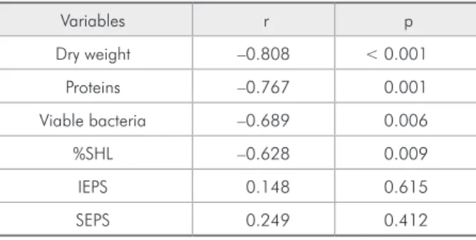
Table 1 - Correlation (r) and significance (p) between chlorhexidine concentration (0 – 0.12%) and the response variables.
Variables r p
Dry weight –0.808 < 0.001
Proteins –0.767 0.001
Viable bacteria –0.689 0.006
%SHL –0.628 0.009
IEPS 0.148 0.615
SEPS 0.249 0.412
*% SHL =Percentage of surface hardness loss. IEPS = Insoluble extrapolys-sacharides. SEPS = Soluble extrapolysextrapolys-sacharides.
48 3.00
3.50 4.00 4.50 5.00 5.50 6.00 6.50 7.50
7.00
pH
72
Time (h)
96 120
Control 0.012% CHX
0.12% CHX
0.05% NaF
0.024% CHX
Graph 1 - pH of the culture medium after 48 of biofilm growth in the absence of treatments and at each 24 h after beginning the treatments (72, 96 and 120 h data). The statistical significance among the treatments is described in the Results section (n = 4).
Surface hardness loss (SHL) was used as indica-tor of enamel demineralization.11
Statistical analysis
The assumptions of equality of variances and normal distribution of errors were checked for all the response variables tested and those that did not satisfy these conditions were transformed.24 The
relationship between CHX concentrations and the variable evaluated was estimated by regression anal-ysis. When signiicant correlation was found, the data were submitted to ANOVA followed by Tukey’s test, with the exception of acidogenicity, which was analyzed by repeated measures. Original data were used with the exception of viable bacteria counts, which were transformed to log10. The software SPSS for Windows 15.0 (SPSS, Chicago, IL, USA) was used and the signiicance level was ixed at 5%.
Results
A statistically signiicant linear effect was found between CHX concentration and bioilm dry weight, total soluble proteins and viable bacteria, and enamel demineralization (Table 1), showing dose-response effect for these variables. No effect
differ from those of the control (p = 0.209 and 0.758, respectively) but were higher than the baseline val-ues (p = 0.001 and 0.045, respectively) (Graph 2).
The viable bacteria count in the bioilm (Graph 3), normalized by bioilm dry weight, was
signiicantly lower (p < 0.001) for the treatment with 0.12% CHX when compared with the baseline and the control group. However, the viable bacteria counts of bioilm treated with 0.05% NaF did not differ from those of the control and the baseline
val-2.50 2.00 1.50 1.00 0.50 A mo u nt 0.00 Groups Control Baseline a A b B 0.012% CHX c B 0.024% CHX C ac 0.05% NaF AB bc 0.12% CHX A ac Dry weight (mg) Soluble proteins (µg× 10− 3)
0 .6 7 0 .1 1 6 2 .0 7 0 .5 8 5 1 .6 3 0 .5 1 1 .1 7 0 .3 7 1 1 .1 3 0 .1 5 1 1 .4 2 0 .4 4 8
Graph 2 - Means of biofilm dry weight (mg) and amount of soluble
proteins (µg x 10-3) for the baseline
and according to the treatments (n = 4).
C FU /mg dry weight bio film
0.00E + 00 1.00E + 08 2.00E + 08 3.00E + 08 4.00E + 08 5.00E + 08 6.00E + 08 7.00E + 08 8.00E + 08 9.00E + 08
Groups Control
Baseline 0.012% CHX 0.024% CHX 0.12% CHX 0.05% NaF
A AB AB C B AB 2 .2 7 E + 0 8 4 .3 9 E + 0 7 3 .2 0 E + 0 8 4 .4 9 E + 0 5 2 .1 0 E + 0 8 5 .7 1 E + 0
8 Graph 3 - Means of viable bacteria
(CFU/mg dry weight) in the biofilms grown for 48 h in the absence of the treatments (baseline) and after 3 days of treatments described (n = 4). Data
were transformed by log10.
%SH L 0.00 10.00 20.00 30.00 40.00 50.00 60.00 70.00 Groups Control A Baseline AB 0.12% CHX AB 0.012% CHX B 0.05% NaF B 0.024% CHX AB 3 6 .2
0 56
.1 0 3 7 .7 0 3 2 .0 0 3 0 .1 0 2 7 .2 0
Graph 4 - Means of enamel demineralization (%SHL) after 48 h of biofilm growth in the absence of the treatments (baseline) and after 3 days of the treatments described. (n = 4).
ues (p = 0.998 and 0.511, respectively).
As regards enamel demineralization (Graph 4), 0.12% CHX and 0.05% NaF signiicantly re-duced the %SHL when compared with the control (p = 0.036 and 0.017, respectively), but these treat-ments did not differ between them (p = 0.99).
Discussion
Bioilm models are important tools to evaluate the biochemical and microbiological composition of bioilm formed under different conditions or the changes caused on the substratum surface on which the bioilm is attached. Therefore, the conditions of bioilm formation and the substratum used must be as close as possible to those of real life.
The improved model of S. mutans bioilm growth was validated and dose response effect of CHX on S. mutans bioilm was shown for most variables (Table 1). Therefore, the model is suficiently sensitive to show bioilm and enamel demineralization changes in the presence of antimicrobial substances. The treatment 2x/day with 0.12% CHX showed a bacte-ricidal effect, killing a large proportion of the viable bacteria in the bioilm (Graph 3), decreasing the bio-ilm capacity to produce acids (Graph 1), avoiding the increase in bioilm mass (dry weight and total proteins) (Graph 2) and consequently, the enamel de-mineralization process was stopped (Graph 4). This effect may be attributed to the ability of the CHX molecule to bind to the negatively charged bacterial cell surface, alter and disrupt the integrity of the cell membrane, causing bacterial death.25,26 The
concen-tration of 0.012% CHX had a bacteriostatic effect, not interfering in the viable bacteria counts, but af-fecting the acid production level, which was lower than that of the control group, but it was not able to prevent enamel demineralization. At this sublethal stage, the effects of CHX are reversible; removal of excess CHX by neutralizers allows the bacterial cell to recover.26 This implies that the structural damage
caused by 0.012% CHX was less than that caused by 0.024% and 0.12% CHX. The results found with the use of this model are supported by a clini-cal trial showing that 0.12% CHX is more effective in reducing S. mutans CFU than lower
concentra-tions.27
As opposed to CHX, luoride did not show any effect on bioilm formation based on biomass (Graph 2) and viable bacteria counts (Graph 3). It also did not inhibit sucrose fermentation since the pH of the media did not differ when compared with the control (Graph 1), nevertheless, it reduced enam-el demineralization (Graph 4). The indings are sup-ported by the knowledge that at least 10 ppm of lu-oride constantly in the media is necessary to inhibit sugar fermentation.28 In this model, simulating the
clinical use of mouthrinse, although the bioilm was treated with 225 ppm of luoride, the duration time of treatment was only 1 min. Although 0.05% NaF showed absence of antimicrobial effect, it reduced enamel demineralization, suggesting that the main effect of F on caries control is physicochemical.29,30
Moreover, the indings suggest that CHX did not have a direct effect on the synthesis of extracellular polysaccharides (Table 1). This result is apparently in disagreement with Koo et al.13 (2003) but it
re-lects the way in which the results were expressed. Koo et al.13 (2003) expressed the results in amount
of polysaccharides found in the bioilm and in the present study the results were normalized by bio-ilm dry weight. Since the biobio-ilm weight increased (Graph 2) but the viable bacteria decreased (Graph 3), the reduction in the amount of EPS should be attributed to bacterial death and not to a speciic ef-fect of CHX inhibiting the synthesis of extracellular polysaccharides.
In conclusion, the results suggest this improved
S. mutansmodel can be used to test the effect of an-timicrobial agents on bioilm growth and on enamel demineralization.
Acknowledgments
References
1. Marsh PD. Are dental disease examples of ecological catas-trophes? Microbiology. 2003 Feb;149(2):279-94.
2. Takahashi N, Nyvad B. Caries ecology revisited: Micro-bial dynamics and the caries process. Caries Res. 2008 Oct;42(6):409-18.
3. Carlsson J. Bacterial metabolism in dental biofilms. Adv Den-tal Res. 1997 Apr;11(1):75-80.
4. Loesche WJ. Role of Streptococcus mutans in a human dental decay. Microbiol Rev. 1986 Dec;50(4):353-80.
5. Marsh PD. Dental as plaque biofilm and a microbial commu-nity: implications for health and disease. BMC Oral Health. 2006 Jun;6 Suppl 1:S1-S14.
6. Rölla G. Why is sucrose so cariogenic? The role of glucosyl-transferases and polysaccharides. Scand J Dent Res. 1989 Apr;97(2):115-9.
7. Schilling KM, Bowen WH. Glucans synthesized in situ ex-perimental salivary pellicle function as specific bindings sites for Streptococcus mutans. Infect Immun. 1992 Jan;60(1):284-95.
8. Koo H, Xiao J, Klein MI. Extracellular Polysaccharides ma-trix- An often forgotten virulence factor in oral biofilm re-search. Int J Oral Sci. 2009 Dec;1(4):229-34.
9. Xiao J, Koo H. Structural organization and dynamics of exopolysaccharide matrix and microcolonies formation by Streptococcus mutans in biofilms. J Appl Microbiol. 2010 Jun;108(6):2103-13.
10. Dibdin GH, Shellis RP. Physical and biochemical studies of
Streptococcus mutans sediments suggest new factors linking the cariogenicity of plaque with its extracellular polysaccha-ride content. J Dent Res. 1988 Jun;67(6):890-5.
11. Cury JA, Rebelo MA, Del Bel Cury AA, Derbyshire MT, Tabchoury CP. Biochemical composition and cariogenicity of dental plaque formed in the presence of sucrose or glucose and fructose. Caries Res. 2000 Nov-Dec;34(6):491-7. 12. Sissons CH. Artificial dental plaque biofilm model systems.
Adv Dent Res. 1997 Apr;11(1):110-26.
13. Koo H, Hayacibara MF, Schobel BD, Cury JA, Rosalen PL, Park YK et al. Inhibition of Streptococcus mutans biofilm accumulation and polysaccharide production by apigenin and tt-farnesol. J Antimicrob Chemother. 2003 Nov;52(5):782-9.
14. Coenye T, Honraet K, Rigole P, Jimenez PN, Nelis HJ. In vitro inhibition of Streptococcus mutans biofilm formation on hydroxyapatite by subinhibitory concentrations of anthraqui-nones. Antimicrob Agents Chemother. 2007 Apr;51(4):1541- 4.
15. Deng DM, Hoogenkamp MA, Ten Cate JM, Crielaard W. Novel metabolic activity indicator in Streptococcus mutans
biofilms. J Microbiol Methods. 2009 Apr;77(1):67-71. 16. Jones CG. Chlorhexidine: Is it still the gold standard?
Peri-odontol 2000. 1997 Oct;15(1):55-62.
17. Marinho VCC. Evidence-based effectiveness of topical fluor-ides. Adv Dent Res. 2008 Jul;20(1):3-7.
18. Emilson CG. Potential efficacy of chlorhexidine against mu-tans streptococci and human dental caries. J Dent Res. 1994 Mar;73(3):682-91.
19. White DJ. Reactivity of Fluoride Dentifrice with Artificial Caries. I. Effects on subsurface lesion: F uptake, distribu-tion, surface, hardening and remineralization. Caries Res. 1987;21:126-40.
20. Aires CP, Del Bel Cury AA, Tenuta LMA, Klein MI, Koo H, Duarte S et al. Effect of sucrose and starch on dental biofilm formation and on dentin demineralization. Caries Res. 2008 Sep;42(5):380-6.
21. Lowry OH, Rosebrough NJ, Farr AL, Randall RJ. Protein measurement with the Folin phenol reagent. J Biol Chem. 1951 Nov;193(1):265-75.
22. Herigstad B, Hamilton M, Heersink J. How to optimize the drop plate method for enumerating bacteria. J Microbiol Methods. 2001 Mar;44(2):121-9.
23. Dubois M, Gilles KA, Hamilton JK, Rebers PA, Smith F. Colorimetric method for determination of sugars and related substances. Anal Chem. 1956 Mar;28(3):350-6.
24. Box GEP, Hunter WG, Hunter JS. Statistics for experimenters. New York: Wiley, c1978. 656 p.
25. Hugo WB, Longworth AR. Some aspects of the mode of action of chlorhexidine. J Pharm Pharmacol. 1964 Oct;16:655-62. 26. Rölla G, Melsen B. On the mechanism of the plaque inhibi-tion by chlorhexidine. J Dent Res. 1975 Jun;54 (Spec Nº B): B57- 62.
27. Clark DC, Guest JL. The effectiveness of three different strengths of chlorhexidine mouthrinse. J Can Dent Assoc. 1994 Aug;60(8):711- 4.
28. Bradshaw DJ, Marsh PD, Hodgson RJ, Visser JM. Effect of glucose and fluoride on competition and metabolism within dental bacterial communities and biofilms. Caries Res. 2002 Mar-Apr; 36(2):81- 6.
29. ten Cate JM. Current concepts on the theories of the mech-anism of action of fluoride. Acta Odontol Scand. 1999 Dec;57(6):325- 9.