Streptococcus mutans in denture stomatitis patients
under antifungal therapy
Streptococcus mutans
em pacientes com estomatite protética
submetidos à terapia antifúngica
Laurylene César de S. Vasconcelos a Fábio Correia Sampaio a
Maria Carméli Correia Sampaio b Maria do Socorro Vieira Pereira c Maria Helena Pereira Peixoto c
a Department of Clinical and Social Dentistry, Federal University of Paraíba, João Pessoa, PB, Brazil
b Department of Pharmaceutical Sciences, Federal University of Paraíba, João Pessoa, PB, Brazil c Department of Molecular Biology, Federal University of Paraíba, João Pessoa, PB, Brazil
Correspondence:
Laurylene César de Souza Vasconcelos R. Maria Helena da Rocha, 113 - Aeroclube João Pessoa, PB – Brasil
58036-823
E-mail: laurylene@uol.com.br
Received: May 20, 2009 Accepted: March 31, 2010
Conflict of Interest Statement: The authors state that there are no financial and personal conflicts of interest that could have inappropriately influenced their work.
Copyright: © 2010 Vasconcelos et al.; licensee EDIPUCRS. This is an Open Access article distributed under the terms of the Creative Commons Attribution-Noncommercial-No Derivative Works 3.0 Unported License.
Abstract
Purpose: To assess the number of Streptococcus mutans in saliva of patients with denture stomatitis before and after antifungal therapy.
Methods: After examining 93 patients, 47 were selected for fungal test. Then, from this sample, thirty patients were selected: 15 with positive and 15 with negative diagnosis for candidiasis that were evaluated for S. mutans counting, salivary flow and buffer capacity evaluation. Oral hygiene and prosthesis hygiene, period using prosthesis, lesion type and salivary data were related with clinical laboratorial characteristics of the patients with Candida.
Results: The most frequent lesions were type I (43.5%) and II (53.5%). The amount of S. mutans was six times higher in patients with candidiasis and it was associated with low salivary flow and poor oral hygiene. After therapy, a reduction of S. mutans was verified particularly in patients with normal salivary flow. The values ranged from 0.01 to 3.88 x 10⁴ cfu/mL.
Conclusion: The data suggest that Streptococcus spp collaborates with Candida spp in the etiology and pathogenesis of denture stomatitis. The use of oral antimicrobial agents may provide a beneficial effect for denture stomatitis patients that are under antifungal therapy and that have poor oral hygiene and unfavorable salivary parameters.
Key words: Dental prosthesis; antifungal agents; Candida; Streptococcus mutans
Resumo
Objetivo: Verificar o número de Streptococcus mutans em saliva de pacientes com estomatite protética antes e após a terapia antifúngica.
Metodologia: Após exame clínico de 93 pacientes, 47 foram selecionados para exame micológico e desta amostra foram selecionados trinta pacientes: 15 com diagnóstico positivo e 15 com diagnóstico negativo de candidose foram avaliados para contagem de S. mutans, determinação de fluxo salivar e capacidade tampão. Higiene bucal e da prótese, tempo de confecção, tipo de lesão e dados salivares foram relacionados com características clínicas e laboratoriais de Candida.
Resultados: As lesões frequentes foram dos tipos I (43,5%) e II (53,5%). A quantidade de S. mutans foi seis vezes maior em pacientes com candidose e foi associada com baixo fluxo salivar e higiene oral deficiente. Após a terapia, a redução de S. mutans foi verificada particularmente em pacientes com fluxo salivar normal. Os valores variaram de 0,01 a 3,88 UFC/ml x 104. Conclusão: Os dados sugerem que os Streptococcus colaboram com Candida spp na etiopatogenia da estomatite protética. O uso de agentes antimicrobianos orais pode propiciar efeito benéfico para pacientes com estomatite protética submetidos à terapia antifúngica e que apresentam higiene oral deficiente e parâmetros salivares desfavoráveis.
Introduction
The surface of acrylic dentures usually host several
microorganisms (fungi and bacteria) that are dificult to
remove even by vigorous mechanical cleaning or chemical agents (1). This environment produced by the prosthesis can promote yeast proliferation with or without predisposing local factors (2).
Denture stomatitis is an oral pathology of multifactorial etiology that affects a large number of patients using complete or partial dentures. The main etiologic factors related to denture stomatitis are trauma, poor oral hygiene and infection with Candida species. In general, the patients with denture stomatitis complain of edema, hyperemia, pain in the affected areas and burning mouth when a low salivary
low is taking place (3,4).
The adhesion of Candida cells to oral surfaces is a key initial event in pathogenesis of oral candidiasis, and the complex
structure of oral lora with mixed species communities can
intermediate the predisposition for many oral conditions
including candidiasis (3). There are evidences that denture
stomatitis is not a result of C. albicans solely, but rather it
is an outcome of multispecies bioilms that may include
Streptococcus mutans and Staphylococcus aureus (5,6). In fact, it has been already observed that co-adhesion between C. albicans and many Streptococcal spp. promotes oral colonization by yeast cells (6). Streptococcus mutans is a frequent member of acrylic dentures surfaces and if incubated simultaneously with Candida albicans may compete for biding sites but it can also promote yeast adhesion (5). This bacteria-yeast interaction has been a matter of investigation of many in vitro and in vivo studies (5,7-10). The analysis of the in vitro adherence of S. mutans and C.albicans can contribute to the understanding of the behavior of these organisms in the dental plaque. The interaction of these microorganisms in a combined culture can be understood as mutualistic, once both seem to be favored (11).
The adhesion is regarded as the irst step for oral bioilm
formation and distinct mechanisms of adherence can contribute for candidiasis resistance to antifungal therapeutic
agents (8). In complex bioilms such as those found in the
oral cavity, the ability of yeast to agglutinate with bacteria
can be mediated by species interaction within the bioilm as well as by external factors like saliva, oral hygiene and
exposure to antimicrobial agents (8,9). Despite the in vitro evidences of the Candida-bacteria interaction, few studies have investigated the S. mutans in patients with candidiasis or yeast related diseases (10,12). The aim of this study was to assess the number of Streptococcus mutans in saliva of patients with diagnosis of denture stomatitis before and after antifungal therapy.
Methods
The study protocol was approved by the Local Ethical Committee of the Federal University of Paraíba, João Pessoa, PB, Brazil. Only patients who were assisted at
the dental clinic and signed the consent were selected for the research. The criterion for participating in the research was dental prosthesis users. The exclusion criteria were: patients with systemic predisposing or confounding factor (diabetes, hyperthyroidism, immune diseases, radiotherapy and chemotherapy treatments), use of medicines that could affect any Candida infection such as antidepressive, immunosuppressive and anti-bacterial agents (2).
A total of 93 patients were examined and forty seven
presented clinical signs of denture stomatitis. The clinical
signs were classiied in three stages described by Newton
as pin-point hyperemia, diffuse hyperemia and nodular
hyperemia of the entire denture area (13).
Swabs were used for collecting material from prosthesis (P1) and palatine lesions (P2) of patients with candidiasis. The material was subsequently placed on CHROMagar Candida for culture, isolation and identiication of fungi species. CHROMagar is a selective and differential medium that allows selective isolation of yeasts and simultaneously.
identiies (by color reactions and colony morphology)
colonies of C. albicans ,C. tropicalis and C. Krusei with a
high degree of accuracy (14).
Mycological exams as described previously were carried out
in this sub-sample (4). The presence of Candida infection
was conirmed in 32 patients, but only 15 presented more than
20 cfu (Colony Forming Units), a level which characterizes
infection (1). Finally, the sample was composed of 30
patients with denture stomatitis: 15 with candidiasis and 15 without candidiasis.
Antifungal therapy was applied for the 15 patients who were positive for candidiasis. The patients received a tube
of a commercial antifungal medicine (Daktarin® gel oral,
Janssen Cilag Farmacêutica, 40 g) and instruction for using 3 times a day for two weeks. They were instructed to use a suficient amount of gel that could cover the affected areas
as recommended by the manufacturer. Denture were cleaned each night with neutral soap and washed with tap water. Patients were strongly requested to avoid using dentures at night during the test period.
Before starting the therapeutic period, oral hygiene parameters and salivary test were carried out before the therapeutic period
of 2 weeks. Mycological and microbiological tests were
carried out twice: before the patients have started using the
medicine and two days after inishing the therapeutic period.
Oral hygiene and prosthesis hygiene parameters were
collected according to the guidelines of Sakki et al. (15). Saliva parameters (stimulated salivary low rate and buffer
capacity) were collected as described by Krasse et al. (16).
The patients were classiied in three levels of salivary low
rate: a) hiposalivation = 0.7mL/min, b) intermediate salivary
low = 0.71 up to 1.0 mL/min and c) normal salivary low
when above 1.1 mL/min. For buffer capacity, the reference levels were considered as normal for pH above 5 and low
for pH below or equal to 4. The pH measurements were
determined using indicate paper but intermediate values
were checked in pH electrode for inal classiication (below
The S. mutans counting was carried out for all 30 patients at baseline and for 15 patients after using the antifungal
medicine. Non-stimulated saliva samples were placed
in Brain Heart Infusion (BHI, Difco) and later on MSB
medium: mitis salivarius agar (Difco) supplemented with 440 mM sucrose, 39 mM potassium tellurite and 0.2 units/mL
of bacitracin, as recommended by Gold et al. (17). All procedures were in duplicate.
Statistical analysis was carried out using SPSS (Statistical
Package for Social Sciences) v.10.0. The assumptions of
equality of variances and normal distribution of errors were
checked for all numerical variables. Since the distribution of
the errors was homogeneous, data were tested by Student’s
T test with a signiicance level of 5%.
Results
Most patients were female (n=24) with an age range between
27 and 69 years-old. Within this range, the most frequent
observations were between 49 up to 59-years-old. The lesions types observed were type I (43.5%) and type II (53.5%). Type III composed 3% of the lesions.
Candida albicans was the predominant yeast isolated
(86.6%) followed by. C. tropicalis, C. parapisilosis, C. glabrata e C. krusei.
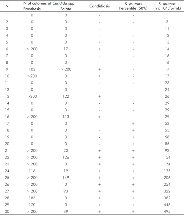
Table 1 shows the mean fcu/mL of S. mutans and Candida spp of all patients. Ten subjects who showed high counts of Candida in the prosthesis were positive for candidiasis and were also with high number of S. mutans in saliva.
N N of colonies of Candida spp Candidiasis S. mutans Percentile (50%)
S. mutans
(n x 104 cfu/mL)
Prosthesis Palate
1 0 0 - - 1
2 0 0 - - 3
3 0 0 - - 11
4 0 0 - - 12
5 0 0 - - 13
6 > 200 17 + - 14
7 0 0 - - 16
8 0 0 - - 16
9 103 > 200 + - 17
10 >200 0 + - 17
11 0 0 - - 23
12 0 0 - - 24
13 >200 122 + - 26
14 0 0 - - 29
15 0 0 - - 29
16 > 200 113 + - 29
17 0 0 - + 53
18 0 0 - + 55
19 0 0 - + 58
20 0 0 - + 85
21 > 200 20 + + 92
22 > 200 126 + + 154
23 > 200 0 + + 174
24 116 19 + + 175
25 > 200 149 + + 206
26 > 200 0 + + 254
27 > 200 93 + + 322
28 183 0 + + 382
29 170 0 + + 446
30 > 200 39 + + 495
Table 1. Mean cfu/mL of S. mutans and Candida spp of 30 patients (15 patients positive and 15 negative
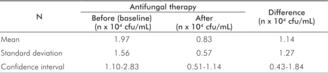
Table 2 shows the reduction in salivary S. mutans (cfu/mL) after the antifungal therapy. In percentages, the differences
range from 3.8 up to 82% with a mean reduction of 45%. Table 3 presents the mean (SD) values (cfu/mL) of S. mutans of patients before the antifungal therapy (baseline) according
to other parameters. Table 4 presents the distribution of
patients categorized in low and high S. mutans counting (cfu/mL) of patients who with and without candidiasis. The estimate amount S.mutans of in saliva among patients with
deicient hygiene was ten fold higher compared to those with good hygiene. Among those patients with deicient hygiene (n=13), the mean (SD) values (cfu/mL) of S.mutans were
2.24(1.50) and 0.93(0.50) before and after the antifungal
therapy, respectively. For those with good hygiene (n=2) there values were 0.21(0.10) and 0.12(0.01), respectively.
Table 2. Mean, standard deviation and confidence interval of
S. mutans (cfu/mL) in saliva of patients (n=15) before and after the use of antifungal medicine.
N
Antifungal therapy
Difference (n x 104 cfu/mL)
Before (baseline) (n x 104 cfu/mL)
After (n x 104 cfu/mL)
Mean 1.97 0.83 1.14
Standard deviation 1.56 0.57 1.27
Confidence interval 1.10-2.83 0.51-1.14 0.43-1.84
Table 3. Mean (SD) of fcu/mL of S. mutans (baseline), time of use of prosthesis, salivary flow rate, buffer capacity of patients who
with and without candidiasis.
Candidiasis N S. mutans
(n x 104 cfu/mL)
Period of using prosthesis (months)
Salivary flow rate (mL/min)
Buffer capacity (pH)
Negative 15 0.3 (0.22)a 69.1 (49.3)a 1.2 (0.3)a 6.6 (0.7)a
Positive 15 1.97 (1.56)b 95.6 (83.5)b 0.7 (0.2)b 5.2 (1.2)b
Means in the same column followed by distinct superscripts indicate statistical significance (Student T test, P<0.05).
S. mutans (n x 104 fcu/mL)**
Diagnosis of candidiasis
Total
Negative Positive
Low 11 5 16
High 4 10 14
Total 15 15 30
* Chi-square = 4.82, d.f.=1, p<0.05.
** cut-off point of 29 x 104 cfu/mL of S. mutans (percentile 50%). Table 4. Relationship between
patients who showed low or high
S. mutans in saliva and patients
diagnosed as Candida-positive
(presence of C. albicans on
prosthesis or palate).*
Discussion
C. albicans was the major isolated yeast in the selected subjects. This was also observed in other studies (2,9). The predominant site for colonies was the prosthesis indicating the importance of the mechanism of adherence of yeast cells
on a non-shedding surface (6,15). Adhesion is the irst step
for yeast colonization on the oral epithelium, and dentures may become an important source for reinfection (18).
For denture stomatitis patients the risk of candidiasis is
high since porous acrylic surfaces can provide more biding sites than natural teeth and shedding surfaces as oral mucosa (1).
Shinada et al. (8) observed that the presence of Candida was highest on the mucosal denture surfaces followed by clasp, tongue, and remaining teeth. Therefore, it is not a surprise
S. mutans can inluence re-infection, this is still a matter of debate (7).
Recently, many investigations have focus more on bioilm formation than in planktonic cells to explain the bacterial
behavior in the in vivo oral environment (19). Regarding candidiasis, few studies have investigated the bacteria-yeast relationship using clinical parameters that can modulate
the bioilm formation on different surfaces (5,10,12). It
is important to consider bacterial-Candida interactions in the study of fungal virulence. Aspects of the host’s microbial community might normally protect against fungal proliferation or might play an important role in the preparation of the fungus for its role in the infection. Also, mixed bacterial-fungal infections might have properties that are distinct from single-species infections (20). These
co-species communities, probably existing as a mixed bioilm, are dificult to treat with antibiotics and antifungals (6).
Saliva and oral hygiene can be important mediators for oral
bioilm development. Speciic salivary components may act
promoting yeast-bacteria co-aggregation and adherence to
oral surfaces (9,4,17). Low salivary low can also facilitate bioilm development and this is certainly a matter of concern
when treating old subjects prone to dry mouth related problems (8,17).
In our study, Candida spp cfu values were obtained from
bioilm material of dentures or palatal lesions, whereas. S. mutans cfu levels were evaluated from saliva samples as an indication of S. mutans counting. Although S. mutans are
major constituents of cariogenic oral bioilms, the saliva counting can be a good indicator of the major oral bioilms
in the mouth in many oral conditions (21,22). In fact, a variation of S. mutans cells in different sites of the mouth is expected. Therefore, the option of collecting the S.mutans data from saliva was regarded as appropriate. In addition, all procedures were carried out following established guidelines of material collection (16). In any case, considering the cfu counting of S.mutans in the evaluated subjects, one must
estimate that in denture oral bioilms there are at least 5-fold
more bacteria than those found in saliva. Girad Junior et al.
(2) observed that the dental bioilm formed in the presence
of sucrose would be an indirect contributing factor for yeast adherence due to an increase in Streptococcus spp. As shown
in Tables 1 and 4, most of the subjects with high counts
of Candida also had high counts of S. mutans. Moreover, subjects with candidiasis and high amounts of S.mutans in
the oral mouth were the virtually the same with unfavorable salivary parameters and poor oral hygiene. The question at this point is not if a potentialization of the adherence capacity of Streptococcus mutans and Candida albicans
takes place in a bioilm development, but rather if this
synergic effect can be estimated or predicted clinically. The clinical implications of these results are that candidiasis treatment can be more effective when including frequent oral
hygiene measures to reduce bioilm formation and S.mutans
in the oral mouth (23). On the other hand, a risk for dental
caries due to Candida is likely as reported by Moalic et al.
(24) and Nikawa et al. (25). A low bioilm pH may favor S. mutans but Candida can also take advantage of adherence mechanism in such environment.
The observation of lesions types I and II in these subjects indicates that candidiasis was not an acute event in these patients. Poor oral hygiene and unfavorable salivary parameters can probably aggravate this condition by many ways including the Candida-Streptococcus co-aggregation. From the clinical point of view, compliance for oral health instructions can be problematic. Therefore, in addition to oral hygiene instructions, the use of antimicrobials might be an interesting choice to patients with a high S. mutans counting and resistant candidiasis.
Conclusions
It can be concluded that:
1. S. mutans counting was higher in patients with candidiasis
and it was associated with low salivary low and poor oral
hygiene;
2. After therapy, it was veriied a reduction of S. mutans particular in patients with normal salivary parameters; and
3. Streptococcus spp collaborates with Candida spp in the etiology and pathogenesis of denture stomatitis. The use of
oral antimicrobial agents may provide a beneicial effect for
denture stomatitis patients that are under antifungal therapy and that have poor oral hygiene and unfavorable salivary parameters.
Acknowledgments
This study is part of a doctoral thesis and it was supported by Lauro Wanderley University Hospital for its realization.
References
Budtz-Jörgensen E. Etiology, pathogeneses, therapy and prophylaxis 1.
of oral yeast infections. Acta Odontol Scand 1990;48:61-9 Girard Junior B, Landry RG, Giasson L. La stomatite prothétique: 2.
etiologie et condérations cliniques. J Can Dent Assoc 1996;62:808-12. Könsberg R, Axéll T. Treatment of Candida – infected denture 3.
stomatits with a miconazole lacquer. Oral Surg Oral Med Oral Pathol 1994;78:306-11.
Vasconcelos LC, Sampaio MC, Sampaio FC, Higino JS. Use of Punica 4.
granatum as an antifungal agent against candidosis associated with denture stomatitis. Mycoses 2003;46:192-6.
Linossier A, Vargas A, Villegas R, Chimenos E. Quantitative 5.
Baena-Monroy T, Moreno-Maldonado V, Franco-Martinez F, Aldape-6.
Barrios B, Quindos G, Sanchez-Vargas LO. Candida albicans, Staphylococcus aureus and Streptococcus mutans colonization in patients wearing dental prosthesis. Med Oral Patol Oral Cir Bucal 2005;1:E27-39.
Holmes AR, Gopal PK, Jenkinson HF. Adherence of
7. Candida albicans
to a cell surface polysaccharide receptor on Streptococcus gordonii. Infect Immun 1995;63:1827-34.
Shinada K, Ozaki F, Cordiero JG, Okada S, Shimoyama K, Nagao 8.
M, Ichinose S, Yamashita Y. A morphological study of interactions of Candida albicans and Streptococcus mutans. Kokubyo Gakkai Zasshi 1995;62:281-6.
Jorge AOC, Koga-Ito CY, Gonçalves CR, Fantinato V, Unterkircher 9.
CS. Presença de leveduras do gênero Candida na saliva de pacientes com diferentes fatores predisponentes e de indivíduos controle. Rev Fac Odontol Univ São Paulo 1997;11:279-85.
El-Azizi MA, Starks SE, Khardori N. Interactions of
10. Candida albicans
with other Candida spp. and bacteria in the biofilms. J Appl Microbiol 2004;96:1067-73.
Barbieri DSV, Vicente VA, Fraiz FC, Lavoranti OJ, Svidzinski TIE, 11.
Pinheiro R L. Analysis of the in vitro adherence of Streptococcus mutans and Candida albicans. Braz j Microbiol 2007;38: 624-31.
Shinada K, Teraoka K, Asaka T, Cordeiro JG, Ozaki F, Shimoyama 12.
K et al. Distribution of Candida species and mutans streptococci related to oral conditions in elderly persons. Kokubyo Gakkai Zasshi 1997;64:512-17.
Newton
13. AV. Denture Sore Mouth: a possible Ætiology. Br Dent J 1962;357-60.
Pfaller MA, Houston A, Coffmann S. Application of CHROMágar 14.
Candida for rapid screening of clinical specimens for Candida albicans, Candida tropicalis, Candida krusei and Candida (Torulopsis) glabrata. J Clin Microbiol 1996;34:58-61.
Sakki TK, Knuuttila MLE, Läärä EL, Anttila SS. The association of yeasts 15.
and denture stomatitis with behavioral and biologic factors. Oral Surg Oral Med Oral Pathol Oral Radiol Endod 1997;84:624-9. Krasse B. Risco de cárie: um guia prático para avaliação e controle. 16.
2ª ed. São Paulo: Quintessence Books; 1988.
Gold OG, Jordan HV, Van Houte J. A.selective medium for 17.
Streptococcus mutans. Arch Oral Biol 1973;18:1357-64. Noborikawa E, Silveira FRX, Witzel, AL, Lotufo, MA. Biotypes of 18.
Candida albicans isolates from the oral mucosa of HIV seropositive and control subjects. Rev Odonto Ciênc 2009;24:258-63. Marsh PD. Role of the oral microflora in health. Microb ecol health 19.
dis 2000;12:130-7.
Wargo.MJ, Hogan, DA. Fungal-bacterial interactions: a mixed bag 20.
of mingling microbes. Curr Opin Microbiol 2006;9:359-64. Sullivan A, Borgström MK, Granath L, Nilsson G. Number of mutans 21.
streptococci or lactobacilli in a total dental plaque sample does not explain the variation in caries better than the numbers in stimulated whole saliva. Community Dent Oral Epidemiol 1996;24:159-63. Hintao J, Teanpaisan R, Chongsuvivatwong V, Ratarasan C, Dahlen 22.
G. The microbiological profiles of saliva, supragingival and subgingival plaque and dental caries in adults with and without type 2 diabetes mellitus. Oral Microbiol Immunol 2007;22:175-81. Vasconcelos LCS, Sampaio FC, Sampaio MCC, Pereira MSV, Higino 23.
JS, Peixoto MHP. Minimum inhibitory concentration of adherence of Punica granatum Linn (pomegranate) gel against S. mutans, S. mitis and C. albicans. Braz Dent J 2006;17:223-7.
Moalic E, Gestalin A, Quinio D, Gest PE, Zerilli A, Le Flohic AM. The 24.
extent of oral fungal flora in 353 students and possible relationships with dental caries. Caries Res 2001;35:149-55.
Nikawa H, Egusa H, Makihira S, Nishimura M. Ishida K, Furukawa M, 25.