Peripheral hearing evaluation in children with Down
syndrome
Avaliação auditiva periférica em crianças com síndrome de Down
Barbara Carrico1, Alessandra Giannella Samelli1, Carla Gentile Matas1, Fernanda Cristina Leite Magliaro1,
Renata Mota Mamede Carvallo1, Suelly Cecília Olivan Limongi1, Ivone Ferreira Neves-Lobo1
ABSTRACT
Purpose: This study sought to characterize the peripheral auditory sys-tem of individuals with Down syndrome (DS) using conventional and high-frequency audiometry. Methods: We performed a cross-sectional and observational study. Fifteen individuals with DS, who were of both genders and between 7 and 15 years of age, participated in this study. The following procedures were performed: otoscopy, tympanometry with ipsilateral and contralateral acoustic reflex, pure-tone audiometry, vocal audiometry and high-frequency audiometry. Results: There was a predominance of mild conductive hearing loss in one or both ears. The mean hearing thresholds for conventional audiometry were below 20 dB HL and between 20 and 40 dB HL for high-frequency audiometry. The Pearson correlation coefficient indicated a moderate positive correlation between the 9-14 kHz thresholds and age. Conclusion: Overall, no significant differences were observed when comparing the right and left ears of individuals with DS, in regards to pure-tone audiometry, immit-tance testing and speech audiometry. Most children showed middle ear abnormalities and conductive hearing loss. Moreover, high-frequency audiometry suggested the onset of impaired cochlear function, which may be associated with frequent otitis media episodes and/or early cochlear degeneration.
Keywords: Hearing; Audiometry; Down syndrome; Auditory threshold; Hearing loss, high-frequency
RESUMO
Objetivo: Caracterizar o sistema auditivo periférico de indivíduos com síndrome de Down, por meio da audiometria convencional e de altas frequências. Métodos: Estudo do tipo transversal e observacional. Par-ticiparam 15 indivíduos com síndrome de Down, de ambos os gêneros, entre 7 e 15 anos de idade. Foram realizados os seguintes procedimentos: Meatoscopia, Timpanometria com pesquisa do reflexo acústico ipsilateral e contralateral, Audiometria Tonal, Audiometria Vocal e Audiometria de Altas Frequências. Resultados: Houve predomínio de perda auditiva condutiva de grau leve, em uma ou ambas as orelhas. As médias dos limiares auditivos para a audiometria convencional ficaram abaixo de 20 dBNA e, para a audiometria de altas frequências, ficaram entre 20 e 40 dBNA. O coeficiente de correlação de Pearson revelou correlação moderada positiva, entre os limiares de 9 a 14 kHz e a idade. Conclusão:
De forma geral, não foram observadas diferenças significativas, quando comparadas as orelhas direita e esquerda de indivíduos com síndrome de Down, na audiometria tonal, imitanciometria e logoaudiometria. A maioria das crianças apresentou alteração de orelha média e perda auditiva condutiva. A audiometria de altas frequências sugere o início de prejuízo da função coclear, que pode estar associado às otites médias frequentes e/ou à degeneração coclear precoce.
Descritores: Audição; Audiometria; Síndrome de Down; Limiar auditi-vo; Perda auditiva de alta frequência
Study conducted at the Laboratory of Speech Therapy Research in Primary Care in Audiology, Department of Physical, Speech and Occupational Therapy, School of Medicine, Universidade de São Paulo – USP – São Paulo (SP), Brazil.
(1) Department of Physical, Speech and Occupational Therapy, School of Medicine, Universidade de São Paulo – USP – São Paulo (SP), Brazil.
Conflict of interests: No
Author’s contribution: BC execution of the research, development of the study, development of the timeline, literature review, data collection and analysis, manuscript writing; AGS advisor, development of the study, development of the timeline, data analysis, correcting the wording of the manuscript, approval of the
final version; CGM development of the study, development of the timeline, data analysis, correcting the wording of the manuscript, approval of the final version; FCLM development of the study, development of the timeline, literature review, data analysis, correcting the wording of the manuscript, approval of the final
version, manuscript submission and procedures; RMMC development of the study, data analysis, correcting the wording of the manuscript, approval of the final
version; SCOL development of the study, data analysis, correcting the wording of the manuscript, approval of the final version; IFNL development of the study, data analysis, correcting the wording of the manuscript, approval of the final version.
Correspondence address: Alessandra Giannella Samelli. R. Cipotânea, 51, Cidade Universitária, São Paulo (SP), Brazil, CEP: 05360-160. E-mail: [email protected]
INTRODUCTION
Hearing loss can be caused by environmental or genetic fac-tors. For instance, some genetic disorders demonstrate isolated hearing loss, whereas others show hearing loss associated with abnormalities of other organs due to a variety of syndromes(1). Down syndrome (DS) is a genetic disorder characterized by the presence of an extra copy of chromosome 21 or excess genetic material present on this chromosome(2). Diagnosis is based on a series of signs and symptoms, and confirmation is established by chromosome analysis. Because not all affected individuals show the same characteristics, cytogenetic analysis is necessary(3,4).
Studies have demonstrated that this syndrome occurs in 1 in every 1,000 live births. The most common clinical charac-teristics include intellectual disability, muscular hypotonia, oblique palpebral fissures, increased vascularity, microcephaly and flat occiput. Additional characteristics include a small and flat nose, low nasal bridge, cardiovascular malformations and respiratory infections due to obstruction of the upper airways(5). Muscular hypotonia affects the muscles of the bronchial tree, impairing the elimination of secretions. The accumulation of mucus can lead to infections of the upper airways and a consequent increase in the incidence of otitis media(5). Another factor that may contribute to the increased incidence of ear infections in DS patients is the dysfunction or impairment of the middle ear, which is frequently observed in this population. This factor is related to anatomical defects such as an abnormal Eustachian tube, persistent mesenchymal tissue in the tympanic cavity, external auditory canal stenosis and hypoplasia of the mastoid(6).
Hearing loss occurs in approximately two-thirds of chil-dren with DS and may present as conductive, sensorineural or mixed hearing loss(7,8). However, the prevalence of conductive hearing loss is greater, occurring in approximately 60-80% of individuals with DS(9-11).
Histopathological studies have shown that in cases of se-cretory otitis media, there may be diffusion of bacterial toxins and cytokines from the middle ear to the cochlea by way of the round window membrane. These events can cause structural injury to the inner ear, such as rupture of cochlear membranes, resulting in sensorineural hearing loss(12-14).
Congenital abnormalities of the inner ear are not frequent, although individuals with DS present with anatomically smaller cochlea than typically developing children(15). Moreover, begin-ning in the second decade of life, individuals with DS present a decline in hearing thresholds with a progressive “presbycusis” type pattern(10,11,16).
Few studies have performed peripheral auditory system eva-luations in subjects with DS. Because hearing loss can impair language development and oral expression, complete audiolo-gical evaluation of these individuals is crucial for differential diagnosis and treatment guidance. This study therefore aimed to
characterize the peripheral auditory system of individuals with DS, using both conventional and high-frequency audiometry.
METHODS
This was a prospective observational study approved by the Ethics Committee for Analysis of Research Projects, School of Medicine, Universidade de São Paulo (USP) (No. 138/11). In addition, parents or guardians signed informed consent forms prior to enrollment in the study.
A total of 15 individuals with DS were studied; these pa-tients were of both genders, between the ages of 7 and 15 years (mean 10 years and 9 months ± 1 year and 6 months) and in-cluded 8 females and 7 males. The presence of other associated impairments, such as neurological and psychiatric disorders that could impede the application of audiological procedures, was considered exclusion criteria. It should be noted that subjects’ handedness was not considered in the present study.
First, an interview was conducted with parents or guardians in which information about medical and otologic history was collected. The external auditory canal and tympanic membrane were then examined to determine the existence of problems that could interfere with the evaluation, such as the presence of cerumen, foreign bodies, etc. A Heine® otoscope was used for this examination.
To investigate the ipsilateral and contralateral acoustic reflexes and mobility of the tympanic-ossicular chain, immit-tance testing was performed using an Interacoustics® AT 235 analyzer. A type A tympanogram with the presence of acoustic reflex was considered a normal result, whereas a type B or C tympanogram and/or the absence of acoustic reflex was con-sidered an abnormal result(17).
Conventional pure-tone audiometry and high-frequency audiometry were performed in a soundproof booth using a GSI-61 audiometer (Grason-Stadler®) and model TDH-50 and HDA 200 supra-aural headphones(18).
Hearing thresholds with air conduction were obtained at frequencies ranging from 250-8,000 Hz and with bone conduction at frequencies from 500-4,000 Hz whenever air conduction thresholds exceeded 15 dB HL. Air conduction thresholds above 15 dB HL were defined as the presence of hearing loss. Hearing losses were classified according to type, either as conductive hearing loss or sensorineural hearing loss, and according to degree, using the mean of the 500-4,000 Hz frequencies(19). It is important to note that classification according to degree was not performed in cases of hearing loss at an isolated frequency.
Next, speech audiometry was performed to obtain the speech reception threshold (SRT) and speech discrimination index (SDI)(20).
All thresholds (obtained in dB HL) were determined using the descending method in steps of 10 dB and the ascending method in steps of 5 dB. The hearing thresholds at high frequencies were classified as normal according to the mean standards for high-frequency thresholds (10, 12.5, 14 and 16 kHz) proposed by a previous study(21) for each age group. The result was classified as abnormal if one or more frequencies in one or both ears was higher than the previously proposed standard(21).
All subjects were evaluated in a maximum of two ses-sions, performed on sequential days, and acoustic impedance measurements were always performed at the beginning of the evaluation.
Data analysis
For data analysis, a quantitative and qualitative description of the audiological profile of individuals with DS was made. The Wilcoxon test, equality of two proportions, ANOVA and the Pearson correlation coefficient were used for statistical analysis. The results were analyzed with a significance level of 5% (0.05).
RESULTS
With regard to the otological history of the 15 children, 10 had a history of otitis media, and the number of episodes in these 10 individuals ranged from 2 to 8 (average 5.3 episodes).
There was a higher percentage of subjects with abnormal immittance testing results (tympanometry and acoustic refle-xes) bilaterally. However, this difference was not significant (Table 1).
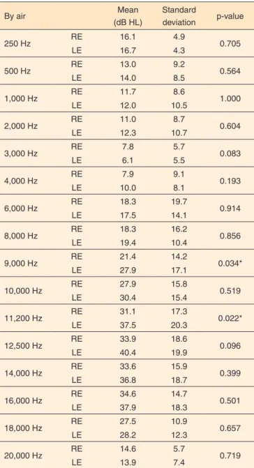
There was no difference between the right and left ears for audiometric thresholds between 250 and 8,000 Hz. However, for high frequencies, differences were observed between right and left ears at frequencies of 9,000 Hz and 11,200 Hz, with worse thresholds for the left ear. Qualitative analysis by indi-vidual revealed the following results:
- Five subjects (33.3%) had mild conductive hearing loss, considering the mean of the 500-4,000 Hz frequencies in one or both ears (accompanied by changes in immittance testing, type B or C tympanograms and/or the absence of acoustic reflexes);
- Three subjects (20.0%) had hearing loss at the 8,000 Hz frequency alone, in one or both ears (with or without a
change in immittance testing);
- Four subjects (26.7%) had hearing thresholds within normal limits in both ears (accompanied by changes in immittance testing, type C tympanograms and the absence of acoustic reflexes);
- Three subjects (20.0%) had hearing thresholds within nor-mal limits in both ears (accompanied by nornor-mal results on immittance testing).
In the classification of hearing thresholds at high frequen-cies, only 2 individuals (13.33%) presented normal results for the frequencies tested, whereas 13 children (86.67%) showed altered hearing thresholds.
Comparisons between right and left ears in pure-tone air conduction audiometry are presented in Table 2.
Table 1. Immittance testing findings in individuals with Down syndrome Immittance
testing n % p-value
Abnormal 10 66.6
0.131
Normal 5 35.7
Test of equality between two proportions (p<0.05)
Table 2. Comparison between right and left ears in pure-tone air con-duction audiometry (n=15)
By air Mean
(dB HL)
Standard
deviation p-value
250 Hz RE 16.1 4.9 0.705
LE 16.7 4.3
500 Hz RE 13.0 9.2 0.564
LE 14.0 8.5
1,000 Hz RE 11.7 8.6 1.000
LE 12.0 10.5
2,000 Hz RE 11.0 8.7 0.604
LE 12.3 10.7
3,000 Hz RE 7.8 5.7 0.083
LE 6.1 5.5
4,000 Hz RE 7.9 9.1 0.193
LE 10.0 8.1
6,000 Hz RE 18.3 19.7 0.914
LE 17.5 14.1
8,000 Hz RE 18.3 16.2 0.856
LE 19.4 10.4
9,000 Hz RE 21.4 14.2 0.034*
LE 27.9 17.1
10,000 Hz RE 27.9 15.8 0.519
LE 30.4 15.4
11,200 Hz RE 31.1 17.3 0.022*
LE 37.5 20.3
12,500 Hz RE 33.9 18.6 0.096
LE 40.4 19.9
14,000 Hz RE 33.6 15.9 0.399
LE 36.8 18.7
16,000 Hz RE 34.6 14.7 0.501
LE 37.9 18.3
18,000 Hz RE 27.5 10.9 0.657
LE 28.2 12.3
20,000 Hz RE 14.6 5.7 0.719
LE 13.9 7.4
Comparisons between right and left ears using the SRT and SDI tests revealed differences that were not considered significant (Table 3).
The Pearson correlation coefficient indicated a moderate positive correlation between age and the 9-14 kHz thresholds and a weak positive correlation for the other hearing threshol-ds evaluated. These results indicated that increased age was associated with a deterioration of hearing thresholds, mainly at the frequencies that demonstrated a moderately positive correlation (Table 4).
DISCUSSION
One of the characteristics observed in DS is the high preva-lence of hearing loss due to middle ear infections(7-11). Abnormal immittance testing results were obtained for 66.6% of subjects
Table 3. Comparison between right and left ears in the speech reception threshold test and speech discrimination index
SRT RE LE SDI RE LE
Mean (dB HL) 16.0 17.3 Mean 96.8% 96.0%
Median 15 20 Median 96% 96%
Standard deviation 10.0 9.2 Standard deviation 3.3% 2.8%
Q1 10 10 Q1 96% 96%
Q3 20 20 Q3 100% 96%
n 15 15 n 5 5
CI 5.1 4.7 CI 2.9% 2.5%
p-value 0.449 p-value 0.655
Wilcoxon test (p≤0.05)
Note: SRT = Speech reception threshold; SDI = Speech discrimination index; RE = right ear; LE = left ear; Q1 = 1st quartile; Q3 = 3rd quartile; CI = confidence interval
Table 4. Pearson correlation between hearing thresholds at frequencies between 250 and 20,000 Hz and age
Threshold intensity x age
(for each frequency) Pearson correlation (r)
250 Hz 0.43#
500 Hz 0.48#
1,000 Hz 0.48#
2,000 Hz 0.28#
3,000 Hz 0.34#
4,000 Hz 0.22#
6,000 Hz 0.33#
8,000 Hz 0.41#
9,000 Hz 0.59*
10,000 Hz 0.66*
11,200 Hz 0.65*
12,500 Hz 0.61*
14,000 Hz 0.64*
16,000 Hz 0.41#
18,000 Hz 0.32#
20,000 Hz 0.18#
# Weak positive correlation; * Moderate positive correlation
in this study, which agrees with the prevalence observed in previous studies.
In addition, 33.3% of subjects showed mild conductive hea-ring loss in one or both ears, and 26.7% had heahea-ring thresholds within normal limits in both ears, but with abnormalities in immittance testing. These results suggest that these children may have been entering or leaving an episode of otitis media, although hearing thresholds were not compromised, confirming other findings reported in the literature(11,16).
The prevalence of otitis media episodes, as evidenced by the otologic history of the study participants, also agrees with the literature(7-11); we found that 66% of DS subjects had a history of otitis, with a mean of 5.3 episodes.
The literature also reports that abnormalities of the inner ear are not common in individuals with DS(15), which is in agreement with the findings of this study, which did not detect sensorineural hearing loss in any case. It is important to note that there is a tendency toward increased sensorineural hearing loss in the second decade of life in individuals with DS(11), al-though this was not the case for participants in the present study.
Regarding conventional audiometry hearing thresholds, there was no difference between left and right ears or for the SRT and SDI speech tests, which is in agreement with a previous study(22). Differences were observed between the right and left ears for high frequencies at 9,000 Hz and 11,200 Hz. In addition, left ears presented air conduction thresholds at high frequen-cies higher than right ears, and this finding was also reported in another study for frequencies of 9 kHz and 11.2 kHz(23). However, most studies demonstrate concordance of results in regard to the absence of differences between the high-frequency thresholds obtained in the left and right ear(21,24-27).
One previous study on this theme(21) determined a mean standard for high-frequency thresholds in individuals without otological disorders between 4 and 60 years of age, distributed according to age group. Comparing the thresholds obtained in this study with those proposed in the previous study, 86.67% of children with DS showed abnormal hearing thresholds.
In another study on this topic, 31 children aged 7 to 12 years were evaluated, with hearing thresholds below 20 dB HL, for conventional audiometry frequencies. The children were divi-ded into 2 groups: one group with up to 3 episodes of otitis and the other with 4 or more episodes of otitis media. The second group showed higher hearing thresholds at high frequencies at all frequencies evaluated. The mean 9-18 kHz thresholds did not exceed 15 dB HL for the first group but were between 13 and 26 dB HL for the second group, suggesting that 4 episodes of otitis media are sufficient to determine differences in hearing thresholds at high frequencies(18).
According to the results of the above-cited study(18), which was performed with children of a similar age to those in the present study as well as a history of frequent otitis media, it can be concluded that the thresholds obtained in the present study were worse for all high frequencies evaluated, suggesting that cochlear function in these children can be affected even in the absence of sensorineural hearing loss in conventional audiometry. It can also be inferred that the presence of hearing loss only at 8,000 Hz, observed in this study in 20% of cases, suggests that cochlear impairment was the result of frequent otitis media in individuals with DS.
It has also been reported that high-frequency audiometry is more sensitive to the effects of otitis media on cochlear function, most likely due to the proximity of the region res-ponsible for the high frequencies in the cochlea to the round window membrane and the middle ear space. In addition, recurrent episodes of otitis media may damage high-frequency thresholds in the long term, even after complete resolution of the condition(27).
It is important to note that individuals with DS may present early cochlear degeneration, which is referred to as “early presbycusis”by some authors(11.16). It is therefore believed that this factor may contribute to reduced hearing thresholds at high frequencies at an earlier stage. In the present study, the Pearson correlation coefficient revealed a moderate positive correlation for 9-14 kHz thresholds and a weak positive correlation for the other hearing thresholds evaluated, indicating a trend for worsening hearing thresholds with increasing age, even in very young study subjects.
It is important to mention that the variable “age” is a factor that must be considered in studies of hearing thresholds at high frequencies, as it has been shown that small children do not perform well in this evaluation. However, in the case of the participants in this study, the mean age was approximately 11 years, an age range described as appropriate for investigating hearing thresholds at high frequencies(29).
Although cognitive development in individuals with DS can affect hearing thresholds, especially with regard to higher frequencies, auditory responses cannot be disregarded because hearing loss can contribute to the worsening of cognitive skills in individuals with DS(16). The need for greater care in beha-vioral hearing evaluations in this population should therefore be stressed.
Despite these limitations, the present study demonstrated the importance of using high-frequency audiometry to evalu-ate individuals with DS while monitoring cochlear function. Indeed, frequent episodes of otitis media and the trend toward early cochlear degeneration may contribute to the development of sensorineural hearing loss, which is not initially evident through conventional audiometry alone.
CONCLUSION
Overall, no significant differences were observed when comparing the right and left ears of individuals with DS, in regards to pure-tone audiometry, immittance testing and speech audiometry. Most children presented middle ear abnormalities and conductive hearing loss, and the high-frequency audiometry results suggest the onset of impaired cochlear function, which may be associated with frequent otitis media episodes and/or early cochlear degeneration.
ACKNOWLEDGEMENTS
The authors would like to acknowledge the Foundation for Research Support of the State of São Paulo (Fundação de Amparo à Pesquisa do Estado de São Paulo - FAPESP) for its support in carrying out this study, case number 2010/18650-3.
REFERENCES
1. Ginsberg IA, White TP. Considerações otológicas em audiologia. In: Katz J. Tratado de audiologia clínica. São Paulo: Manole; 1999. p. 6-23. 2. Limongi SCO. Linguagem na síndrome de Down. In: Ferreira LP, Befi-Lopes DM, Limongi SCO. Tratado de Fonoaudiologia. São Paulo: Rocca; 2004. p. 954-66.
3. Oliveira ACB, Jorge ML, Paiva SM. Aspectos relevantes à abordagem odontológica da criança com síndrome de Down. Rev CROMG. 2001;7(1):36-42.
4. Sommer CA, Henrique-Silva F. Trisomy 21 and Down syndrome: a short review. Braz J Biol. 2008;68(2):447-52. http://dx.doi.org/10.1590/ S1519-69842008000200031
5. Mustacchi Z. Síndrome de Down. In: Mustacchi Z, Peres S. Genética baseada em evidências: sídromes e heranças. São Paulo: CID; 2000. p. 817-94.
7. Roizen NJ. Down syndrome. In: Batshaw ML. Children with disabilities. 5th ed. Baltimore: Brookes; 2002. p. 361–76.
8. Han F, Yu H, Zhang J, Tian C, Schmidt C, Nava C, et al. Otitis media in a mouse model for Down syndrome. Int J Exp Pathol. 2009;90(5):480-8. http://dx.doi.org/10.1111/j.1365-2613.2009.00677.x
9. Werner LA, Mancl LR, Folsom RC. Preliminary observations on the development of auditory sensitivity in infants with Down syndrome. Ear Hear. 1996;17(6):455-68.
10. Tomé DC, Sanchez TG, Bento RF. Síndrome de Down e o otorrinolaringologista: características gerais e aspectos otológicos (Parte I). Arq Int Otorrinolaringol. 1999;3(3):93-8.
11. Hassmann E, Skotnicka B, Midro AT, Musiatowicz M. Distortion products otoacoustic emissions in diagnosis of hearing loss in Down syndrome. Int J Pediatr Otorhinolaryngol. 1998;45(3):199-206. http:// dx.doi.org/10.1016/S0165-5876(98)00106-2
12. Winter AJ, Comis SD, Osborne MP, Tarlow MJ, Stephen J, Andrew PW, et al. A role for pneumolysin but not neuraminidase in the hearing loss and cochlear damage induced by experimental pneumococcal meningitis in guinea pigs. Infect Immun. 1997;65(11):4411-8.
13. Cauwenbege P, Watelet JB, Dhooge I. Uncommon and unusual complications of otitis media with effusion. Int J Pediatr Otorhinolaryngol. 1999;49 Suppl 1:S119-25. http://dx.doi.org/10.1016/ S0165-5876(99)00214-1
14. Tuomanen EI. Pathogenesis of pneumococcal inflammation: otitis media. Vaccine. 2000;19 Suppl 1:38-40. http://dx.doi.org/10.1016/S0264-410X(00)00276-0
15. Harada T, Sando I. Temporal bone histopathologic findings in Down´s syndrome. Arch Otolaryngol. 1981;107(2):96-103.
16. Marcell MM. Relationships between hearing and auditory cognition in Down’s syndrome youth. Downs Syndr Res Pract. 1995;3(3):75-91. http://dx.doi.org/10.3104/reports.54
17. Jerger J. Clinical experience with impedance audiometry. Arch Otolaryngol.1970;92(4):311-24 http://dx.doi.org/10.1001/ archotol.1970.04310040005002
18. Ferreira MS, Almeida K, Atherino CCT. Limiares de audibilidade em altas frequências em crianças com história de otite média secretora bilateral. Rev Bras Otorrinolaringol. 2007;73(2):231-8. http://dx.doi. org/10.1590/S0034-72992007000200014
19. Russo ICP, Pereira LD, Carvallo RMM, Anastásio ART.
Encaminhamentos sobre a classificação do grau de perda auditiva em nossa realidade. Rev Soc Bras Fonoaudiol. 2009;14(2):287-8. http:// dx.doi.org/10.1590/S1516-80342009000200023
20. Santos TMM, Russo ICP. Logoaudiometria. In: Santos TMM, Russo ICP. A prática da audiologia clínica. 3a ed. São Paulo: Cortez; 1991.p. 73-88.
21. Pedalini MEB, Sanchez TG, D’Antonio A, D’Antonio W, Balbani A, Hachiya A, et al. Média dos limiares tonais na audiometria de alta frequência em indivíduos normais de 4 a 60 anos. Pró Fono. 2000;12(2):17-20.
22. Oliveira DCCM, Lima MAMT. Da audiometria tonal limiar em baixa e alta frequência: comparação dos limiares auditivos entre tabagistas e não-tabagistas. Braz J Otorhinolaryngol. 2009;75(5):738-44. http://dx.doi. org/10.1590/S1808-86942009000500021
23. Kotzias SA. Influência na fala das altas frequências em portadores de hipoacusia neurossensorial severa e profunda bilateral pré-lingual [tese de doutorado]. São Paulo: Faculdade de Medicina da Universidade de São Paulo; 2004.
24. Zeigelboim BS, Mangabeira-Albernaz PL, Fukuda Y. High frequency audiometry and chronic renal failure. Acta Otolaryngol. 2001;121(2):245-8. http://dx.doi.org/10.1080/000164801300043686
25. Retamal MCR, Marochi R, Zeigelboim BS, Marques JM. Estudo dos limiares de audibilidade nas altas frequências em indivíduos monitoramento normo-ouvintes de 12 a 19 anos. Distúrb Comun. 2004;16(1):35-42.
26. Figueredo RBS, Corona AP. Influência do zumbido nos limiares auditivos de altas frequências. Rev Soc Bras Fonoaudiol. 2007;12(1):29-33. http://dx.doi.org/10.1590/S1516-80342007000100007
27. Carvallo RMM, Koga MC, Carvalho M, Ishida IM. Limiares auditivos para altas frequências em adultos sem queixa auditiva. Acta AWHO. 2002;21(1):62-6.
28. Wiley TL, Torre III P, Cruickshanks KJ, Nondahl DM, Tweed TS. Hearing sensitivity in adults screened for selected risk factors. J Am Acad Audiol. 2001;12(7):337-47.