539
Brazilian Journalof otorhinolaryngology 77 (4) July/august 2011 http://www.bjorl.org / e-mail: [email protected]
Bilateral antrochoanal polyps in an adult
David Weber Sampaio Sousa
1, Sebastião Diógenes Pinheiro
2, Viviane Carvalho da Silva
3, João Paulo Catunda
Bastos
41 Medical doctor, Ceará Federal University. Medical resident in the Otorhinolaryngology Unit of the Walter Canídio University Hospital, Medical School, Ceará Federal University. 2 Medical doctor, São Paulo University. Associate professor and head of the Otorhinolaryngology Unit of the Walter Canídio University Hospital, Medical School, Ceará Federal
University.
3 Master degree in community health, Medical School, Ceará Federal University. Assintant physician of the Otorhinolaryngology Unit, Medical School, Ceará Federal University. 4 Medical doctor, Ceará Fedral University. Medical resident in the Otorhinolaryngology Unit of the Walter Canídio University Hospital, Medical School, Ceará Federal University.
Send correspondence to: David Weber Sampaio Sousa - Rua Senador Paula Pessoa, 725, Bairro Cambeba, Fortaleza - CE, Brazil. CEP: 60822-200. Paper submited to the BJORL-SGP (Publishing Management System – Brazilian Journal of Otorhinolaryngology) on May 4, 2010;
and accepted on August 25, 2010. cod. 7066
CASE REPORT Braz J Otorhinolaryngol.
2011;77(4):539.
BJORL
Keywords: endoscopy, nasal obstruction, nasal polyps.
.org
INTRODUCTION
Antrochoanal polyps (ACPs) are single benign polypoid tumors that originate in the mucosa of the maxillary sinus; they may cross its ostium and extend to the floor of the nose, reaching the choana and nasopharynx.1 ACPs
are more common in males before age 40 years, mostly children, teenagers, and young adults.1-6
ACPs are almost always unilateral; there are few published cases of bilateral ACPs in the international literature.2-6 This paper presents
bilateral ACP case in an adult.
CASE REPORT
A male patient aged 37 years presen-ted with complaints of progressive nasal block for the past four years. He denied other nasal symptoms such as rhinorrhea, sternutation crises, itching, hyposmia, or pain. The nasal block improved partially when the patient used topical decongestants; he was asked to stop this medication on the first visit. The patient reported no otologic or pharyngolaryngeal symptoms, asthma, or salicilate intolerance.
Nasofibroscopy revealed a polypoid tumor emerging from each maxillary sinus through widened ostia and extending to the nasopharynx.
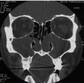
Computed tomography of the paranasal sinuses showed that the maxillary sinuses were filled with soft tissue density material that rea-ched the nasal cavity and the choanae. The other sinuses were normally aerated (Fig. 1).
The tumors were removed surgically - maxillary antrostomy (Caldwell-Luc) with a nasal endoscopy technique. The maxillary sinuses were each filled with a single cystic tumor that was implanted on the lateral wall.
Histology of the two lesions described them as inflammatory polyps. The patient had no postoperative events. The tumors have not recurred six months after the procedure.
DISCUSSION
Gustav Killian first described ACPs in
1906; these tumors comprise about 4% to 6% of all nasal polyps in the general population, and 28% to 33% in children.1-6 Bilateral ACPs
are rare; seven cases have been reported in the English scientific literature by April 2010, two of which in adults.2-6
The etiology of ACPs is unclear.3-6
Chronic rhinosinusitis, cystic fibrosis, and allergy have been often implicated.1-6 Studies have
sho-wn that ACPs usually originate from the lateral wall or the floor of the maxillary sinus.3,6 These
tumors are histologically indistinguishable from intramural cysts in their sinus portion.1,2 Berg
et al. (1988) have suggested that ACPs develop from intramural cysts in the maxillary sinus.2,6
This hypothesis does not explain the rarity of bilateral ACPs, especially because intramural cysts are often bilateral.5
The most common symptom of ACPs is unilateral or bilateral nasal block.1,5 Other
findings include snoring, sleep apnea, oral bre-athing, rhinorrhea with pus, postnasal discharge, epistaxis, dyspnea, hyposmia, dysphagia, and weight loss.1,2 Depending on their volume, ACPs
may obstruct the Eustachian’s tube and cause secretory otitis media.1
The differential diagnosis should be
made with retention mucous cysts, mucoceles, maxillary rhinosinusitis, meningoencephalocele, olfactory stesioneuroblastoma, angiofibroma, and inverted papilloma.1,2
Computed tomography and nasofibros-copy are the gold standard tests for diagnosing ACPs.1,2
Surgical treatment is mandatory.1,2,5
Recurrences are rare when the Caldwell-Luc approach is used; this method provides an ample view and complete removal of involved sinus tissues.2-5 Surgeons tend to avoid this
pro-cedure in children aged below 8 years because of the risk of injuring anterior dental roots and maxillary growth centers.2-5 In this age group,
tumors are removed by avulsion, which has a high recurrence rate.4,5
Another possible approach is nasal en-doscopy. Its advocates highlight the lower recur-rence and complication risks of this approach.2-5
FINAL COMMENTS
Bilateral ACPs are rare; the cases reported thus far have not led to a plausible explanation for this tumor. Studies defining the approach of choice for treating this disease are sparse in the literature.
REFERENCES
1. Freitas MR, Giesta RP, Pinheiro SD, Silva VC. Pólipo antrocoanal: uma revisão de dezesseis casos. Braz J Otorhinolaryngol. 2006;72(6):831-5. 2. Frosini P, Picarella G, De Campora E. Antrocho-anal polyp: Antrocho-analysis of 200 cases. Acta Otorhi-nolaryngol Ital. 2009;29:21-6.
3. Basu SK, Bandyopadhyay SN, Bora H. Bilat-eral antrochoanal polyps. J Laryngol Otol. 2001;115:561-2.
4. Myatt HM, Cabrera M. Bilateral antrochoanal polyps in a child: a case report. J Laryngol Otol. 1996;110:272-4.
5. Yilmaz YF, Titiz A, Ozcan M, Tezer MS, Ozluge-dik S, Ünal A. Bilateral antrochoanal polyps in an adult: a case report. B-ENT. 2007;3:97-9. 6. Jmeian S. Bilateral Antrochoanal polyps
in a child: an extremely rare case. JRMS. 2006;13(2):57-8.