O R I G I N A L A R T I C L E
Fungal microbiota dynamics as a postmortem investigation
tool: focus on
Aspergillus
,
Penicillium
and
Candida
species
J.J.C. Sidrim1,2,3, R.E. Moreira Filho1,2, R.A. Cordeiro1,2,3, M.F.G. Rocha1,2,4, E.P. Caetano1,2, A.J. Monteiro5and R.S.N. Brilhante1,2,3
1 Specialized Medical Mycology Center, Federal University of Ceara´, Fortaleza, Ceara´, Brazil 2 Postgraduate Program in Medical Microbiology, Federal University of Ceara´, Fortaleza, Ceara´, Brazil 3 Postgraduate Program in Medical Sciences, Federal University of Ceara´, Fortaleza, Ceara´, Brazil 4 Postgraduate Program in Veterinary Science, State University of Ceara´, Fortaleza, Ceara´, Brazil 5 Department of Statistics and Applied Mathematics, Federal University of Ceara´, Fortaleza, Ceara´, Brazil
Introduction
Forensic mycology is a relatively new term describing the study of the species of fungi present in cadavers. It can have application in forensic medicine, particularly deter-mination of the fungal groups to help establish the time of death (Carter and Tibbett 2003).
Studies of the relevant role of fungi in postmortem decomposition have been increasing because the corpse is a plentiful source of organic material (Ishii 2006), with a rising number of experimental descriptions and case studies in forensic mycology (Carter and Tibbett 2003; Hitosugi et al. 2006). These studies have shown that certain groups of these micro-organisms can provide valuable clues for estimating the time of death.
Although there have been some descriptions published on the participation of fungi in the postmortem process
(Hitosugi et al. 2006), few have focused on the species that are present at each stage of decomposition and the possible application of this information to forensic medicine. Besides this, the isolation of certain fungal spe-cies in determined geographical areas also helps in the characterization and classification of the typical regional micro-organisms, in view of the variation of species in contact with corpses under different growth conditions (Ishiiet al.2007).
In 2003, Carter and Tibbett demonstrated how and why the field mycology might provide a further tool towards the investigation of crime scenes in forest ecosys-tems. The fruiting structures of certain fungi, particularly the ammonia and postputrefaction fungi, have been recorded repeatedly in association with decomposed mammalian cadavers in different regions of the world (Carter and Tibbett 2003). Based on these reports, the Keywords
decomposition stages, forensic medicine, forensic mycology, fungi, time of death.
Correspondence
Raimunda Samia N. Brilhante, Rua Bara˜o de Caninde´, 210; Montese. CEP: 60.425-540. Fortaleza, CE, Brazil. E-mail: [email protected]
2009⁄0752: received 27 April 2009, revised 23 September 2009 and accepted 24 September 2009
doi:10.1111/j.1365-2672.2009.04573.x
Abstract
Aims:To investigate the presence of fungi during three human decomposition stages: bloated, putrefaction and skeletonization.
Methods and Results:The samples were gathered in the city of Fortaleza, Ceara´, Brazil, from the public morgue and cemeteries. The material was submitted to conventional mycological procedures by direct examination and macro⁄micro morphological and biochemical analyses. The main fungi isolated were Aspergillus spp., Penicillium spp. and Candida spp. in the bloated stage (n= 34 cadavers) and in the putrefaction stage (n= 6 cadavers), while in the skeletonization stage (n= 20 cadavers), the main fungi were Aspergillus spp., Penicilliumspp. andMucorsp.
Conclusions:Aspergillus, Penicillium and Candida species were associated with decomposed human cadavers.
present study investigated the presence of fungi during three decomposition stages: the bloated, putrefaction and skeletonization stages.
Material and methods
Ethical aspects
The present study was previously submitted to evaluation by the research ethics committee of State University of Ceara´ and obtained approval under number 064969333-9.
Sample collection sites
The samples were gathered at the city morgue in Fortaleza, in the state of Ceara´ (northeastern Brazil), and from public cemeteries in that state. All the cadavers were examined between January and December 2007.
Study subjects
The samples were taken from human corpses in three stages of decomposition: bloating stage (n= 34), putre-faction stage (n= 6) and skeletonization stage (n= 20). Each cadaver was evaluated in advance to establish the postmortem interval. All subjects were between the ages of 18 and 60 at death, victims of unnatural death – homi-cide, suihomi-cide, accidental asphyxiation, poisoning, electro-cution, traffic accidents and other mechanical traumas. In the bloating stages, all corpses were found at home. In the putrefaction stage, four corpses were examined after being exhumed and two were found in fields. In the skeletonization stage, all corpses were found in fields.
Biosafety
To preserve the integrity of the material collected and to protect the researchers’ health, biological masks and 0disposable caps, gowns, slippers and gloves were worn at all times (Brunicardiet al.2006).
Gathering the samples
The cadaver material was taken from the sites with the greatest probability of fungal growth: mouth, rectum, vagina, under the foreskin, lungs, skin, scalp hair, clothing and the surrounding area (grave soil and coffin frag-ments). The skin samples, approx. 1Æ0 cm in length, were
taken by scraping with a sterile scalpel, and the hair sam-ples, approx. five units with 1Æ0 cm in length, were
obtained with sterile tweezers. These samples were decon-taminated before collection with 70% isopropyl alcohol. Sterile swabs were used to collect material from the
muco-sas of the mouth, rectum, vagina and under the foreskin, using a rotating movement. At least two samples were collected from each site, one for direct examination and the other for culturing (Balows et al.1992). In the case of the samples from the pulmonary sites, a biopsy was performed with a sterile scalpel, and a single fragment 1Æ0 cm in length was retrieved from the main bronchial
tube and one from the peripheral bronchial tube. For characterization of the cadaver environment in the skelet-onization stage, when organic material is scanty, samples of approx. 1Æ0 cm in length from clothing and coffins and
1Æ0 g of grave soil were collected. The samples were placed
in a test tube with 3 ml of saline solution at a temperature of 25–28C and sent to the Specialized Medical Mycology Center (CEMM) of the Department of Pathology and Legal Medicine of Ceara´ Federal University.
Laboratory processing
Each specimen was clarified in 40% potassium hydroxide (KOH) and placed between slides and slide covers for microscopic examination at 100· and 400·. Along with direct examination, an aliquot of the material was inocu-lated at three points in dishes containing 2% glucose Sabouraud agar, Sabouraud agar with vancomycin plus polymixin B (to inhibit the growth of Gram-positive and Gram-negative contaminating bacteria, respectively) and Sabouraud agar with vancomycin plus polymixin B and cycloheximide (to inhibit contaminating airborne fungal strains). The dishes were incubated at 25–28C in the dark for up to 20 days, with daily observations until fungal growth was detected. The fungi were identified by phenotypical analyses, comprising macromorphology (texture, surface and diameter of the colony as well as the presence of pigmentation), micromorphology (size, surface and pigmentation of conidia and morphology of conidiogenic cells) and biochemical tests (e.g. nitrogen assimilation test and carbohydrate fermentation test) (De Hooget al., 2000).
Results
conidial heads, uni- and biseriate conidiogenous cells, conidiophore hyaline, echinulate conidia, 3Æ5-lm-diameter,
yellowish-green colonies; Aspergillus niger – radiating conidial heads, smooth-walled conidiophores,
50–100-lm-diameter vesicles, biseriate conidiogenous cells,
metu-lae twice as long as the phialides, brown and ornamented conidia with warts, 3Æ5–5Æ0lm in diameter; Penicillium piceum– colonies growing moderately, rapidly in Czapek Dox agar, velutinous to floccose, pale to bright yellow, with olivaceous green conidiation and orange-brown to dark brown reverse, thin- and smooth-walled conidio-phores 15–22 um long, biverticillate penicillin cells, metu-lae in dense whorls, 7–12·3–4lm, each metulae with
3–8 phialides, smooth-walled conidia, ellipsoidal 3Æ0–3Æ5·2Æ2–2Æ5lm; Penicillium rugulosum – restricted
velutinous colonies, yellow-green to dark green in colour, attaining <12-mm diameter in 7 days, conidiophores with 70–100-lm-long stipes and with smooth walls, usually
biverticillate penicillin cells, metulae 10–15 lm long,
ellipsoidal conidia, 3Æ0–3Æ5·2Æ5–3Æ0lm; Penicillium verruculosum – velutinous or floccose colonies, attaining over 18-mm diameter in 7 days, white to bright yellow mycelium, green conidial mass, smooth-walled conidio-phores with stipes 150–250lm long, phialides with 7–10
whorls, 8–15-lm long, spherical conidia with roughened
walls, 3Æ0–3Æ5lm in diameter, good growth at 37C; Candida albicans – cream-coloured colonies, pseudo-mycelium emerging from cellular clump and Chlamydo-conidia on Tween-80 agar and germ tubes with human serum and growth at 37C;Candida parapsilosis – cream-coloured colonies, pseudomycelium present, mostly abundant with giant cell, with width 8–10lm and length
15–20lm (De Hooget al.2000).
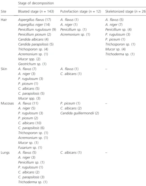
From the 34 cadavers in the bloated phase, we collected 234 samples for laboratory analysis, distributed among mucosas (n= 102), hair (n= 64), skin (n= 34) and
Table 1Species of fungi isolated from the hair, skin, mucosas and lungs of corpses in the bloated, putrefaction and skeletonization
stages Site
Stage of decomposition
Bloated stage (n= 143) Putrefaction stage (n= 12) Skeletonized stage (n= 26)
Hair Aspergillus flavus(17) A. flavus(1) A. flavus(5)
Aspergillus niger(14) A. niger(1) A. niger(7)
Penicillium rugulosum(9) Penicilliumsp. (1) Penicilliumsp. (4)
Penicillium piceum(2) Acremoniumsp. (1) P. rugulosum(3)
Candida albicans(4) P. piceum(1)
Candida parapsilosis(5) Trichosporonsp. (1)
Trichosporonsp. (4) Mucorsp. (4)
Acremoniumsp. (1) Trichodermasp. (1)
Mucorspp. (2)
Geotrichumsp. (1)
Skin A. flavus(7) A. flavus(1) –
A. niger(3) C. albicans(1)
P. rugulosum(3)
P. piceum(1)
C. albicans(5)
C. parapsilosis(5)
Mucorspp. (3)
Mucosas A. flavus(11) P. piceum(1) –
A. niger(5) C. albicans(2)
P. rugulosum(3) Candida guilliermondii(2)
P. piceum(2)
C. albicans(10)
C. parapsilosis(6)
Trichosporonsp. (1)
Acremoniumsp. (1)
Mucorsp. (1)
Fusariumsp. (1)
Lungs A. flavus(5) C. albicans(1) –
A. niger(3)
Penicilliumsp. (1)
P. rugulosum(1)
C. albicans(2)
C. parapsilosis(3)
lungs (n= 34). The quantitative association between the fungal genera and external collection sites (hair and skin) in the bloated stage was as follows: hair – A. flavus= 17, A. niger= 14, P. rugulosum= 9, P. piceum= 2, C. albi-cans= 4 and C. parapsilosis= 5, Trichosporon sp.= 4, Mucor sp. = 2, Geotrichum sp. = 1 and Acremonium sp. = 1; skin – C. albicans= 5 and C. parapsilosis= 5, A. flavus= 7, A. niger= 3, P. rugulosum= 3, P. pice-um= 1 andMucorsp. = 3. The results in the oral, genital and rectal mucosas were as follows: A. flavus= 11, A. niger= 5, C. albicans= 10 and C. parapsilosis= 6, Penicilliumsp. = 5P. rugulosum= 3,P. piceum= 2, Acre-monium sp. = 1, Fusarium sp. = 1, Trichosporon sp. = 1 and Mucor sp. = 1. The following fungi were found in the lungs: A. flavus = 5, A. niger= 3, C. albicans= 2, C. parapsilosis= 3, Penicillium sp. = 1, P. rugulosum= 1 andTrichodermasp. = 1 (Table 1).
The samples taken from the external sites (hair and skin) of the six corpses examined in the putrefaction stage yielded the following genera: hair – A. flavus= 1, A. niger= 1, Penicillium sp. = 1 and Acremonium sp. = 1 and skin – A. flavus= 1 and C. albicans= 1. The fungi present in the oral, genital and rectal mucosas were as follows: Candida guilliermondii= 2 and C. albicans= 2; P. piceum= 1. Only one C. albicans strain was found in the lungs (Table 1). Finally, the samples taken from the 20 corpses evaluated in the skeletonization stage yielded 90 isolates of filamentous fungi and three isolates of yeasts. The sites of the corpses themselves (hair and bone) produced the following genera: hair – A. niger= 7, A. flavus= 5, P. rugulosum= 3, P. piceum= 1, Mucor sp. = 4; Trichoderma sp. = 1 and Trichosporon sp. = 1
and bone – Penicillium spp. = 10 e P. verruculosum = 1, A. flavus= 6,A. niger= 3 andMucorsp. = 3 (Table 1).
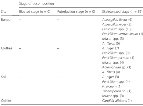
The nearby sites examined (clothing, soil and coffins) yielded the following genera: clothing – A. niger= 7, A. flavus= 5, Penicillium sp. = 9, P. piceum= 1; Mucor sp. = 4 and Acremonium sp. = 1; soil – A. flavus = 4, A. niger= 3, Penicillium sp. = 4, P. piceum= 1; Mucor sp. = 3 and Trichosporon sp. = 1; and coffins – C. albi-cans= 1 (Table 2). After identification, the fungi were deposited in the collection of the Specialized Medical Mycology Center (CEMM, Ceara´ Federal University).
Discussion
Because of the many factors – environmental and indivi-dual characteristics of corpses – that affect the growth of fungi, it is not surprising that there are differences between the few reports available to forensic mycology researchers (Ishii et al. 2007) and the findings described in regions with peculiar characteristics, such as north-eastern Brazil.
One point stands out in results of fungal isolation in all the stages evaluated (bloated, putrefaction and skeletonization), namely that direct mycological examina-tion is not fruitful. The bacterial kinetics promotes faster division of the prokaryotic cells in comparison with the eukaryotic fungal cells. The latter also require a narrower range of temperature, pH, moisture and luminosity conditions for adequate division and population dynam-ics (Murray et al. 1990). Therefore, direct mycological examination by optical microscope is of negligible value to identify the postmortem fungi.
Table 2Genera of fungi isolated from nearby sites examined (clothing, soil and coffins)
Site
Stage of decomposition
Bloated stage (n= 0) Putrefaction stage (n= 0) Skeletonized stage (n= 67)
Bones – – Aspergillus flavus(6)
Aspergillus niger(3)
Penicilliumspp. (10)
Penicillium verruculosum(1)
Mucorspp. (3)
A. flavus(5)
Clothes – – A. niger(7)
Penicilliumspp. (9)
Penicillium piceum(1)
Mucorspp. (4)
Acremoniumsp. (1)
A. flavus(4)
Soil – – A. niger(3)
Penicilliumspp. (4)
P. piceum(1)
Trichosporonsp. (1)
Mucorspp. (3)
Instead, it is essential in forensic mycology studies to use culture media and⁄or other complementary labora-tory methods to identify the presence of fungi. Regard-ing the form of asexual fungal division, the filamentous form prevailed in the bloated and skeletonization stages. As known, the majority of fungi reproduce asexually in nature, and many of their representatives are airborne strains and can easily grow on practically any substrate (e.g. Penicillium spp. and Aspergillus spp.) (Sharma 1988).
The results found in this study may not reflect the diversity of fungi in each stage of decomposition. We believe that refinement of the method employed can have a great impact on the recovery of some fungal species. In this respect, it is possible that the inclusion of other substances to the isolation media, such as benomyl, capa-ble of inhibiting the growth of Aspergillus sp. (Luzet al. 2007), will permit isolating species with less competitive power, such as the dematiaceous fungi.
The abundant growth of Aspergillus spp. and Penicil-lium spp. under the conditions employed might have impeded the isolation of typical soil species, such as the hyphomycetes, which were not recovered even with the addition of the inhibitor cyclohexamide to the culture media. To permit the growth of other fungal species not isolated in this work, we also suggest incubation of the material at a temperature of 37C, because of the heat sensitivity of various species ofPenicilliumspp. (De Hoog et al.2000).
In contrast, yeasts predominated in the putrefaction stage. In the oral, genital and rectal mucosas, the isolation of yeasts is more frequent because they are part of the normal microbiota of these areas. Until the present study, there were only a limited number of reports of the isolation of the fungal micro-organisms cited here under conditions of corpse putrefaction (Ishii 2006; Ishii et al. 2007). As a rule, this absence of information has been because of the failure to collect and investigate material under those conditions. Although the results demon-strated here are only descriptive, the mere presence of these fungi in these conditions is the reason for greater interest in fungi in forensic medicine, as a way to estab-lish the time of death.
The external collection sites were more propitious than the internal ones for fungal growth for both airborne fungi and yeasts, especially the genera Aspergillus and Candida, respectively. This corroborates other works on the postmortem alterations caused by fungi (Collier 2005). The yeasts also grew more on the skin than in the hair region. This fact can be explained by the after-death rupture of the skin barriers and greater contact with mucous secretions, besides their possible presence in the skin microbiota.
Another interesting observation regarding our postmor-tem isolation of fungi is the absence of dematiaceous fungi (able to produce pigment, generally melanin) among those isolated. This fact may be because of the growth characteristics common to various representatives of this group, which usually grow slowly, producing lesions with chronic evolution. Also, the intense competi-tion with bacteria, hyaline fungi and insects could have contributed to their absence in this study.
Although observation of the presence of fungi on the surface of corpses by forensic medical practitioners is not recent, the isolation of these organisms is not carried out routinely. Because their description can even give indica-tions of the place of death, additional studies are neces-sary for the use of fungi as a forensic tool (Ishii 2006).
The present study is only a starting point, and the results are not yet sufficient to allow the presence of fungi to act as effective biological markers of the time of death. However, it does demonstrate that there are differences in the fungi isolated during the process of corpse decompo-sition, especially by the isolation of fungi such as Aspergil-lus spp., Penicillium spp. and Candida spp. These results indicate that the presence of fungi on, in and around cadavers can provide additional information to determine the time of death as accurately as possible. Further research with larger samples and more detailed description of conditions is necessary to ratify the find-ings presented here and to establish the real importance of mycology as a tool to assist with forensic medicine, as is already the case of forensic entomology (Menezes et al. 2007).
Conclusion
These findings already permit tracing out a horizon for deeper understanding of the subject. However, much more research will be necessary to develop this new segment of mycology, enabling the frequent use of its findings in forensic science, as is already the case with entomology.
Acknowledgements
This work was supported by the National Research Council (CNPq) and by the Ceara´ State Foundation for Development of Science and Technology (FUNCAP), Process 9053⁄08.
References
Brunicardi, F.C., Anderson, D.K. and Brandt, M.L. (2006) Schwartz’s Principles of Surgery Self-assessment and Board Review. New York: McGraw Hil, 123–153.
Carter, D.O. and Tibbett, M. (2003) Taphonomic mycota: fungi with forensic potential.J Forensic Sci48, 168–171. Collier, J.H. (2005)Estimating the Postmortem Interval in
Forensic Cases through the Analysis of Postmortem Deterio-ration of Human Head Hair. Master of Arts Dissertation, Louisiana: Louisiana State University.
De Hoog, G.S., Guarro, J., Gene, J. and Figueras, M.J. (2000) Atlas of Clinical Fungi, 2nd edn. Spain: Central Bureau voor Schimmelcultures, Utrecht, the Netherlands and 0Rovira i Virgili University.
Hitosugi, M., Ishii, K., Yaguchi, T., Chigusa, Y., Kurasa, A., Kido, M., Nagai, T. and Tokudome, S. (2006) Fungi can be a useful forensic tool.Leg Med (Tokyo)8, 240–242.
Ishii, K. (2006) Analysis of fungi detected in human cadavers. Leg Med (Tokyo)8, 188–190.
Ishii, K., Hitosugi, M., Yaguchi, T. and Tokudome, S. (2007) The importance of forensic mycology.Leg Med (Tokyo)9, 287.
Luz, C., Netto, M.C. and Rocha, L.F. (2007)In vitro suscepti-bility to fungicides by invertebrate-pathogenic and sapro-bic fungi.Mycopathol164, 39–47.
Menezes, A.J., Kanchan, T., Monteiro, F., Manipady, S. and Rao, P. (2007) Forensic mycology.Leg Med (Tokyo)9, 48. Murray, P.R., Drew, W.L., Kobayashi, G.S. and Thompson,
J.H. Jr (1990)Medical Microbiology. St Louis, MO: The C. V. Mosby Company.