J Bras Pneumol. 2008;34(10):885-888 885
Case Report
In some cases, there is a family history of the disease, suggesting an autosomal recessive inheritance pattern.(4)
Most patients are asymptomatic. In symptomatic patients, symptoms typically appear in the advanced phase
Introduction
Pulmonary alveolar microlithiasis (PAM) is a rare disease that affects both lungs, characterized by the presence of small calculi (calcium phosphate) within the alveolar spaces. (1) The etiology remains unknown.(2,3)
Pregnancy in a patient with severe pulmonary alveolar microlithiasis*
Gravidez em paciente com microlitíase alveolar pulmonar grave
José Osmar Bezerra de Souza Filho1, Cristiane Maria Cavalcante Silveira1, Aline Barreto da Cunha2,
Valéria Goes Ferreira Pinheiro3, Francisco Edson de Lucena Feitosa4, Marcelo Alcântara Holanda5
Abstract
Pulmonary alveolar microlithiasis (PAM) is a rare disease that affects both lungs. It is characterized by the presence of small calculi (calcium phosphate) within the alveolar spaces. We report the case of a 26-year-old female whose diagnosis was based on characteristic findings on chest X-rays and high-resolution computed tomography scans. The patient, 28 weeks pregnant, was rehospitalized 10 months after the diagnosis, presenting hypoxemic acute respiratory failure and severe restrictive ventilatory defect on spirometry. After 32 completed weeks of gestation (228 days), she was submitted to cesarean section, and the outcome was successful for mother and newborn. PAM has a vari-able clinical course. It is suggestive of an autosomal recessive inheritance pattern and has been associated with positive family history. The etiology of PAM is unclear, and many authors speculate that there is a local enzymatic defect responsible for the intra-alveolar accumulation of calcium. Reports of patients with PAM who become pregnant are exceptional, and this is the first case described in Brazil. The course of this disease is usually slow and progressive, and patients typically die of cardiorespiratory failure. The present case illustrates the need to offer female patients, especially those with advanced disease, genetic counseling and orientation regarding the risks of pregnancy. Currently, the only effective therapy is lung transplantation.
Keywords: Pulmonary alveoli; Pregnancy; Respiratory insufficiency.
Resumo
A microlitíase alveolar pulmonar (MAP) é uma doença rara que atinge ambos os pulmões, caracterizada pela presença de pequenos cálculos (fosfato de cálcio) nos espaços alveolares. Relatamos o caso de uma paciente do sexo feminino, de 26 anos, cujo diagnóstico foi confirmado com base nos achados marcantes na radiografia de tórax e tomografia computadorizada de alta resolução. A paciente, gestante de 28 semanas, retornou ao hospital 10 meses após o diagnóstico apresentando insuficiência respiratória hipoxêmica e com distúrbio ventilatório restritivo grave à espirometria. Após completadas 32 semanas e 4 dias de gestação, foi submetida aparto cesariano, com sucesso para mãe e filha. A MAP tem evolução clínica variável. Tem provável caráter autossômico recessivo e associação com história familiar positiva. A etiologia é incerta, e muitos autores especulam que haja um defeito enzimático local responsável pelo acúmulo intra-alveolar de cálcio. Relatos de pacientes com MAP que engravidaram são excepcionais, sendo o presente caso o primeiro descrito no Brasil. O curso dessa doença costuma ser lentamente progressivo, e os pacientes geralmente falecem devido à insuficiência cardiorrespiratória. O presente caso ilustra a necessidade de se oferecer aconselhamento genético e orientações sobre o risco de gravidez às pacientes, especialmente em casos de doença avançada. Atualmente, a única terapia efetiva é o transplante pulmonar.
Descritores: Alvéolos pulmonares; Gravidez; Insuficiência respiratória.
* Study carried out at the Universidade Federal do Ceará – UFC, Federal University of Ceará – School of Medicine, Fortaleza, Brazil. 1. Medical Student. Universidade Federal do Ceará – UFC, Federal University of Ceará – Fortaleza, Brazil.
2. Resident in Clinical Medicine. Universidade Federal do Ceará – UFC, Federal University of Ceará – School of Medicine Walter Cantídio University Hospital, Fortaleza, Brazil.
3. Adjunct Professor in the Department of Clinical Medicine. Universidade Federal do Ceará – UFC, Federal University of Ceará – School of Medicine, Fortaleza, Brazil. 4. Supervisor of the Medical Residency Program in Gynecology and Obstetrics. Universidade Federal do Ceará – UFC, Federal University of Ceará – Assis Chateubriand Maternity Teaching Hospital, Fortaleza, Brazil.
5. Adjunct Professor in the Clinical Medicine Department. Universidade Federal do Ceará – UFC, Federal University of Ceará – School of Medicine, Fortaleza, Brazil. Correspondence to: Cristiane Maria Cavalcante Silveira. Rua Nogueira Acioli, 1080, apto. 801, Aldeota, CEP 60110-141, Fortaleza, CE, Brasil.
Tel 55 85 3366-8101. E-mail: [email protected] Financial Support: None.
886 Souza Filho JOB, Silveira CMC, Cunha AB, Pinheiro VGF, Feitosa FEL, Holanda MA
J Bras Pneumol. 2008;34(10):885-888
and instructed to use oxygen at home— 1 L / min. Ten months after diagnosis, the patient was rehos-pitalized at 28 weeks and 2 days of pregnancy, with worsening of the cyanosis and dyspnea. She was admitted to the hospital, and oxygen therapy by nasal catheter was initiated (5 L/ min). The blood gas analysis results were as follows: pH, 7.34; arte-rial carbon dioxide tension, 43 mmHg; artearte-rial oxygen tension, 49 mmHg; arterial oxygen satu-ration, 81%; and bicarbonate, 22.7 mEq/L. After stabilization of hypoxemia, using a Venturi mask at 35%, and improvement of dyspnea, the patient was transferred to the high risk prenatal clinic of the Assis Chateubriand Maternity Teaching Hospital. The patient remained hospitalized throughout pregnancy, with nasal oxygen administration (1 L/ min) and oxygen saturation of approximately 94%. After 32 weeks and days of gestation, she underwent cesarean section. During the procedure, she remained in use of the Venturi mask at 50%, without hypoxia. The newborn was responsive, crying at birth, with hydrated and normotensive fontanelle, weighting 1,960 g, measuring 42 cm, with a cephalic perimeter of 31.5 cm and a thoracic perimeter of 28.5 cm, all of which are appropriate for the gestational age, presenting zone III jaun-dice and a favorable response to phototherapy. The one-minute Apgar score was 8, and the two-minute of the disease. The diagnosis is based on
radiolog-ical findings of radiopaque micronodular infiltrates in both lungs, occasionally with a superimposed diffuse reticular pattern, resulting in a “sandstorm” appearance, a term employed to describe diffuse microcalcifications.(3,5-7)
At diagnosis, some patients present symptoms such as dyspnea, productive cough, hemoptysis and, less frequently, chest pain.(3)
We report the case of a patient diagnosed with PAM who became pregnant and evolved success-fully, being the second such case reported in the literature to date and the first in Brazil.(4)
Case report
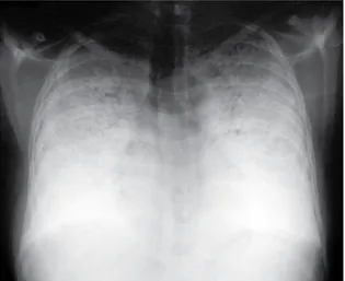
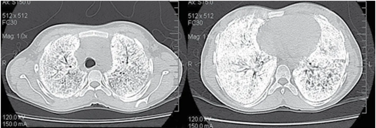
A 26-year-old female, an agriculturist, was referred to the Pulmonology Department of the Federal University of Ceará School of Medicine Walter Cantídio University Hospital for diagnostic investigation in March of 2005. She reported having had dyspnea on extreme exertion for 10 years. She also reported occasional wheezing. In the previous 4 years, there had been progressive worsening of the dyspnea, which began to occur on mild exertion, principally at night, accompanied by a productive cough, with whitish or yellowish secretion. For the preceding 2 years, she had awoken at night due to dyspnea and, in the previous year, she had presented attacks on a daily basis. She reported no fever, hemoptysis or chest pain. Upon physical examina-tion, the patient presented tachycardia, tachypnea, pallor and cyanosis (central and peripheral). Arterial oxygen saturation was 80%, without the use of supplemental oxygen. Pulmonary auscultation revealed reduced breath sounds throughout, with fine crackles in the lower thirds and in the anterior region of both hemithoraces. She presented digital clubbing. Routine chest X-ray revealed pronounced bilateral diffuse pulmonary infiltrate, with calcium density (Figure 1). High-resolution computed tomography (HRCT) revealed diffusely distributed calcified micronodules, with calcifications in the pleural space and the interlobular septa (Figure 2). Transthoracic echocardiogram was performed, revealing mild pulmonary hypertension, with a pulmonary artery systolic pressure of 43 mmHg and an ejection fraction of 58%. A diagnosis of PAM was therefore established. After stabilization of the acute status, the patient was discharged
Pregnancy in a patient with severe pulmonary alveolar microlithiasis
J Bras Pneumol. 2008;34(10):885-888 887
enzymatic defect is responsible for the intra-alve-olar accumulation of calcium.(2) Clinical signs and
symptoms typically appear in the advanced stage of the disease, when the only treatment available to the patient is lung transplantation.(9)
In a review of 576 cases of PAM reported in the literature, it is reported that there is a positive family history in approximately 50% of cases.(2)
In relation to the clinical status, the most common symptom is dyspnea on exertion and at rest. Other reported symptoms are productive cough, fatigue, chest pain, hemoptysis, palpitations, weight loss, stridor and headache. In a study involving 10 patients diagnosed with PAM, 2 presented no complaints at the onset of the condition. Of those same 10 patients, 4 presented restrictive lung disease and 6 presented normal pulmonary function.(4,5)
As a diagnostic method, HRCT of the chest has greater sensitivity than does chest X-ray, better characterizing typical alterations, such as inter-lobular septa thickening and subpleural interstitial thickening, which are common in patients with PAM, as well as more clearly showing the extent of the disease.(6-8)
In the abovementioned study of 10 patients with MAP, it was suggested that the number and the extent of the alterations found on the HRCT be proportional to the degree of loss in pulmonary function. Radiological alterations varied in relation to extent and to forms of presentation, justifying the inhomogeneous and unpredictable clinical course.(10)
Apgar score was 9. The newborn presented normal heart and lung auscultation. Reflexes were present and symmetric. Approximately 1 year and 8 months after delivery, the patient returned to the outpatient clinic bringing the child, who presented normal psychomotor development. The patient evolved with progressive worsening of the dyspnea in this period. Spirometry was performed, with the following results: forced vital capacity (FVC), 2.80 L (25.9% of predicted); forced expiratory volume in one second (FEV1), 2.59 L (13.9% of predicted); FEV1/ FVC ratio, 91.1% (75.6% of predicted); and forced expiratory flow between 25% and 75% of FVC, 3.63 L/s (65% of predicted) The patient was therefore diagnosed with severe restrictive respiratory disorder.
The patient complained of dizziness and pain in the lower limbs at 4 min into the examination, which was consequently interrupted.
Discussion
The present case report shows that PAM can reach an advanced stage in young patients. In addi-tion, the fact that the pregnancy was successfully carried to term in a patient with extensive disease emphasizes the importance of offering appropriate family planning to patients of childbearing age who present with this disease. This is a rare clinical entity, characterized by the presence of small intra-alveolar calcium calculi (calciferites, calcospherites or microlites).(8) In case-series studies, there are no
reports of gender predominance.(8) The etiology is
uncertain, and many authors speculate that a local
Figure 2 - High-resolution tomographic axial slices of the lung apices and bases. Length and width of the
window: −486 and 1800 Hounsfield units, respectively. Bilateral, diffuse acinar opacities, with high calcium density, in
888 Souza Filho JOB, Silveira CMC, Cunha AB, Pinheiro VGF, Feitosa FEL, Holanda MA
J Bras Pneumol. 2008;34(10):885-888
2. Mariotta S, Ricci A, Papale M, De Clementi F, Sposato B, Guidi L, et al. Pulmonary alveolar microlithiasis: report on 576 cases published in the literature. Sarcoidosis Vasc Diffuse Lung Dis. 2004;21(3):173-81.
3. Wang HY, Shen HH, Jiang ZN. Pulmonary alveolar microlithiasis: report of four familial cases. Chin Med J (Engl). 2004;117(6):950-2.
4. Al-Alawi AS. Familial occurrence of pulmonary alveolar microlithiasis in 3 siblings. Saudi Med J. 2006;27(2):238-40.
5. Barbolini G, Rossi G, Bisetti A. Pulmonary alveolar microlithiasis. N Engl J Med. 2002;347(1):69-70.
6. Wurche KD, Kubale R, Vallèe D, Ostertag H. Quantification of pulmonary alveolar microlithiasis in the CT [Article in German]. Rofo. 1987;147(1):36-8.
7. Julhl J, Crummy A. Interpretação Radiológica. Rio de Janeiro: Guanabara Koogan, 2000.
8. Marchiori E, Gonçalves CM, Escuissato DL, Teixeira KI, Rodrigues R, Barreto MM. Pulmonary alveolar microlithiasis: high-resolution computed tomography findings in 10 patients. J Bras Pneumol. 2007;33(5):552-7.
9. Bonnette P, Bisson A, el Kadi NB, Colchen A, Leroy M, Fischler M, et al. Bilateral single lung transplantation. Complications and results in 14 patients. Eur J Cardiothorac Surg. 1992;6(10):550-4.
10. Deniz O, Ors F, Tozkoparan E, Ozcan A, Gumus S, Bozlar U, et al. High resolution computed tomographic features of pulmonary alveolar microlithiasis. Eur J Radiol. 2005;55(3):452-60.
The principal differential diagnoses include miliary tuberculosis, pulmonary hemosiderosis, sarcoidosis and amyloidosis, as well as metastatic pulmonary calcification associated with chronic renal insufficiency and hemodialysis.(2,3)
The distribution of PAM is worldwide. According to a review of the literature, cases have been reported in 51 countries, in 12 of which—France, Bulgaria, Germany, India, Italy, Turkey, Japan, Russia, Poland, Spain, the United States and Yugoslavia—at least 10 cases have been described.(2)
The first reported case of a woman with PAM who became pregnant was in a 36-year-old woman, who also carried the pregnancy to term.(8)
The course of this disease is typically slow, and patients with PAM generally die due to cardiorespi-ratory failure. Currently, the only effective therapy is lung transplantation.(9)