MARGINAL BONE LOSS AND SURVIVAL RATE OF CLINICAL
STUDIES WITH ZIRCONIA DENTAL IMPLANTS –
SYSTEMATIC REVIEW AND META-ANALYSIS
Dissertação apresentada à Universidade Católica Portuguesa
para obtenção do grau de Mestre em Medicina Dentária
Por:
Higor Borges
MARGINAL BONE LOSS AND SURVIVAL RATE OF CLINICAL
STUDIES WITH ZIRCONIA DENTAL IMPLANTS –
SYSTEMATIC REVIEW AND META-ANALYSIS
Dissertação apresentada à Universidade Católica Portuguesa
para obtenção do grau de Mestre em Medicina Dentária
Por:
Higor Borges
Orientador: Prof. Doutor Gustavo V. O. Fernandes
Co-orientador: Prof. Doutor André Correia
V AGRADECIMENTOS
Agradeço a Deus por iluminar e guiar meus passos ao longo dessa jornada, que envolve o desenvolvimento deste projeto, permitindo que eu tenha um aprimoramento profissional e pessoal
Aos meus pais Pedro e Cláudia e ao meu irmão Hugo por sempre acreditarem no meu potencial e me apoiarem. Sei que minha jornada até aqui não foi nada fácil, mas sempre que fraquejei, os senhores me ergueram.
À minha amada Marina e ao nosso príncipe Enzo por cada dia de tanto amor e alegria. Vocês são e sempre serão minhas maiores motivações. Obrigado por estarem comigo nesta caminhada.
À minha avó Ivanilde que sempre presou e apoiou os estudos dos seus filhos e netos.
À minha sogrinha querida Mari, que não se encontra mais entre nós, por sempre cuidar de mim com tanto carinho e tratar-me como um filho. Obrigado por tudo que fizestes por mim e por nossa família.
Aos Professores Gustavo e André pelo aceite em guiar-me durante todo esse processo. Aos senhores atribuo grandemente o meu amadurecimento científico e profissional.
À Dra. Helena Donato pelas recomendações e por possibilitar a pesquisa através de um dos bancos de dados adotados neste trabalho.
Ao meu amigo e colega Alejandro por compartilhar sua experiência e conhecimento no que diz respeito ao desenvolvimento de trabalhos científicos.
Agradeço à Universidade Católica Portuguesa e seus coordenadores pela oportunidade de hoje estar aqui concretizando esse sonho. Fui recebido com muita atenção e respeito, e ao longo de todo esse tempo, tratado com muita dedicação e carinho.
VII
RESUMO
Objetivo: Avaliar a taxa de sobrevivência cumulativa e a perda óssea marginal peri implantar de implantes dentários de zircónia submetidos a um acompanhamento de pelo menos 12 meses após a reabilitação protética. Materiais e Métodos: A procura sistemática eletrónica através dos bancos de dados PubMed (MEDLINE) e EMBASE foi realizada independentemente por dois revisores a fim de identificar estudos clínicos publicados entre janeiro de 2005 e abril de 2019 com no mínimo 10 pacientes e 12 meses de acompanhamento após carga funcional. As referências dos artigos selecionados foram revisadas manualmente à procura de estudos adicionais. Resultados: A remodelação marginal óssea apresentou perdas médias de 0.80 mm (95% CI 0.60-1.00 mm) e 1.01 mm (95% CI 0.72-1.29 mm) em 1 ano e após 2 anos de funcionalização respetivamente. Não foi possível realizar a meta-análise para a taxa de sobrevivência uma vez que a maioria dos estudos não forneceram valores de intervalo de confiança ou desvio padrão. A taxa de falha foi reportada para um período de 2.75 anos de acompanhamento, onde a prevalência de falha precoce, falha tardia e fratura foi de 3.4%, 1.7% e 1.7% respetivamente. Conclusão: Os implantes de zircónia apresentaram resultados comparáveis aos dos implantes de titânio no que diz respeito à perda óssea marginal em períodos de observação de curto prazo após a restauração protética. No entanto, mais estudos clínicos a longo prazo bem planejados são necessários antes seja dada recomendação para adoção de implantes de zircónia na prática diária.
Palavras-chave: implantes dentários, óxido de zircónio, cerâmicas, taxa de sobrevivência, revisão sistemática.
IX
ABSTRACT
Purpose: To evaluate cumulative survival rate and peri-implant marginal bone loss of zirconia dental implants subjected to a follow-up of at least 12 months after prosthetic rehabilitation. Materials and Methods: A systematic electronic search through the databases PubMed (MEDLINE) and EMBASE was performed by two independent reviewers to identify clinical studies published between January 2005 and April 2019 with a minimum of 10 patients and 12 months of follow-up after functional loading. References from the selected articles were manually reviewed for further studies. Results: From the initial 1225 articles retrieved, only 19 met all the inclusion criteria. The marginal bone remodelling accounted mean losses of 0.8 mm (95% CI 0.60-1.00 mm) and 1.01 mm (95% CI 0.72-1.29 mm) at 1-year and after 2-year post-loading respectively. Failure rate of 6.8% was calculated for a mean follow-up period of 2.75 years, where the prevalence of early failure, late failure and implant fracture was 3.4%, 1.7% and 1.7% respectively. The meta-analysis regarding the survival rate of one- and two-piece zirconia dental implants was not possible due to the lack of information about confidential interval or standard deviation on most of the included articles. Conclusion: Zirconia implants presented values comparable to titanium implants with respect to marginal bone loss at short-term observation periods following prosthetic delivery. However, more long-term well-designed clinical studies are required before giving the recommendation on the adoption of zirconia implants in daily practice.
Keywords: dental implants, zirconium oxide, ceramics, survival rate, systematic review.
ÍNDICE GERAL
1. INTRODUCTION ... 1
2. MATERIALS AND METHODS ... 5
2.1. FOCUSED QUESTION ... 5
2.2. INFORMATION SOURCES AND SEARCH STRATEGY ... 5
2.3. INCLUSION CRITERIA ... 5
2.4. EXCLUSION CRITERIA ... 7
2.5. SELECTION OF STUDIES ... 7
2.6. RISK OF BIAS AND QUALITY ASSESSMENT ... 7
3. RESULTS ... 9
3.1. STUDY SELECTION ... 9
3.2. STUDY CHARACTERISTICS AND QUALITY ASSESSMENT ... 17
3.3. PERI-IMPLANT MARGINAL BONE LOSS ... 20
3.4. BIOLOGICAL COMPLICATIONS ... 22
3.5. MECHANICAL COMPLICATIONS ... 23
3.6. CUMULATIVE IMPLANT SURVIVAL RATE ... 23
4. DISCUSSION ... 27
5. CONCLUSION ... 34
1
1. INTRODUCTION
Owing to its well established good osseointegration, biocompatibility, strength and corrosion resistance, titanium is currently described as the gold standard biomaterial for dental implants.(1) Its mechanical and biological properties have been extensively studied in several experimental and clinical applications developed over the past decades.(2) Moreover, in a recently published systematic review, the ten years overall survival rate of titanium implants was calculated at 96.4%, which demonstrates its good performance at long-term replacement of missing teeth.(3)
Nevertheless, titanium implants might be associated with discoloration of peri-implant soft tissue, leading to a dull greyish background in case of thin gingival biotype (4) or eventual mucosal recession.(5) Besides the aesthetics concerns, potential hypersensitivity responses reported due to metallic ions released by Ti implant degradation (6) have contributed to the increasing demand for completely metal-free dental reconstructions. Although titanium allergy has a low estimated prevalence of 0.6%, the appearance of some form of acute reaction or post-implant surgery complication cannot be disregarded, particularly in predisposing patients.(7)
The continuous heightening of the aesthetic patterns relating to implant-supported rehabilitations as well as the health concerns associated to the use of titanium implants have boosted a constant investigation for alternative materials that could better comply with this objective. The development and use of ceramics as implant bulk material has been subjected to extensive research in order to assess their clinical performance.(2,8,9)
The first generation of ceramic implant was clinically introduced more than 30 years ago. The crystalline bone screw, as described by its developer S. Sandhaus, was made of aluminium oxide (Al2O3).(10) However it is no longer available on the market possibly due to increased risk of fracture when loaded extra-axially, which was attributed to its unsatisfactory biomechanical properties.(5)
Nowadays, zirconium dioxide (zirconia) is the material adopted by dental industry for the fabrication of ceramic implants. Since its introduction for medical application in the 80s, ceramics have been extensively used as the bulk material of orthopaedic prosthesis for total hip replacement surgery.(11) Initially used in dentistry for crowns and implant abutments, (12,13) zirconia had its application scope amplified
2
due to superior physical and mechanical properties compared to other ceramics in terms of flexural strength, modulus of elasticity and fracture toughness.(5,12)
Currently, most of the commercially available ceramic implants are fabricated in yttria stabilized tetragonal zirconia polycrystalline (Y-TZP). Y-TZP is a yttria-dopped zirconia ceramic composed of tetragonal crystal grains with mean size of hundreds of nanometers, obtained by the addition of 2 to 3% mol yttrium oxide (yttria, Y2O3) to zirconium oxide (zirconia, ZrO2) at room temperature.(14) The zirconia-yttria ceramic is characterized by optimal physical and mechanical properties including low porosity (<0.1%), high density (>6 gcm3), a favourable bending strength (900 – 1200 MPa) and compression strength (2000 MPa), a high fracture toughness (7 – 10 MPa m-1) and an appropriate Young’s modulus (210 GPa),(14) which makes it suitable to be applied in dental implantology.
Zirconia presents a polymorphic structure that at room temperature assumes the monoclinic (M) crystalline form, then tetragonal (T) form at 1170ºC followed by cubic (C) phase at temperatures above 2300ºC. (14) Forces applied to the surface of the zirconia causes transitions between T and M phases, resulting in volumetric changes, i.e., modification on the crystalline phase of the zirconia provokes a structural expansion at the transformed zone (e.g. vicinity of a crack), inducing compressive forces that seal the crack. This phenomenon leads to an increased crack propagation resistance, which is referred by phase transformation toughening.(15) However, the metastable yttria-zirconia is susceptible to low-temperature degradation (LTD) or ageing, a progressive water induced transformation of the tetragonal pattern into the monoclinic one. This mechanism, trigged by the exposure of zirconia to the wet conditions of oral cavity, entails consequences over performance, reliability and lifetime of zirconia devices due to resulting formation of micro and macrocracking followed by surface roughening and reduced strength, toughness and density.(16,17)
Zirconia demonstrates good biocompatibility (18,19) and induces less extensive inflammatory reaction and bone resorption than titanium.(20) Histological analysis of soft tissue surrounding titanium implants revealed a keratinized oral gingival epithelium, a non-keratinized sulcular epithelium and a junctional epithelium attached to the implant surface. The latter was separated from the subjacent crestal bone by a scar-like gingival connective tissue, characterized by collagen fibres bundles oriented parallel to the implant long axis.(21) Moreover, zirconia implants presented a
peri-3
implant biological width structure similar to that of titanium implants and a comparable level of soft tissue integration was observed between both types of materials.(22)
An investigation in vivo suggested similar level of blood flow on the peri-implant soft tissue compared to that seen in the mucosa surrounding natural teeth. This increased microcirculatory dynamic was considered beneficial for the preservation of immune defence against external pathogens (23), considering that the supracrestal connective tissue lateral to the implant was reported to be poorly vascularized (24).
Bacterial colonization by periodontal pathogens around the implant surface causes peri-implant gingival inflammation, leading to peri-implantitis, which in turn is appointed as one of the major contributing factors for dental implant failure (25) together with occlusal overload.(26) In vivo studies revealed a reduced plaque accumulation potential of zirconia in comparison to titanium.(27,28)
Studies have shown that zirconia stimulates bone formation by regulating translation mechanisms related to osteogenesis and bone remodelling in osteoblast-like cells exposed to the ceramic surface. (29,30). Investigation (31) evaluating the effect of surface topography on osseointegration showed improved bone response on surface-modified zirconia implants as the bone remodelling process appeared to be sensitive to the level of ceramic roughness. The authors also reported a statistically significant lower removal torque on the machined surface zirconia implants compared to that of surface-modified zirconia implants, which in turn was similar to the one observed on oxidized titanium implants. Additionally, the torque forces caused the bone-implant interface to rupture instead of simply separate, implying in the capacity of surface-modified zirconia implants to achieve firm bone stability.(31)
Two recently published systematic reviews (22,32) involving animal studies reported comparable mean bone-implant contact values for zirconia and titanium implants. Both the quantitative surface roughness values and the procedure adopted for the surface characterization appears to influence the osseointegration process of zirconia implants.(22)
Taking all these points into account, it seems zirconia implants present the ability to replace missing teeth with preservation of a healthy peri-implant hard and soft tissue following its integration with the referred surrounding tissues. Since the publication of the latest systematic review on zirconia implants outcomes, newer clinical studies with longer follow-up periods have been released, which in turn can
4
enable a better evaluation of the performance of this material in comparison to the titanium.
Therefore, the purpose of the present review was to systematically evaluate the available evidence on the outcomes of zirconia dental implants in clinical studies with respect to survival rate and marginal bone loss (MBL). Such a review would be essential before any recommendations could be made on treatment with ceramic implant and would contribute to the current debate on adoption of ceramic as a bulk material for dental implants.
5
2. MATERIALS AND METHODS
This systematic review was conducted following the Preferred Reporting Items for Systematic reviews and Meta-Analysis (PRISMA) guidelines (33) with the focused question being determined according to the Population, Intervention, Comparison and Outcome (PICO) strategy.(34) The protocol for this systematic review was registered on PROSPERO (identification number to be provided by the Centre for Reviews and Dissemination/ CRD – University of York).
Focused Question
The focused question for the present review was as follows: “In clinical studies with partially and fully edentulous patients (P), do the oral rehabilitations with zirconia implants (I), when compared with titanium implants (C), exhibit differences in clinical outcomes (O)?
Information sources and search strategy
An extensive electronic search was conducted through MEDLINE (PubMed) and EMBASE databases with a platform-specific search strategy combining controlled terms (MeSH and Emtree) and text words, detailed in Table 1. An additional manual search was performed on the references of included articles to identify relevant publications. Only articles published in the English-language dental literature from January 2005 until and including April 2019 were included. Two reviewers (H.B. and G.F.) independently performed the electronic and manual search.
The publications obtained from the search through all mentioned databases were imported into a reference management software (EndNote X9/ Thomson Reuters, Philadelphia, USA) and subsequently screened.
Inclusion criteria
This systematic review was based on randomized controlled clinical trial (RCT), prospective (PS) and retrospective (RS) clinical studies.
6
Table 1 – Search strategy carried out and filters applied
MEDLINE (PubMed) Embase
#1
P – Fully or partially edentulous patients treated with dental implants
((“Dental Implants" [MeSH Terms]) OR (“Dental Implants, Single-Tooth" [MeSH Terms]) OR (Dental Implant* [Supplementary Concept]))
(‘tooth implantation’/exp OR ‘tooth implant’/exp)
#2
I – Rehabilitation with zirconia dental implants
((“Zirconium” [MeSH Terms]) OR (Zirconium Oxide [Supplementary Concept]) OR (Zirconia [Supplementary Concept]) OR (Yttria Stabilized Tetragonal Zirconia [Supplementary Concept]) OR (“Ceramics” [MeSH Terms]))
(‘zirconium oxide’/exp OR ‘zirconium’/exp OR ‘ceramics’/exp OR ‘yttria stabilized tetragonal zirconia’/exp)
#3 C – Rehabilitation with titanium dental implants #4 O – Clinical outcomes
Search Combination
(#1 AND #2)
No combination was done with #3 and #4, since the majority of the papers on dental implants are about titanium, and the combination with keywords related to outcome would limit even more the search.
Filters English, Humans
7
The additional inclusion criteria for study selection were:
• Human studies published in English-language dental literature from January 2005 until April 2019 with at least 10 patients treated.
• Partially or fully edentulous patients rehabilitated with zirconia dental implants. • A follow-up of at least 12 months after functional loading.
• Detailed information on the implant used.
• Reported details regarding survival and/or failure rates.
• Only the publication with the longest follow-up was included in case of multiple studies involving the same patient cohort (population).
Exclusion criteria
Clinical studies that didn’t meet the entire inclusion criteria were excluded. Reports based on questionnaires, interviews and case reports/series were also rejected as well as systematic reviews, publications investigating individually designed zirconia implants or involving patients with significant health problem (ASA Physical Status 3 and above).
Selection of studies
Duplicates were excluded and the remaining articles screened by tittle and abstract for eligibility. Further examination with regard to inclusion and exclusion was subsequently made by full-text analysis. The full-text of any title or abstract that did not provide sufficient information regarding the inclusion criteria was also obtained. Any disagreement between the reviewers was discussed with a third author (A.C.). Cohen’s kappa test was adopted to evaluate reviewers’ agreement on both title and abstract selection.
Risk of bias and quality assessment
The assessment of risk of bias and study quality of the included investigations was performed independently by two reviewers (H.B. and G.F.), where randomization
8
process, groups similar at baseline, blinded group allocation, random housing, blinded interventions, random and blinded outcome assessment, reporting of drop-outs and other biases (funding) domains were addressed.
Data extraction and method of analysis
The reviewers extracted the data independently from the selected articles for further analysis using data extraction tables, which included the following parameters:
• Author(s), year of publication and study design (RCT/PS/RS); • Mean observation period
• Number of patients and implants at the initial stage of the research, location of the implant (maxilla/mandible), mean age of patients and age range;
• Implant bulk material (yttria-stabilized tetragonal zirconia polycrystal [Y-TZP], alumina toughened zirconia [ATZ]), titanium [Ti]), implant design (1-piece/ 2-piece), implant system, implant surface treatment and surface roughness; • Use of bone augmentation procedure;
• Type of prosthetic reconstruction (single crown [SC], fixed partial denture [FPD], implant-supported overdenture [ISO]) and prosthesis retention mode;
• Loading mode after implant placement (immediate/conversional) and time period between implant placement and final prosthetic reconstruction (weeks); • Number of patients and implant drop outs, number of early and late implant
failure and number of implant fracture;
• Cumulative implant survival rate (%), implant success rate (%), and peri-implant marginal bone loss (mm).
The meta-analysis involved the comparison of the data obtained for the MBL at a mean observation period of 1 year of functional service and after 2 years. The meta-analysis for the cumulative survival rate was not feasible. All meta-analysis was performed using the software Excel (Microsoft, Redmond, USA), where random effect model at a 5% significance level was used. Heterogeneity across the studies was quantified using the I2 inconsistency test. Values above 75% were considered an indication of substantial heterogeneity. For those studies that confidential interval (CI) was not provided, standard deviation (SD) value was used for the calculation of a CI.
9
3. RESULTS
Study selection
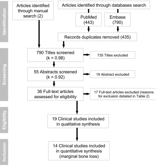
1223 studies were identified from the electronic database search (MEDLINE: 443; EMBASE: 780). A further two publications were considered from the manual search through the references of the included articles. Of the 1225 articles initially found, 435 duplicates (310 electronically; 125 manually) were removed, and the remaining 790 were reviewed by tittle. The title screening resulted in 55 articles to be evaluated by abstract, which subsequently yield 36 studies to be considered for full-text assessment for further evaluation on the inclusion and exclusion criteria. Seventeen full-texts were excluded based on exclusion criteria, detailed in Table 2. Finally, a total of 19 studies met the inclusion criteria and were included in the current review (Fig.1, Table 3–6).
The kappa values for the inter-examiner agreement between the reviewers were 0.98 for the title screening and 0.92 for the abstract screening.
Table 2 – Excluded studies and reason for exclusions
Author/Year Reason for exclusion
Blaschke & Volz, 2006 Data not clear for evaluation
Oliva et al., 2007 Individually designed zirconia implant investigated Pirker & Kocher, 2009 Individually designed zirconia implant investigated
Borgonovo et al., 2010 Publication on the same patient cohort of Borgonovo et al., 2013 Oliva et al., 2010 Individually designed zirconia implant investigated
Borgonovo et al., 2012 Sample size (8 patients)
Kohal et al., 2012 Publication on the same patient cohort of Kohal et al., 2018 Gahlert et al., 2013 Publication on the same patient cohort of Roehling et al., 2016 Osman & Ma, 2014 Publication on the same patient cohort of Osman et al., 2014 Siddiqi et al., 2015 Publication on the same patient cohort of Osman et al., 2014 Borgonovo et al., 2016 Sample size (6 patients)
Gahlert et al., 2016 Publication on the same patient cohort of Bormann et al., 2018 Jung et al., 2016 Publication on the same patient cohort of Balmer et al., 2018 Hollander et al. 2016 Publication on the same patient cohort of Lorenz et al., 2019 Spies et al., 2016 Publication on the same patient cohort of Spies et al., 2015 Kniha et al, 2018 Data not clear for evaluation
10
Table 3 – Detailed data of the included studies
Study Patient Implant
Author/year Study Design
Mean obs.
per. (months) Setting N
Age (years)
N Location Material Mean Range
One-piece design
Lorenz et al., 2019 PS 93.6 Univ./Priv. 28 63.5 39-80 83 Max: 38 Man: 45 YTZP
Balmer et al., 2018 PS 36.6 Univ. 60 48.1 20-70 71 Max: 23 Man: 48 YTZP
Bormann et al., 2018 PS 36 Priv. 44 48 18-78 44 Max: 40 Man: 4 YTZP
Kniha et al., 2018a PS 36 Univ. 87 55 NR 117 NR YTZP
Kniha et al., 2018b RS 12 Priv. 86 55 25-67 92 (period. healthy) Max: 93 Man: 30 YTZP
31 (period. compr.)
Kohal et al., 2018 PS 36 Univ. 65 NR 18-70 66 Max:18 Man: 48 YTZP
Kniha et al., 2017 PS 12 Priv. 78 55 NR 82 NR YTZP
Roehling et al., 2016 RS 71.28 Priv. 71 54.9 18-85 161 Max: 85 Man: 77 YTZP
Grassi et al., 2015 PS 61.2 Univ./Priv. 17 52.3 35–70 16 (fresh socket) Max: 26 Man: 6 YTZP
16 (healed socket)
Spies et al., 2015 PS 36 Univ. 40 NR NR 53 NR ATZ
Osman et al., 2014 RCT 12 Univ. 12 62 46–80 73 Max: 40 Man: 33 YTZP
12 56 Max: 32 Man: 24 Ti
Kohal et al., 2013 PS 12 Univ. 28 NR NR 56 Max: 12 Man: 44 YTZP
Payer et al., 2013 PS 24 Univ. 20 44.4 27–71 20 Max: 11 Man: 9 YTZP
Borgonovo et al., 2013 PS 48 Univ. 13 60 38–75 35 Max: 20* Man: 8* YTZP
Cannizarro et al., 2010 RCT 12 Priv. 20 38 18–54 20 (occ load.) Max: 12 Man: 8 YTZP 20 39 26–55 20 (non-occ load.) Max: 17 Man: 3
11
Table 3 – Detailed data of the included studies (continued)
Study Patient Implant
Author/year Study Design
Mean obs.
per. (months) Setting N
Age (years)
N Location Material Mean Range
Two-piece design
Becker et al., 2017 PS 25.5 Univ. 52 47.6 NR 52 Max: 13 Man: 35 YTZP
Payer et al., 2015 RCT 24 Univ. 22 46 24–77 16 Max: 3 Man: 13 YTZP
15 Max: 4 Man: 11 Ti
Cionca et al., 2015 PS 19.38 Univ. 32 51.9 24–75 49 Max: 24 Man: 25 ATZ
One- and Two-piece design
Brüll et al., 2014 RS 18.4 Priv. 74 51 18-72 121 NR YTZP
Mean obs. per.: Mean observation period; RCT: Randomized controlled trial; PS: Prospective clinical study; RS: Retrospective clinical study; Univ.: University; Priv.: Private Practice; YTPZ: yttria-stabilized zirconia; ATZ: alumina-toughened zirconia; Ti: titanium; NR: not reported; Max: Maxilla; Man: Mandibula; Period. healthy: periodontally healthy; Period. compr.: periodontally compromised; occ. load.: occlusally loaded; non-occ. load.: non-occlusally loaded.
12
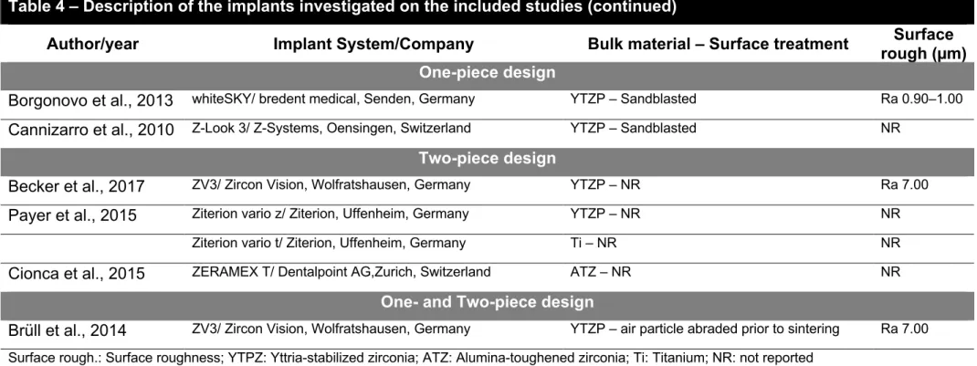
Table 4 – Description of the implants investigated on the included studies
Author/year Implant System/Company Bulk material – Surface treatment Surface
rough (µm)
One-piece design
Lorenz et al., 2019 Z-Look 3/ Z-Systems, Oensingen, Switzerland YTZP – Sandblasted NR Balmer et al., 2018 ceramic.implant/ VITA Zahnfabrik, Bad Säckingen,
Germany
YTZP – Sandblasted, acid-etched Ra 1.20
Bormann et al., 2018 PURE Ceramic Implant/ Straumann AG, Basel, Switzerland YTZP – Sandblasted, large-grit, acid-etched Sa 0.70 Kniha et al., 2018a PURE Ceramic Implant/ Straumann AG, Basel, Switzerland YTZP – Sandblasted, large-grit, acid-etched Sa 0.70 Kniha et al., 2018b PURE Ceramic Implant/ Straumann AG, Basel, Switzerland YTZP – Sandblasted, large-grit, acid-etched Sa 0.70 Kohal et al., 2018 ZiUnite/Nobel Biocare, Göteborg, Sweden YTZP – Slurry containing zirconia powder and
burnable pore former applied onto surface
Sa 1.24
Kniha et al., 2017 PURE Ceramic Implant/ Straumann AG, Basel, Switzerland YTZP – Sandblasted, large-grit, acid-etched Sa 0.70 Roehling et al., 2016 Z-Look 3/ Z-Systems, Oensingen, Switzerland YTZP – Sandblasted NR Grassi et al., 2015 whiteSKY/ bredent medical, Senden, Germany YTZP – Sandblasted NR Spies et al., 2015 Ziraldent FR1/ Metoxit AG, Thayngen, Switzerland ATZ – Sandblasted, ceramic slurry applied onto
surface
Ra 1.80
Osman et al., 2014 Southern Implants, Irene, South Africa YTZP – Acid-etched Ra 0.50–0.80 Ti – Sandblasted, acid-etched Ra 1.00–2.00 Kohal et al., 2013 ZiUnite/Nobel Biocare, Göteborg, Sweden YTZP – Slurry containing zirconia powder and
burnable pore former applied onto surface
Sa 1.24
13
Table 4 – Description of the implants investigated on the included studies (continued)
Author/year Implant System/Company Bulk material – Surface treatment Surface
rough (µm)
One-piece design
Borgonovo et al., 2013 whiteSKY/ bredent medical, Senden, Germany YTZP – Sandblasted Ra 0.90–1.00 Cannizarro et al., 2010 Z-Look 3/ Z-Systems, Oensingen, Switzerland YTZP – Sandblasted NR
Two-piece design
Becker et al., 2017 ZV3/ Zircon Vision, Wolfratshausen, Germany YTZP – NR Ra 7.00 Payer et al., 2015 Ziterion vario z/ Ziterion, Uffenheim, Germany YTZP – NR NR
Ziterion vario t/ Ziterion, Uffenheim, Germany Ti – NR NR
Cionca et al., 2015 ZERAMEX T/ Dentalpoint AG,Zurich, Switzerland ATZ – NR NR
One- and Two-piece design
Brüll et al., 2014 ZV3/ Zircon Vision, Wolfratshausen, Germany YTZP – air particle abraded prior to sintering Ra 7.00 Surface rough.: Surface roughness; YTPZ: Yttria-stabilized zirconia; ATZ: Alumina-toughened zirconia; Ti: Titanium; NR: not reported
14
Table 5 – Detailed information of the prosthetic rehabilitation
Author/year Type of prosthetic reconstruction Retention mode abutment/crown Time for Recons.(weeks) One-piece design
Lorenz et al., 2019 SC, FPD CR, CR Max: 24 Man: 16
Balmer et al., 2018 SC, FPD CR, CR Max: 16 Man: 8
Bormann et al., 2018 SC CR 24 to 28
Kniha et al., 2018a NR NR NR
Kniha et al., 2018b NR NR 12 to 20
Kohal et al., 2018 SC CR Max: 14 Man: 6
Kniha et al., 2017 SC CR 12
Roehling et al., 2016 SC, FPD, ISO CR, CR, RM At least 12
Grassi et al., 2015 SC CR 12–16
Spies et al., 2015 SC, FPD CR, CR Max: 14 Man: 6
Osman et al., 2014 ISO RM 12–16
Kohal et al., 2013 FPD CR Max: 14 Man: 6
Payer et al., 2013 SC CR 16
Borgonovo et al., 2013 SC, FPD CR, CR 24
Cannizarro et al., 2010 SC CR 16–20
Two-piece design
Becker et al., 2017 SC CR Max: 12 Man: 10
Payer et al., 2015 SC CR Max: 24 Man: 16
Cionca et al., 2015 SC CR 27.57 ± 11.29
One- and Two-piece design
Brüll et al., 2014 SC, FPD CR, CR 18.4 ± 12–68
Time for Recons.: Period between implant placement and final prosthetic reconstruction; SC: single crown; FDP: fixed partial denture; ISO: implant-supported overdenture; CR: cement-retained; RM: removable; Max: maxilla; Man: mandibula; NR: not reported
15
Table 6 – Detailed data on the outcomes of the included studies
Author/year Loading
mode
Drop-out (N)
Success rate (%) Mean MBL (mm) Survival
rate (%)
Patient Implant
One-piece design
Lorenz et al., 2019 NR 0 0 NR 1.20 ± 0.76 100
Balmer et al., 2018 NR 5 5 NR 0.70 ± 0.72 98.5
Bormann et al., 2018 Conventional 13 13 97.7 0.97 ± 0.88 97.7
Kniha et al., 2018a NR 6 12 95.4 0.78 100
Kniha et al., 2018b NR 0 (period. healthy) 0 95 0.58 100
0 (period. compr.) 0 94 0.11 100
Kohal et al., 2018 Conventional 4 4 66 (grade I), 79 (grade II) 1.45 ± 1.96 90.8
Kniha et al., 2017 NR 0 0 100 NR 100
Roehling et al., 2016 NR 0 0 77.6 0.97 ± 0.07 77.3
Grassi et al., 2015 Immediate 1 (fresh socket) 1 NR 1.29 ± 0.25 93
0 (healed socket) 0 NR 1.17 ± 0.33 100
Spies et al., 2015 Conventional 1 1 96.5 (grade I) 100 (grade II) 0.79 94.2
Osman et al., 2014 Conventional 1 (YTZP) 7 NR 0.42 ± 0.40 71.2
4 (Ti) 28 NR 0.18 ± 0.47 82.1
Kohal et al., 2013 NR 0 0 60 (grade I), 72 (grade II) 1.95 ± 1.71 98.2
Payer et al., 2013 Conventional 0 0 95 1.29 95
Borgonovo et al., 2013 Conventional 3 7 100 1.63 100
Cannizarro et al., 2010 Immediate 0 (occ load.) 0 NR 0.90 ± 0.48 85
16
Table 6 – Detailed data on the outcomes of the included studies (continued)
Author/year Loading
mode
Drop-out (N)
Success rate (%) Mean MBL (mm) Survival
rate (%)
Patient Implant
Two-piece design
Becker et al., 2017 Conventional 4 4 NR NR 95.8
Payer et al., 2015 Conventional 0 (YTZP) 0 93.3 1.48 ± 1.05 93.3
0 (Ti) 0 100 1.43 ± 0.67 100
Cionca et al., 2015 Conventional 2 2 NR NR 87.3
One- and Two-piece design
Brüll et al., 2014 Conventional 0 0 NR 0.13 ± 0.60 96.5
Period. healthy: periodontally healthy; Period. compr.: periodontally compromised; occ. load.: occlusally loaded; non-occ. load.: non-occlusally loaded; YTZP: yttria-stabilized zirconia; Ti: titanium; NR: not reported
17
Figure 1 – Flow diagram for the search strategy and selection process for the included studies
Study characteristics and quality assessment
Within the 19 studies selected for analysis, only 3 were randomized controlled trials (2,8,9), whereas 13 were prospective clinical trials (35–47) and 3 were retrospective controlled trials (48–50), all published between 2010 and 2019. Of these, fourteen studies were included in the meta-analysis for evaluation of MBL and had the risk of bias and quality assessed (Figure 2–3).
Id en tif ic at io n Sc re eni ng El igi bi lit y In clu sio n PubMed (443) Embase (790) Articles identified through manual search (2)
Articles identified through databases search
Records duplicates removed (435)
790 Titles screened (k = 0.98) 55 Abstracts screened (k = 0.92) 735 Titles excluded 19 Abstract excluded 36 Full-text articles
assessed for eligibility 17 Full-text articles excluded (reasons for exclusion detailed in Table 2)
19 Clinical studies included in qualitative synthesis
14 Clinical studies included in quantitative synthesis
18
The studies included a total of 881 patients with a mean age of 44.2 (range 18– 85) years that were treated with 1294 ceramic implants and 71 titanium implants. All the investigations described the participants as systemically healthy and 11 included current smoking patients. The vast majority of the publications (15) studied one-piece ceramic implants (n = 1055 implants), 3 examined 117 implants of the two-piece ceramic design and 1 study investigated a total of 121 implants of one and two-piece ceramic systems. Most of the studies involved ceramic implants made up of Y-TPZ (n = 1192), whereas implants produced in ATZ (n = 102) were examined in two investigations.(43,51) The distribution of the zirconia implants was described in 16 studies (n = 910), being 465 in the maxilla and 445 in the mandible. Only two studies (2,9) involved direct comparison of zirconia implants (n = 89) with titanium implants (n = 71, maxilla: 36; mandible: 35), both of them RCTs. One publication evaluated the influence of conventional non-occlusal loading over the immediate occlusal loading of zirconia implants on reduction of early failure.(8) Another study investigated immediate loaded implants installed either in healed and post-extraction sites.(42)
Most of the evaluated implants (n = 679) were placed in academic settings, whilst 571 implants were performed in private practice. Rehabilitation treatment involving both types of premises was reported in two publications and accounted for 115 implants.
Among the selected studies, ten different ceramic implants systems were described. Although appropriately registered to be used, only five of them are commercially available (ceramic. implant, whiteSKY, PURE Ceramic Implant, Ziraldent FR1 and ZV3).
Seven out of the fourteen studies that reported simultaneous bone augmentation provided information on the bone grafting material adopted, which included either xenogenic bone mineral (Bio-Oss, Geistlich Pharma AG, Wolhusen, Switzerland), synthetic bone (macroporous biphasic calcium phosphate, MBCP+, Leone, Firenze, Italy) or autogenous bone.
The evaluated studies presented a follow-up period varying between 1 and 7.8 years, with overall mean observation period of 2.75 years. Moreover, the average of patients who could not be followed for the entire study period was of 4.99% (drop-out range 0–29.55%).
19
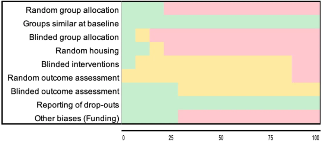
Figure 2 – Reviewers’ judgments for each risk of bias parameter evaluated for each of the 14 studies assessed in the meta-analysis.
Gr as si et al . 2015 Pa ye r et al . 2015 Ca nn iz ar ro et al . 2010 Pa ye r et al . 2013 Ko ha l et al . 2013 Bo rg on ov o et al . 2013 Bo rm an n et al . 2018 Os ma n et al . 2014 Sp ie s et al . 2015 Ko ha l et al . 2018 Ba lm er et al . 2018 Kn ih a et al . 2018b Kn ih a et al . 2018a Br üll et al . 2014
random group allocation - + + - - - - + - - - - - -
groups similar at baseline + + + + + + + + + + + + + +
blinded group allocation - NR + - - - - - - - - - - -
random housing - + + - - - - - - - - - - - blinded interventions - - + NR NR NR NR NR NR NR NR NR NR - random outcome assessment - NR - NR NR NR NR NR NR NR NR NR NR - blinded outcome assessment + NR + NR + NR NR NR NR + NR NR NR - reporting of drop-outs + + + + + + + + + + + + + +
other biases (Funding) NF F F F F NF F F F F F NF NF F
20
Figure 3 – Plot of percentage distribution of the reviewers’ judgments on each risk of bias parameter across the evaluated studies.
Peri-implant marginal bone loss
Radiographic marginal bone remodelling between implant placement and follow-up was evaluated in 17 studies, with the mean value varying between 0.13mm(48) and 1.95 mm.(40) Three publications were excluded from the analysis of MBL as two assessed orthopantomograms (39,49) and one provided no detailed value for the MBL.(43) All the 14 evaluated studies made the peri-implant bone level measurements based on periapical radiographs (conventional and digital) taken using some sort of standardized paralleling technique. Most of the selected studies considered the distance between implant shoulder or base of abutment and most coronal level of bone-to-implant contact on both mesial and distal surfaces as references for the MBL measurement. However, other reference points such as top of ball abutment (9) and transition zone between straight abutment part and implants threads (40) were also cited.
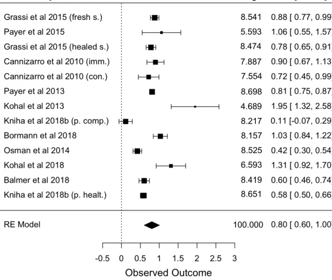
The marginal bone remodelling process accounted a mean loss of 0.80 mm (95% CI 0.60-1.00 mm) at 1-year post-loading period (Figure 2) and was achieved based on the date of 10 studies. Borgonovo et al.(35) and Spies et al.(41) were not included in this analysis as no value or clear information on MBL at 1-year of service was provided. Overall mean bone gain of 0.2 mm instead of loss at 1-year observation period was encountered in the investigations of Brüll et al.(48) and Kniha et al.(47). These findings contradict from the values reported in the literature for the first year of
21
service, which justified the exclusion of these studies from this analysis. A high heterogeneity among the studies was observed (I2 = 96.109%).
Figure 2 – Forest plot for peri-implant marginal bone loss at one year of functional loading of 10 studies. (RE Model: Random Effect Model)
Most of the resorption observation was reported to occur during the osseous healing phase, prior to the final prosthetic rehabilitation. Bormann et al.(37) accounted minimal mean bone gain of 0.06 mm between 12 and 36 months. The marginal bone changes values described in the text of Kniha et al.(47), were different from those observed in the table with descriptive measurements provided in the study. In the table, instead of reducing 0.12 mm between 3 months and 1 year as informed in the text, the authors added 0.12 mm and computed this change as a bone gain and not as a loss. And because of that, this error was carried forward, giving a wrong value for mean crestal bone level at 3-year follow-up.
Author/year Mean [95%CI] Weight
8.541 5.593 8.474 7.887 7.554 8.698 4.689 8.217 6.593 8.419 8.651 8.157 8.525 100.000
22
Crestal bone changes were assessed for observation period of two years and above (Figure 3). Data regarding the MBL at the latest follow-up recorded in 10 investigations were analysed, giving an overall mean MBL of 1.01 mm (95% CI 0.72-1.29) and high heterogeneity value (I2 = 97.046%).
Figure 3 – Forest plot for peri-implant marginal bone loss after two years of functional loading of 10 studies. (RE Model: Random Effect Model)
Biological complications
Biological complications in the peri-implant tissue were assessed by 15 of the 19 studies. The most cited parameters evaluated were probing pocket depth, plaque index (regular and modified), sulcus bleeding index (regular and modified), clinical attachment level, marginal soft tissue level (mucosal/ gingival recession), implant
Author/year Mean [95%CI] Weight
9.924 5.742 9.801 10.065 9.865 9.256 8.174 7.786 9.747 9.658 9.981 100.000
23
mobility, pink aesthetic score, soft tissue inflammation, presence or absence of suppuration and pain.
Mechanical complications
This review classified failures according to their timing into early and late failures. Although a type of failure, implant fracture was evaluated separately, being identified in three investigations, totalling 22 Y-TPZ implants (1.7%). In the study (49) where most of the fractures were observed (n = 18), 15 were narrow implants (3.25 mm in diameter) and three had diameter of 4.0 mm. Of these, 14 were recorded in the maxilla and 4 in the mandible. None of the implants with diameter of 5.0 mm fractured. All the events occurred following prosthetic restoration, between 0.8 and 69.7 (mean period of 15.3) months after implant placement, at the threading part of the coronal portion of the implant body. Another study (9) reported two fractured implants in the maxilla and one in the mandible, placed in three patients rehabilitated with implant-supported overdenture. The third study (48) was the only among the three investigations involving implant fracture that evaluated marked implants (n = 1). However, information on design, time-point and location of the fractured implant were not specified.
Biomechanical complications related to the prosthesis were no assessed in this review, as the dental implant was aimed rather than the entire implant-prosthesis complex.
Cumulative Implant Survival Rate
All the 19 publications provided information on the cumulative implant survival rate. From 1294 zirconia implants placed in 881 participants, 14 studies reported failure of a total of 66 implants, where 44 were lost before final prosthetic reconstruction (3.4%). The remaining (n = 22) were considered late failure as the implants were lost after being subjected to functional loading, accounting to 1.7% of all zirconia implants installed. Most of the failures were recorded within the first year of service. The location of the failed implants was specified in 9 investigations (maxilla: 38; mandible: 19). When considering the region of the jaws where the failures occurred, only 4 studies provided this information, totalling 4 failures in the anterior zone of the maxilla and 8 in
24
the posterior region (maxilla: 4; mandible: 4). The calculated failure rate is summarized in Table 7, where failure timing (early and late) and implant fracture values were both considered. When combining the figures informed in 14 studies for early and late failure and implant fracture with respect to arch location, 54 of 387 implants placed in upper jaw were lost (13.9%), whereas the failure value for the implants inserted in lower jaw was 6.1% (24 of 391 implants). Ten failed implants did not have the location informed by their respective studies.
Table 7 – Calculated failure rate – early failure, late failure and implant fracture prevalence in the included studies.
Author/year Implants N
Early failure Late failure Fracture Failure rate
N % N % N % %
One-piece design
Lorenz et al., 2019 83 0 0.0 0 0.0 0 0.0 0.0
Balmer et al., 2018 71 1 1.4 0 1.4 0 0.0 1.4
Bormann et al., 2018 44 1 2.3 0 2.3 0 0.0 2.3
Kniha et al., 2018a 117 0 0.0 0 0.0 0 0.0 0.0
Kniha et al., 2018b 123 0 0.0 0 0.0 0 0.0 0.0 Kohal et al., 2018 66 3 4.5 3 9.1 0 4.5 9.1 Kniha et al., 2017 82 0 0.0 0 0.0 0 0.0 0.0 Roehling et al., 2016 161 14 8.7 4 22.4 18 2.5 22.4 Grassi et al., 2015 32 1 3.1 0 3.1 0 0.0 3.1 Spies et al., 2015 53 3 5.7 0 5.7 0 0.0 5.7 Osman et al., 2014 73 12 16.4 6 28.8 3 8.2 28.8 Kohal et al., 2013 56 1 1.8 0 1.8 0 0.0 1.8 Payer et al., 2013 20 1 5.0 0 5.0 0 0.0 5.0 Borgonovo et al., 2013 35 0 0.0 0 0.0 0 0.0 0.0 Cannizarro et al., 2010 40 5 12.5 0 12.5 0 0.0 12.5 1056 42 3.98 13 1.23 21 1.99 7.20 Two-piece design Becker et al., 2017 52 0 0.0 2 3.8 0 3.8 3.8 Payer et al., 2015 16 0 0.0 1 6.3 0 6.3 6.3 Cionca et al., 2015 49 1 2.0 5 12.2 0 10.2 12.2 117 1 0.85 8 6.84 0 0 7.69
One- and two-piece design
Brüll et al., 2014 121 1 0.83 1 0.83 1 0.83 2.5
121 1 0.83 1 0.83 1 0.83 2.48
25
Different reasons concerning the early failure were pointed by the studies including placement of implant in previously periodontally compromised site, immediate provisionalization of the implant (placed in fresh extraction socket), immediate prosthetic loading, low patient compliance, loss of primary stability due to problems in the osseointegration process, smooth implant surface, para-functional habits and failed osseointegration due to “aseptic loosening”. With respect to late failure, the main causes highlighted were occlusal overloading, reduced osseointegration due to fibro-osseous integration of the implant with the surrounding, peri-implantitis and loss of osseointegration without specific reasons.
Implant survival was defined as implants remaining in situ at the examinations during the observation period, irrespective to their conditions. The survival rate derived from the data of the included articles ranged from 71.2% at 1 year (9) to 100% at 7.8 years (39). However, a meta-analysis of the survival rate was not feasible due to the lack of information about confidential interval and standard deviation on most of the included studies.
Implant success was evaluated in 12 studies (Table 6) by means of different proposed criteria involving various clinical and radiographic parameters. The criteria proposed by Albrektsson et al.(52), adopted in two studies (42,50), preconised the absence of radiographic peri-implant radiolucencies, implant mobility, clinical pain, infection in the peri-implant soft tissues and annual marginal bone loss inferior to 0.2 mm after first year of service.
Two investigations (37,49) cited the criteria previously described by Buser et at.(53) that assessed implant mobility, continuous radiolucency around the implant, peri-implant infection with suppuration, pain, foreign body discomfort and/or dysesthesia and possibility for restoration. According to Jahn and d’Hoedt (54) a successful treatment involved MBL <0.5 mm, peri-implant vertical bone loss lower than 30% of the implant length, implant mobility <1 mm, pocket depth measuring less than 4 mm and patient satisfaction. This criteria was used in two studies (44,47). Apart from some of the parameters previously pointed, the criteria proposed by Naet et al.(55) and Snauwaert et al.(55) also considered periotest value (<+8) and was applied in further two studies.(2,36) The implant success grading recommended by Östman et al.(56), applied in three investigations, defined as grade I implants showing ≤ 2 mm of
26
bone loss with no clinical and radiographic signs of peri-implant pathologies and grade II the implants with no further pathology and bone resorption ≤ 3 mm. Cannizzaro et al.(8) evaluated implant success using a self-defined criteria that considered implant stability and infection.
27
4. DISCUSSION
The purpose of the current systematic review and meta-analysis was to examine the peri-implant marginal bone loss, the cumulative survival rate and the behaviour of zirconia dental implants investigated in clinical studies with minimum follow-up of 12 months. Due to the limited availability of well-controlled investigations evaluating clinical performance of ceramic dental implants, such as RCTs, a lower level of clinical evidence (PSs and RSs) was included in this review in order to summarize the available information on outcomes.
The survival rate derived from the data of the included articles ranged from 71.2% at 1 year (9) to 100% at 7.8 years (39). However, a meta-analysis of the survival rate was not possible due to the lack of information about confidential interval and standard deviation on most of the included studies.
Only two studies (2,9) compared the zirconia implant and titanium implant, which made it difficult to systematically evaluate these materials by means of a direct comparison. Payer et al. (2) presented an overall survival rate of 100% for titanium implants and 93.3% for zirconia implants, with the latter being attributed to one zirconia implant lost within the first year of service. However, these results should be interpreted with caution due to the reduced sample of both zirconia (n = 16) and titanium (n = 15) dental implants exanimated in the study. The investigation carried out by Osman et al. (9) reported survival rate of 71.2% and 82.1% for zirconia and titanium implants respectively, which in turn was not statistically different (P = 0.15). The reduced survival for both types of material was attributed to the novel distribution of the implants adopted, which involved high prevalence of implant failure, especially those placed in the mid-palate. When arch location was considered, statistically significant differences were observed. Zirconia implants achieved survival rates of 55% and 90.9% at the maxilla and mandible respectively (P = 0.001), whereas the values for titanium implants were 71.9% and 95.8% respectively. Again, there was no statistical discrepancy between the survival outcome of zirconia and titanium with respect to upper (P = 0.14) or lower (P = 0.47) jaws. Based on the findings, the authors (9) speculated a better performance of zirconia implants in rehabilitations of partial edentulism, rather than in fully edentulous cases involving implant-supported overdentures.
28
The majority of the zirconia implants evaluated by the included articles was single-piece design. This system involves a supra-mucosal part (abutment) inherent to the implant body, overcoming the bacterial accumulation and consequent crestal bone resorption associated to the presence of a microgap experienced by two-piece design due to the interface between abutment and its implant platform.(2,36) On the other hand, the presence of the abutment part (two-pieces) penetrated into the oral cavity is a problematic encountered by this type of implant as it will be subjected to loading forces attributed to masticatory activity and tongue movements throughout the healing period.(2,36)
The included studies involving conventional loading protocol reported the use of protective barrier by means of removable splint appropriately fitted or relined dentures immediately after surgery procedure, aiming to shield the implants from premature loading until the permanent prosthetic restoration was delivered. However, both prosthetic approaches depend on the compliance of the patients on the use of the apparatus, thus leading to the uncertainty whether an unloaded bone remodelling environment will be provided.(2,36) A stress-free healing is achievable with a non-removable appliance adhesively attached to the neighbouring teeth, also adopted among some studies. Nevertheless, this approach would not be applicable with posterior implants in situations of Kennedy type I and II. Additionally, provisional bridging would be subjected to patients’ interference as the maintenance of an accurate oral hygiene around the implants is highly important for peri-implant outcomes, which includes the marginal bone remodelling.(2,36)
Apart from the healing concerns on the use of one-piece implants, aesthetic needs and difficulty on cementation of restoration is also challenging, especially in the anterior section.(41) These implants are not available with different abutment angulations,(36) meaning that the emergency profile would have to be altered in case of implant being inappropriately positioned. However, the preparation of the implant, if permitted by the implant system, has to be carried following strictly the protocol recommended by the manufacturer and is limited to a certain level of the initial height of the abutment portion.(42)
The behaviour in vitro (57) of zirconia showed that the grinding process may compromise its fracture strength, which can culminate in the fracture of the implant. This finding, that initiates at the surface and proceeds into the bulk, was attributed to
29
the aging of the material due to flaws or temperature variation induced by the mechanical preparation.(57) Yttria-zirconia is susceptible to aging, process where the content of monoclinic phase increases as the transformation of the metastable tetragonal occurs, resulting in micro/macrocracking and surface roughness associated to reduced toughness and density respectively.(58)
In the aesthetic zone, single-piece implants are commonly inserted in a deeper position in relation to the peri-implant soft tissue, aiming to reach an appropriate emergency profile and a submarginal restoration, which can make removal of luting cement excess far more difficult.(51) However, even after removal of cement remnant, a residual cement film may remain in the peri-implant sulcus, leading to peri-implant inflammatory diseases or implant failure.(59,60) Although not all the studies provided information on the cementation material adopted, the SCs and FPDs were reported to be cemented either with resin or glass ionomer cements.
Currently, a few ceramic dental implant systems are available in two-piece design, and in this review, only four studies involved this type of implant design. All the investigations reported that both abutment connections and definitive prosthetic restorations were cemented (Table 5). In either way, the use of two-piece implants may encounter the same problematic related to cementation. Thus, the importance of the thorough removal and cleaning cement excess during cementation procedure and thereafter perform follow-up examinations to evaluate any tissue adverse response, once cement remnant may be undetectable even in a radiographic image.(61)
The overall mean MBL at 1-year of prosthetic functioning for zirconia implants of the current study is 0.80 mm (95% CI 0.60–1.00). However, for this analysis, two studies that presented overall mean bone gain instead of loss were not include in the assessment at 1-year of service as their marginal remodelling outcome would increase the risk of bias. The divergent results would lower the mean MBL considerably should they were included. The results observed in the present study are in accordance with the figures reported in other systematic reviews involving zirconia implants. Pieralli et al.(62) reported 0.79 mm for 398 ceramic implants inserted in 326 patients. In a further systematic review (63), zirconia implants were divided according to their market availability and evaluated separately. The authors (63) reported a reduced, but not statistically significant different, marginal bone level for the commercially available group (0.69 mm) compared to the non-commercially available zirconia implants (0.95
30
mm). This sort of distinction was not applied in this review due to the limited number of studies retrieved. The values obtained corroborate with those observed for titanium implants after follow-up varying between 1 and 5 years (0.41-0.89 mm).(64)
The lowest zirconia implant survival rate of all included studies was noticed in a RCT (9), which involved high prevalence of early failure and implant fracture. The most probable reason for the fractures was accounted by the authors to the unfavourable bending moments related to peri-implant marginal bone resorption and consequent reduced bone support. Deficiency of the macroscopic design and reduced diameter (ø 3.75 mm) of the implants used were also pointed as contributing factors, suggesting improvement needs on the implant system design and on its biomechanics. Besides parafunctional habits (bruxism) and aspects related to implant design such as diameter and thread design, Roehling et al.(49) also linked fracture to the sandblasting surface preparation, which may alters the fracture strength of zirconia. Additionally, implant overloading and micromotions exceeding the critical limits associated to failure of osseointegration were also listed by the authors.(49)
Moreover, four investigations (40,43,46), including the one carried by Roehling et al.(49) reported that implants suddenly became mobile with no sign of peri-implant soft and hard tissue infection or inflammation. These observations were described to be associated either to mechanical rupture of bone-implant interface (43) caused by premature loading or to failed osseous integration due to reduced surface roughness of the studied implants.(49) The reported failures were not derived by bacterial infection, so referred as “aseptic loosening” by one of the studies.(43) This contradicts with results of an investigation on titanium implants, where neither failed osseointegration nor premature loading but inflammatory process was appointed as the major cause for early failure.(65)
The study carried by Kohal et al.(46) also involved implants failed due to peri-implant infection accompanied by progressive bone resorption, all reported after osseous healing period. Histological analysis of the bone harvest from the apical region of the sites of these removed implants revealed portions with osteointegration patterns similar to those seen around titanium implants. Authors (46) concluded that reduced osteoconductivity capacity of the material could not be appointed as a possible cause for the increased bone loss observed.
31
In summary, a higher failure percentage for zirconia implants was found in the maxilla (13.9%) compared with the mandible (6.1%). Difference in bone quality between both arches can partially justify this result. The cortical component tends to be denser and thicker in the mandible than in the maxilla, and it normally reduces in thickness and density towards the posterior region of both jaws. The trabecular bone is also denser in the lower jaw.(66) So the denser composition of the mandible may provide a better osseous support and favour a primary stabilization of the implant, which is essential for its osseointegration.(67) In addition, the interpretation of this result is further complicated as implants of different designs and surface topographic characteristics were used in various locations of both maxilla and mandible in a non-standardized manner.
Rehabilitation of periodontally compromised patients with titanium dental implants demonstrated worse outcomes, yielding lower survival rates when compared to treatments involving implant sites with history of a healthy periodontium.(68) Many investigations reported significantly higher reduction on peri-implant bone level,(69) more biological complications,(1) and increased failure rate.(70) The evidence available for titanium implants contradicts the results revealed by Kniha et al.(50), as patients with compromised tissue condition did not present significant longitudinal bone loss around the zirconia implants compared to that of periodontally healthy patients. The study also reported similar pocket depth in both groups, which in turn was not statistically different from the measurements obtained 1-year post-loading on the contralateral natural teeth. Both investigated groups accounted no implant loss,(50) opposing the results of a systematic review that reported a survival rate ranging between 79.22 and 100% for the periodontitis subjects.(68)
Many of the included studies assessed implant success, however various criteria adopted were reported, which precludes a comparison between them with respect to this outcome. Moreover, this observation demonstrate a lack of consensus regarding a set of criteria to evaluate success that is universally accepted.(68) Most of the criteria adopted by the included studies considered clinical and radiographic parameters such as implant mobility (52–54), pocket depth (54), peri-implant radiolucency (52,53,55,71), recurrent infection (52,55,71) with suppuration (53) and pain (52,53,55,71). For marginal bone loss, one criteria allowed 0.2 mm of annual loss after the first year of service,(52) other considered success when reductions did not
32
exceed 0.5 mm. (54) Some studies applied a success grading that allowed maximum bone loss of 2 mm and 3 mm for grade I and grade II respectively, in the absence of peri-implant pathological manifestations.(56)
The meta-analysis conducted in this systematic review recorded a mean MBL of 0.80 mm at 1-year following prosthetic restoration, which is in accordance with the recommendations of the consensus report of the First European Workshop on Periodontology that considered successful outcome when bone reductions inferior to 1.5 mm are observed within the first year of functional loading.(72) Moreover, the same consensus suggested further 0.2 mm of annual bone loss after 1-year of service. (72) This review also reported 1.01 mm of loss accounted from implant insertion up to observation periods ranging between 2 and 5 years. Thus, once again, treatment with zirconia implants was considered successful with regard to MBL criterion after 2-year of service.
The assessment of risk of bias and study quality revealed that most of investigations (n = 10) were financially supported, even if reported to be partially funded, by a grant from the manufacturer of the respective studied implant system. Only one of the RCTs fully described a randomized and blinded selection and assessment of patients. In general, information on selection, distribution and examination of patients was not provided at all. None of the studies reported a calculation for the sample size. The included articles did not report calibration of surgeons with respect to the surgery procedure for implantation, causing possible bias in the analysis. Additionally, many important data required for a meta-analysis were not provided by many studies. For the evaluation of MBL, many of the CI values had to be calculated based on the SD. Some studies reported CI in the form of graphs. However, a precise value was not given, and an approximation based on plots would not be appropriated.
Although a considerable number of surgeries for implant installation were performed in private practices, the majority of the procedures reported in studies included in this review were conducted in academic settings. Therefore, the outcomes here observed might not necessarily reflect the clinical results of implantology services provided in private office settings.
All the studies included in this review calculated implant survival rate with regard to the total amount of implants rather than the number of patients evaluated. A less
33
favourable result would possibly be achieved by the investigations if the analysis were made with respect to patient-based data, as the survival rate was obtained from the implant-based data that becomes diluted from the large number of implants installed in the patient sample.(73)
34
5. CONCLUSION
Even with the limitations of this study, the results suggest that the MBL found in the zirconia dental implants is comparable to that reported for titanium implant. In addition, most of the loss during bone remodelling and failures occurred within the first year of service, especially during the healing period, before definitive prosthetic rehabilitation Nonetheless, these results should be interpreted with caution due to the reduced number of RCTs involving direct comparison between zirconia e titanium implants. More controlled, blinded and randomized studies must be conducted, in long-term evaluation, to achieve more predictable results. Hence, the long-long-term effectiveness of zirconia dental implants remains to be further investigated.