I
Faculdade de Ciências
Departamento de Química e Bioquímica
Mariana Nogueira Pinto
Mestrado em Química Tecnológica
2014
Organometallic Complexes of Tc(I) and Re(I) for
radiometalation of biologically active peptides
Faculdade de Ciências
Departamento de Química e Bioquímica
Organometallic Complexes of Tc(I) and Re(I) for
radiometalation of biologically active peptides
Mariana Nogueira Pinto
Dissertação de Mestrado orientada pela Prof.
aDoutora
Maria Helena Garcia
Mestrado em Química Tecnológica
2014
III
Esta tese foi realizada no âmbito do Mestrado em Química Tecnológica, ministrado pela Faculdade de Ciências da Universidade de Lisboa. O Mestrado foi aprovado pela deliberação nº 1068/2009, publicado em Diário da República, 2.ª série — N.º 69 — 8 de Abril de 2009.
O trabalho experimental foi efectuado no Grupo de Ciências Radiofarmacêutica, no Centro de Ciências e Tecnologias Nucleares (C2TN) sediado nas instalações do Campus
Tecnológico e Nuclear (CTN), do Pólo de Loures do Instituto Superior Técnico (IST), sob supervisão da Doutora Maria Paula Cabral Campello.
IV
AGRADECIMENTOS
Os agradecimentos que tenho a fazer são genuínos e pequenos demais para esta página, uma vez que se estendem para além desta folha.
Primeiramente quero agradecer à minha Orientadora de coração, à Dra. Paula Campello, pela sua ajuda incessante, pela disponibilidade, pela simpatia e compreensão, pela motivação e por todos os ensinamentos. Sem ela, esta Tese seria praticamente impossível de concretizar.
À minha Orientadora, Prof. Dra. Helena Garcia, pelo acompanhamento, conselhos e interesse demonstrado.
Sem esquecer o apoio e incentivos importantes por parte da coordenação do Mestrado, nomeadamente à Prof. Dra. Maria José Lourenço, que sempre exigiu dos seus alunos o melhor que eles podem dar.
Ao Prof. Dr. António, agradeço por me ter introduzido o gosto pela área Radiofarmacêutica e ter sugerido o Tema desta Tese. Para além disso, por me ter
apresentado ao Grupo de Ciências Radiofarmacêuticas do C2TN (IST-UL) e ainda por ter
seguido de perto o meu trabalho, com discussões científicas importantes para o desenvolvimento do mesmo.
À Prof. Dra. Isabel Santos, agradeço por me ter recebido no Grupo de Ciências Radiofarmacêuticas
À Dra. Célia Fernandes agradeço a disponibilidade para resolver questões relacionadas com o HPLC e o facto de ter realizado todos os espectros de massa envolvidos neste trabalho.
À Dra. Paula Raposinho, estou grata pela realização dos estudos de biodistribuição e pelas incalculáveis discussões científicas acerca dos estudos de internalização e de uptake celular relativos aos péptidos.
Tenho de agradeçer à Inês Rodrigues a constante ajuda e companhia no laboratório, e também toda a paciência para me explicar as bases essenciais de todos os laboratórios e principalmente as dicas para poupar tempo e neurónios.
Agradeço ao Grupo de Ciências Radiofarmacêuticas (C2TN, IST-UL), em geral pela
forma agradável como me receberam e pela constante ajuda ao longo do ano. De salientar a Susana Cunha, uma caixinha de surpresas, cheia de conhecimentos e experiência para partilhar, além de saber sempre onde encontrar tudo no laboratório.
Ainda quero destacar os momentos de trabalho, mas também de pausa, partilhados com a Leticia Quental, o Filipe Vultos e a Vera Ferreira.
À Maria Belo quero agradecer tudo. Algo difícil de colocar em palavras. Todas as conversas e desabafos, os incentivos e a boa companhia. No fundo, agradeço a amizade demonstrada e que certamente não acabará aqui.
Gostaria de agradeçer aos meus colegas do Mestrado de Química Tecnológica, ao Diogo Magalhães e { Rita Rosado, os “monos mais fixes” que eu conheço e com quem partilhei as experiências dos trabalhos de Tese e do Mestrado.
E por fim, os agradecimentos mais importantes. À minha família, pais e irmão, que me apoiaram sempre incondicionalmente e me proporcionaram manter o meu bem-estar emocional ao longo desta etapa.
Concluo de igual forma ao agradeçer aos meus amigos e ao Nuno, que me ajudaram na parte da descontração, na diversão e muito mais ao longo da vida.
VI
Resumo
O desenvolvimento de Radiofármacos específicos capazes de detectar e/ou tratar
neoplasias continua a ser uma das mais importantes áreas de investigação, uma vez que o cancro tem grandes taxas de incidência na população mundial. A utilização de péptidos biologicamente activos é uma estratégia para direccionar os radiofarmacos aos tumores de uma forma específica, já que os receptores que os reconhecem estão sobreexpressos numa grande variedade de tumores.
A introdução dos precursores organometálicos fac-[M(H2O)3(CO)3]+ (M=Re, 99mTc)
permitiu explorar novas metodologias na marcação de péptidos com radioisótopos de Tc ou Re, com base numa química bem definida e facilmente adaptavél a diferentes estratégias de bioconjugação. Apesar desta vantagem, o uso de complexos de tricarbonilo de Tc(I)/Re(I) na marcação de péptidos tem conduzido a radiopéptidos com uma farmacocinética pouco favorável. Mais do que uma limitação intrinseca da aproximação tricarbonilo, esta tendência reflecte a utilização de ligandos bifuncionais (LBF) que originam péptidos radiomarcados com caracter lipofílico, com uma excreção hepatobiliar indesejável. Assim, a concepção de novos LBF para marcação de péptidos com a unidade fac-[M(CO)3]+ ainda se reveste de grande importância de modo a aproveitar a elevada
estabilidade in vitro/in vivo dos complexos tricarbonilo.
Nesta tese, estudou-se a utilidade e influência de diferentes LBF tridentados na radiometalação de péptidos, com base na aproximação tricarbonilo. O péptido avaliado é um análogo de um Antagonista da Bombesina designado por AR. Este péptido é reconhecido pelos receptores do péptido libertador da Gastrina (GRP-r; Gastrin releasing peptide-receptor), sobreexpressos em vários cancros, nomeadamente no cancro da próstata.
Neste contexto, o objetivo a longo prazo do trabalho descrito nesta tese foi contribuir para a concepção de novos radiofármacos específicos baseados em [99mTc(CO)3]+ para a visualização in vivo dos receptores da GRP, nomeadamente nas
neoplasias da próstata. Para alcançar este objetivo, foram sintetizados três quelantes bifuncionais diferentes e caracterizados pelas técnicas usuais em química, incluindo ESI-MS, RMN e HPLC, tendo sido posteriormente conjugados ao AR. Os ligandos bifuncionais estudados contêm pirazolilo (L1 e L2) e imidazolilo (L3) como grupos coordenantes e têm o mesmo número e tipo de átomos coordenantes (N,N,N). Esperava-se que conduzissem a
complexos estáveis de Re(I)/Tc(I), cuja farmacocinética fosse facilmente optimizada por introdução de grupos hidrofílicos nos anéis de pirazolilo e imidazolilo.
Os dois LBF derivados do pirazolilo que foram estudados contêm espaçadores diferentes entre o grupo carboxílico terminal a usar para ligação ao péptido e a amina central do ligando: L1 contém um braço do tipo propilo e o L2 um braço metilo. Desta forma, esperava-se avaliar a influência da distância do péptido biologicamente activo ao centro metálico no perfil biocinético dos radioconjugados resultantes. O L1 é um quelante do tipo pirazolilo que já se encontra bastante estudado pelo Grupo de Ciências Radiofarmacêuticas (C2TN-IST) e tem excelentes propriedades de coordenação face ao
centro metálico tricarbonilo, bem como fácil conjugação a biomoléculas, nomeadamente a péptidos.
Foi avaliada a capacidade de coordenação de um novo ligando (L5), contendo um grupo coordenante do tipo imidazolilo, face à unidade [M(CO)3]+(M = Re, 99mTc). No caso
do 99mTc, este estudo conduziu a um complexo hidrofílico que foi obtido com rendimento e
pureza radioquímica elevados. Este complexo apresenta ainda uma elevada estabilidade in vitro e in vivo e estudos de biodistribuição em ratinho mostraram que tem um perfil biocinético favorável. Assim, concluimos que este ligando tem características favoráveis para conjugação com péptidos biologicamente activos.
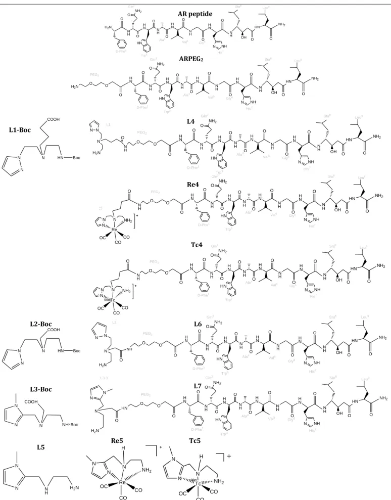
Os análogos peptidicos sintetizados basearam-se na seguinte sequência: DPhe1
-Gln2-Trp3-Ala4-Val5-Gly6-His7-Sta8-Leu9-NH2 (AR), aqual foi acoplada a um espaçador de
polietilinoglicol (PEG) por forma a aumentar a distância entre o centro metálico e a BM e conferir um carácter mais hidrofílico aos conjugados. Para a síntese destes péptidos foi utilizada a estratégia de sintese em fase sólida (resina) e aminoácidos protegidos com o grupo protector Fmoc. Após cada síntese e/ou conjugação a derivados dos ligandos L1-L3, os péptidos e conjugados peptídicos (AR, ARPEG2, L4, L6 e L7) foram purificados e
caracterizados por ESI-MS e HPLC.
No caso do conjugado peptídico L4, resultante da conjugação de ARPEG2 a um
derivado de L1, iniciou-se o estudo da sua radiomarcação com 99mTc. São necessários mais
estudos para melhorar as condições de marcação e continuar com os necessários estudos de captação celular em células tumorais de cancro da próstata e de biodistribuição e farmacocinética em ratinhos com tumores induzidos. Esses estudos deverão ser alargados aos restantes bioconjugados de modo a investigar quais são os mais promissores para a concepção de novos radiofármacos para detecção de cancro da próstata.
VIII
Abstract
The development of radiopharmaceuticals for imaging/therapy of cancer remains an important issue. Biologically active peptides are used as specific carriers for the targeting of tumors, as many peptide receptors are overexpressed in tumors.
The organometallic precursors fac-[M(H2O)3(CO)3]+ (M=Re, 99mTc) allow the
labeling of peptides based on a well-defined and easily adaptable chemistry. However, the resulting complexes are often lipophilic and show unfavorable pharmacokinetics. Improving this issue is still crucial, which requires the design of new BFC’s (bifunctional chelator) for coordination of fac-[M(CO)3]+ and aiming to obtain radiopeptides with more
favorable in vivo profile.
In this context, the goal of this thesis was to contribute for the development of radiopharmaceuticals based on [99mTc(CO)3]+ for in vivo imaging of tumors overexpressing
the Gastrin Releasing Peptide-receptor (GRP-r,), particularly prostate cancer. To accomplish this goal, three different BFC’s were synthesized, characterized and conjugated to a bombesin antagonist peptide, with known ability to target GRP-r.
The investigated BFC's contain pyrazolyl (L1, L2) and imidazolyl (L3) coordinating groups and can lead to hydrophilic complexes of Re(I)/Tc(I). Pyrazolyl-diamine chelators like L1 and L2 are known to have excellent properties to stabilize the [M(CO)3]+ core.On
contrary, imidazolyl-diamine chelators like L3 were not explored so far. Therefore, a model imidazolyl-diamine ligand, L5, was synthesized its coordination capacity towards the [99mTc(CO)3]+ evaluated. L5 forms a hydrophilic complex, in high radiochemical yield
and purity, which presents high stability in vitro/in vivo and a favorable biokinetic profile.
The synthesized peptide analogs were based on the following sequence: DPhe1
-Gln2-Trp3-Ala4-Val5-Gly6-His7-Sta8-Leu9-NH2 (AR). A spacer of polyethyleglycol (PEG) was
conjugated to AR, to impart a more hydrophilic character. For the synthesis of peptides, a solid phase (resin) and Fmoc strategy were used. These studies led to the synthesis of several peptide conjugates (AR, ARPEG2 and L4). It was inititated the study of the
radiolabeling of L4 with[99mTc(CO)3]+. This preliminary results indicated that more studies
are needed to improve the labeling and proceed with the evaluation of cellular uptake and biodistribution/pharmacokinetics in mice with induced prostate tumors.
Palavras-Chave
Radiofármacos
Complexos de Tc(I) e Re(I) Péptidos biológicamente activos Antogonista da bombesina
Keywords
Radiopharmaceuticals Tc(I) and Re(I) Complexes Biologically active peptides Bombesin antagonist
X
TABLE OF CONTENTS
AGRADECIMENTOS ... IV Resumo ... VI Abstract ...VIII Palavras-Chave ... IX Keywords ... IX TABLE OF CONTENTS ... X Table of content of Figures ... XIII Table of content of Schemes...XV Table of content of Tables ... XVI Symbols and Abbreviations ... XVII Scope and Aim ... XX 1. Introduction ... XXIV1.1 Nuclear Medicine and radiopharmaceuticals ... 2
1.1.1 General Considerations ... 2
1.1.2. Radionuclides ... 2
1.1.3. Diagnosis Vs. Therapy in Nuclear Medicine ... 2
1.1.4. Nuclear Imaging ... 3
1.2. Technetium and Rhenium Coordination Chemistry Relevant for Nuclear Medicine…. ... 11
1.2.1. Radiochemistry of Technetium ... 12
1.2.2. Coordination chemistry of 99mTc ... 13
1.2.2.1. The [M(CO)3]+ core (M = 99mTc, Re)... 14
1.2.3. 99mTc Radiopharmaceuticals ... 16
1.3. Peptides in Molecular imaging ... 17
1.4. Objective... 23
2. Materials and Methods... 25
2.1 Solvents and Reagents ... 26
2.2 Purification and Characterization Techniques ... 28
2.2.1. Gravity Column chromatography (GCC) ... 28
2.2.2. Reversed-Phase High Performance Liquid Chromatography (RP-HLPC) ... 28
2.2.3. Thin-layer chromatography (TLC) ... 29
2.2.4. Nuclear Magnetic Resonance (NMR) Spectroscopy ... 29
2.2.6. Spectrophotometry ... 30
2.2.7. Measurements of Radioactivity ... 30
2.2.8. Partition coefficient ... 30
2.2.9. Biodistribution studies ... 30
2.2.10. In Vivo stability studies ... 31
2.3. Synthesis of Ligands ... 32
2.3.2. Synthesis and characterization of N-2-bromoethyl-pyrazole - c1 ... 32
2.3.3. Synthesis and characterization of Synthesis and characterization of [2-(2-Pyrazol-1-l-ethylamino)-ethyl]-carbamic acid tert-butyl ester - c2 ... 32
2.3.4. Synthesis and characterization of 4-[(2-tert-Butoxycarbonylamino-ethyl)-(2-pyrazol-1-yl-ethyl)-amino]-butyric acid ethyl ester - c3 ... 33
2.3.5 Synthesis and characterization of 4-[(2-tert-Butoxycarbonylamino-ethyl)-(2-pyrazol-1-yl-ethyl)-amino]-butyric acid – L1-Boc ... 34
2.3.6. Synthesis and characterization of (2-Amino-etil)-carbamic acid tert-butyl ester – c4 ... 35
2.3.7. Synthesis and characterization of (2-(1-Methyl-1H-imidazol-2-ylmethyl)-amino)-ethyl)-carbamic acid tert-butyl ester – c5 ... 35
2.3.8. Synthesis and characterization of N-(1-metyl-1H-imidazol-2ylmethyl)-ethane-1,2-diamine – L5 ... 36
2.3.9. Synthesis and characterization of benzyl 2-(2-(tert-butoxycarbonylamino)ethylamino)acetate – c6 ... 37
2.3.10. Synthesis and characterization of [(2-tert-Butoxycarbonylamino-ethyl)-(1-methyl-1H-imidazol-2-ylmethyl)-amino]-acetic acid benzyl ester – c7 ... 38
2.3.11. Synthesis and characterization of {(2-tert-Butoxycarbonylamino-ethyl)-[2-(2H-pyrazol-1-yl)-ethyl]-amino}-acetic acid benzyl ester – c8 ... 39
2.3.12. Synthesis and characterization of 4-(2-tert-Butoxycarbonylamino-ethylamino)-butyric acid ethyl ester – c9 ... 39
2.3.13. Synthesis and characterization of [(2-tert-Butoxycarbonylamino-ethyl)-(1-methyl-1H-imidazol-2-ylmethyl)-amino]-acetic acid – L3.3-Boc ... 40
2.3.14. Synthesis and characterization of [(2-tert-Butoxycarbonylamino-ethyl)-(2-pyrazol-1-yl-ethyl)-amino]-acetic acid – L2-Boc ... 41
2.4. Synthesis and characterization of Bombesin Antagonist Peptides and Conjugates….. ... 42
2.4.1. Synthesis of Bombesin Antagonist (AR) peptide ... 42
2.4.2. Synthesis of AR peptide conjugated with polyethyleneglycol (PEG2) (ARPEG2) ... 43
2.4.3. Synthesis of L4 ... 43
2.4.4. Attempted synthesis of L6 ... 44
XII
2.4.6. Cleavage of Bombesin Antagonist and Resin ... 45
2.4.7. Precipitation of Bombesin Antagonist Derivatives ... 45
2.4.8. HPLC analysis and Purification of Bombesin Antagonist Derivatives... 45
2.4.9. Kaiser Test ... 46
2.4.10. Handling and Storage of Peptides ... 47
2.5. Synthesis and Characterization of the Re(CO)3-Complexes ... 48
2.5.1. Rhenium complex fac-[Re(CO)3(κ3-L4)]+ (Re4) ... 48
2.5.2. Rhenium complex fac-[Re(CO)3(κ3-L5)]+ (Re5) ... 48
2.6. Synthesis and characterization of 99mTc-complexes ... 49
2.6.1. fac‐[99mTc(H2O)3(CO)3]+ precursor ... 49
2.6.2. 99mTc complex fac-[99mTc(CO)3(κ3-L5)]+ (Tc5) ... 50
2.6.3. 99mTc complex fac-[99mTc(CO)3(κ3-L4)]+ (Tc4) ... 50
3. Results and Discussion... 51
3.1. Synthesis of Ligands and Bifuncional Chelators (BFC’s) ... 53
3.1.1. Pyrazolyl-Based BFC’s ... 53
3.1.2 Imidazolyl derivatives ... 56
3.2. Synthesis, characterization and biological evaluation of M(CO)3L5 (M= Re, 99mTc) 59 3.2.1. Synthesis and Characterization of the Re surrogate: Re(CO)3L5 ... 59
3.2.2. Synthesis and characterization of the 99mTc(CO3)L5 Complex ... 62
3.2.3 Biodistribution Studies ... 63
3.2.4 In vivo Stability Studies ... 65
3.3. Synthesis and Purification of Bombesin antagonist derivatives ... 65
3.4. Conjugation of the Bombesin Antagonist to the BFC’s ... 70
3.5. Synthesis and Characterization of Metalated Peptides: fac-[M(CO)3(L4)]+ (M=Re, 99mTc) ... 72
4. Concluding Remarks and Outlook ... 76
Table of content of Figures
Figure A.1 - Representation of peptide conjugates studied in this Thesis. ... XXII Figure 1.2 - Multiple imaging modalities are available for small-animal molecular imaging. There are views of typical instruments available, and illustrative examples of the variety of images that can be obtained with these modalities.(A) microPET whole-body coronal image of a rat injected with 18F-FDG; (B) microCT coronal
image of a mouse abdomen after injection of intravenous iodinated contrast medium. (C) microSPECT coronal image of a mouse abdomen and pelvic regions after injection of 99mTc methylene diphosphonate, (D) Optical
reflectance fluorescence image of a mouse (E) microMRI coronal T2-weighted image of a mouse brain. (F)
Optical bioluminescence image of a mouse. (*adapted from(13)). ... 4
Figure 1.3 – SPECT device from Philips. ... 5
Figure 1.4– PET principle of detection. (108) ... 5
Figure 1.5– Representation of the integrated approach. ... 9
Figure 1.6 – Exemples of the Integrated approach. Rhenium complexes which mimic the structure of dihydrotestoterone, progesterone and estradiol. (36) ... 9
Figure 1.7 – Molecular configuration of NeoTect®. (38) ... 10
Figure 1.8 – Representation of the Bifuntional Approach (BFA); BM= biomolecule... 10
Figure 1.9 – 111In-OctreoScan. In this case, the BFC used is 2-[Bis[2-[bis(carboxymethyl)amino]ethyl]amino]acetic acid (DTPA). ... 11
Figure 1.10 – Left: illustration of the contents of a 99Mo/99mTc generator;Right: Decay scheme of 99Mo. There is a 2-keV isomeric transition from the 142-keV level to the 140-keV level, which occurs by internal conversion. Approximately 87% of the total 99Mo ultimately decays to 99mTc and the remaining 13% decays to 99Tc. (7) ... 13
Figure 1.11 - Synthesis of the organometallic precursor [99mTc (H2O)3(CO)3]+. ... 14
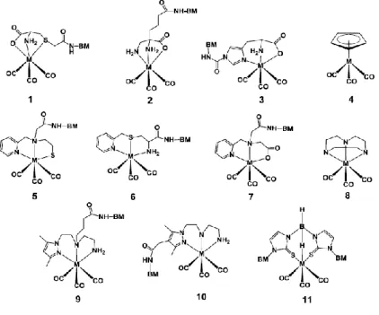
Figure 1.12 - Examples of M(CO)3-complexes stabilized by various types of chelating agents (BM = biomolecule; M = Re/99mTc). 1 - Functionalized cysteine (53); 2 - Funcionalized 2,3-diamino propionic acid derivatives (51); 3-Functionalized histidine derivatives (54,55); 4 – Cyclopentadienyl (56) ; 5, 6 and 7 - Functionalized picolinic acid derivatives (57,58); 8 – Triazacyclononane(59) ; 9 and 10 - Functionalized pyrazolyl-diamine containing ligands (60–64); 11 – Functionalized bis(mercaptoimidazolyl)borates.(65) ... 15
Figure 1.13 - 99mTc-based radiopharmaceuticals for diagnosis in clinical use. (MDP = methylenediphosphonate, MAG3 = mercaptoacetyl-triglycine, HMPAO = hexamethylpropyleneamine oxime, EDC = cysteinate dimer). .... 16
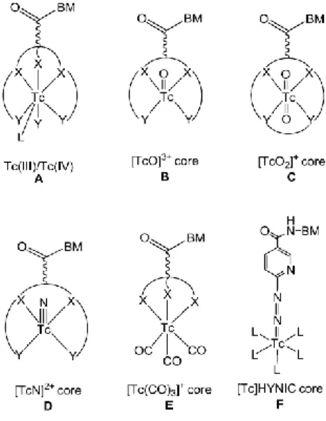
Figure 1.14 – Examples of Tc cores useful fort the labeling of biomolecules (BM) (6) ... 17
Figure 1.15 - 99mTc-Apcitide (Acutec®) ... 17
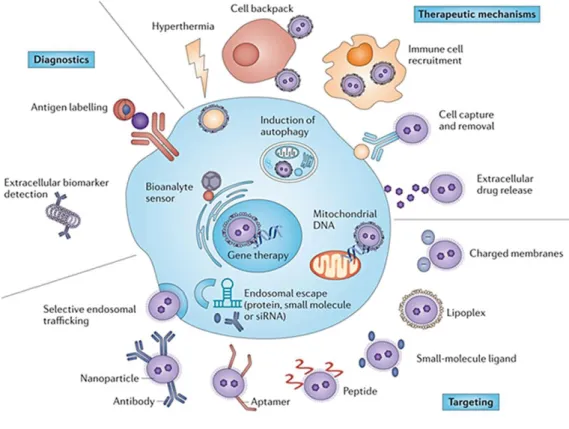
Figure 1.16 – Summary of all unique targeting, diagnostic and therapeutic mechanisms as they relate to cancer cells.(67) ... 18
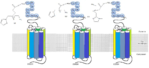
Figure 1.17 - G-Protein-coupled receptor conformation in the cellular membrane.(73) ... 19
Figure 1.18 – Difference of agonists(a) and antagonists(b) of GRP-r. (80)... 21
Figure 1.19 - Struture of DTMA-(X)-BBN(7-14)NH2, where X=GGG(top), GSG, SSS and β-Ala (bottom). *GGG= AA Gly-Gly-Gly; GSG= Gly-Ser-Gly; SSS= Ser-Ser-Ser; ... 22
Figure 2. 1 – RP-HPLC method 1 graphic profile. ... 28
Figure 2.2 – RP-HPLC method 1 graphic profile. ... 29
Figure 3.1 –Boc-protected pyrazolyl and imidazolyl bifuncional chelators. ... 53
Figure 3.2 - 1H-NMR spectrum of L2-Boc in CDCl3. (S= residual CHCl3) ... 55
Figure 3.3 - 1H-NMR spectrum of L5 in CD3OD or D2O. (S= residual Water from CD3OD) ... 57
XIV
Figure 3.5 - 1H-NMR spectrum (Top) and 13C-NMR spectrum (bottom) of Re5 in CD3OD. (S= solvent peak;
S1= Residual Water from CD3OD; S2=Residual MeOH; S3= CD3OD) ... 61
Figure 3.6 – ESI-MS spectrum of Re5. ... 61
Figure 3.7 – RP-HPLC chromatogram of the tricarbonyl precursor. (Method 2) ... 62
Figure 3.8 - RP-HPLC chromatograms of Tc5(γ - detection) and Re5 (UV detection). ... 63
Figure 3.9– Graphic representation of Tc5 Biosdistribution ... 64
Figure 3.10 - RP-HPLC chromatograms of Tc5 (injected preparation), blood serum and urine samples collected at 4 h p.i. (γ - detection/Method 2)... 65
Figure 3.11 – Principle of peptide synthesis in Solid Phase. ... 66
Figure 3.12 - Electromagnetic effect of microwave in Peptide synthesis. ... 67
Figure 3.13 - Structural configuration of AR peptide after resin deprotection. ... 68
Figure 3. 14 - ESI-MS spectrum of AR peptide... 68
Figure 3.15 - ESI-MS spectrum of ARPEG2 peptide. ... 69
Figure 3.16 - Structural configuration of L4 peptide conjugate after resin deprotection. ... 70
Figure 3.17 - ESI-MS spectrum of L4 peptide conjugate. ... 71
Figure 3.18 – HPLC chromatogram of L4 peptide conjugate. (Method 1, section 2.2.2) ... 71
Table of content of Schemes
Scheme 3.1 - Synthesis of L1-Boc; TBAB= tetrabutylammonium bromide ... 54
Scheme 3.2 - Synthesis of L2-Boc (strategy 1). ... 54
Scheme 3. 3 - Synthesis of L2-Boc (strategy 2). ... 56
Scheme 3.4 - Synthesis of L5... 56
Scheme 3.5 – Synthetic pathway to attain 3-phenylpropyl 2-((2-tert-butoxycarbonylamino)ethyl)(1-metyl-1H-imidazol-2-yl)methyl)amino)acetate ... 58
Scheme 3.6 - Synthesis of L3-Boc. ... 58
Scheme 3.7 - Synthesis of the complex Re5. ... 60
Scheme 3.8 – Synthesis of Tc5 ... 63
Scheme 3.9 – Synthesis of ARPEG2 ... 69
Scheme 3.10 - Synthesis of L4, the conjugate of ARPEG2 with L1-Boc. ... 70
Scheme 3.11 – Attempts to synthesize L6 and L7 by conjugation of ARPEG2 with the desired BFC. ... 72
Scheme 3.12 - Synthesis of the metallated peptide Re4. ... 73
XVI
Table of content of Tables
Table 1.1 - Relevant characteristics of the imaging modalities used in clinical set. ... 5
Table 1.2 - Sequences of various bombesin analogs. (76) ... 20
Table 2.1 - List of main solvents, reagents, AAs, and chemicals used in this work. ... 26
Table 2.2 - List of main devices used in this project. ... 27
Table 2.3 – RP-HPLC method 1. ... 28
Table 2.4 – RP-HPLC method 2. ... 29
Table 2.5 – Summary of procedures in Solid Phase Peptide Synthesis (SPPS)... 42
Table 2.6 – Color patterns of Kaiser test... 47
Symbols and Abbreviations
A GRP-r – Gastrin realesing peptide receptor
AA – amino acid Gly – Glycine
ACN – Acetonitrile H
ALA – Alanine
HATU - 1-[Bis(dimethylamino)methylene]-1H-1,2,3-triazolo[4,5-b]pyridinium 3-oxid hexafluorophosphate, N- [(Dimethylamino)-1H-1,2,3-triazolo-[4,5-b]pyridin-1-ylmethylene]-N-methylmethanaminium
hexafluorophosphate N-oxide AR – Bombesin antagonist peptide HCL – Hidrocloridric acid
ARG – Arginine His – Histidine
ASN – Asparagine I
ASP – Aspartic acid Ile – Isoleucine
Avg - Average K
B K2CO3 – Potassium carbonate
BFC - bifunctional chelator KCN - Potassium cyanide
BM - Biomolecule KeV - kiloelectron volt
BN - Bombesin L
Boc - t-Butyloxycarbonyl Leu – Leucine
C LET – linear energy transfer
CT – computed tomography Lys – Lysine
Cys – Cysteine M
D mRNA – menssager ribonucleic acid
d – Doublet MeOH - Methanol
DCM – Diclomethane Met – Methionine
dd – doublet of doublets
MBHA -
4-methylbenzhydrylamineDMF - N,N-Dimethylformamide MS – Mass spectroscopy DNA - Deoxyribonucleic acid m – multiplet
dt- doublet of triplets mTOR – mammalian target of rapamycin
E m – metastable isotope
ESI – MS Electrospray ionization–Mass
spectroscopy MRI – magnetic resonance imaging
EtOAc – Ethyl acetate MHz – megahertz
G N
GCC - gravity column chromatography nat – Natural isotope
Gln – Glutamine N2 – Nitrogen
XVIII
NaOH – Sodium hydroxide TLC - Thin-layer chromatography NMR - Nuclear Magnetic Resonance
Spectrometry Tyr – Tyrosine
P Trp – Tryptophan
PC – Prostate Cancer Trt – Tripheylmethyl
PRRT - peptide receptor radionuclide
therapy U
PEG – Polyehtilene Glycol US - Ultrasons
Phe – Phenylalanine UV – Ultraviolet
ppm – parts per million V
PSA- Prostate-Specific Antigen Val – Valine
Pro – Proline Vis – Visible
p.i. – post injection others
Pd/C – Palladium on carbon α – Alpha
Q β – Beta
q – quartet γ – Gama
QIT – quadrupole η – Yield
quint – quintet
λ
- wave lenghR
y - years
R.T. – Room Temperature Rf- Retention fraction
RP-HLPC - Reversed-Phase High
Performance Liquid Chromatography
S
s – Singlet
SCLCs - Small cell lung carcinomas Ser – Serine
SPECT - Single Photon Emission Computed Tomography
SPPS - Solid Phase Peptide Synthesis T
t – triplet T1/2 – Half-life
TFA - Trifluoroacetic acid THF – Tetrahydrofurane Thr – Threonine TIS - Triisopropylsilane
XIX NH2 O HN O OH N H O N NH H N O N H O H N O N H O HN H N O N H O H2N Trp3 His 7 Gly6 Val5 Ala4 D-Phe1 NH2 O HN O OH N H O N NH H N O N H O H N O N H O HN H N O N H NH2 O O H N Trp3 His 7 Sta8 Leu9 Gly6 Val5 Ala4 Gln2 D-Phe1 O O O H2N PEG2 N N N HN COOH Boc NH2 O HN O OH N H O N NH H N O N H O H N O N H O HN H N O N H NH2 O O H N Trp3 His 7 Sta8 Leu9 Gly6 Val5 Ala4 Gln2 D-Phe1 O O O N H PEG2 N N N H2N O L1 NH2 O HN O OH N H O N NH H N O N H O H N O N H O HN H N O N H NH2 O O H N Trp3 His 7 Sta8 Leu9 Gly6 Val5 Ala4 Gln2 D-Phe1 O O O N H PEG2 N N N NH2 O L 1 Re OC CO CO NH2 O HN O OH N H O N NH H N O N H O H N O N H O HN H N O N H NH2 O O H N Trp3 His 7 Sta8 Leu9 Gly6 Val5 Ala4 Gln2 D-Phe1 O O O N H PEG2 N N N NH2 O Tc OC CO CO 99m L2-Boc N N N HN COOH Boc NH2 O HN O OH N H O N NH H N O N H O H N O N H O HN H N O N H NH2 O O H N Trp3 His7 Sta8 Leu9 Gly6 Val5 Ala4 Gln2 D-Phe1 O O O NH PEG2 L2 N N H2N N O L3-Boc COOH NH N N N Boc NH2 O HN O OH N H O N NH H N O N H O H N O N H O HN H N O N H NH2 O O H N Trp3 His 7 Sta8 Leu9 Gly6 Val5 Ala4 Gln2 D-Phe1 O O O HN PEG2 O H2N N N N L3.3 L5 H2N N H N N Re5 Tc5 NH2 N N N Re H CO CO OC NH2 N H Tc CO CO OC 99m N N
Figure A.1 - Main compounds described in this thesis. ARPEG2 L1-Boc L4 Re4 Tc4 L6 L7
XX
Scope and Aim
Cancer remains one of the most devastating diseases with more than 10 million new cases each year world-wide, which is estimated to increase to 20% by 2020. However, due to a better understanding of cancer, and improved diagnostics equipment and treatment strategies, the mortality is decreasing.(1)
Prostate cancer (PC) has been the most frequently diagnosed noncutaneous cancer and is the second cause of cancer-related mortality after lung and bronchus cancers in men. (2,3) An optimal PC treatment is guided by a clinically staging of the cancer and selecting treatment options based on the stage. The treatment options for men with prostate cancer may include an expectant management (watchful waiting) or an active surveillance, that included surgery; radiation therapy; cryosurgery (cryotherapy); hormone therapy; chemotherapy; vaccine treatment and bone directed treatment. These treatments are generally used one at a time, although in some cases, they may be combined. (1)
Prostate cancer continues to be the second leading cause of cancer-related deaths for men in developed countries. Although the 5-year disease-specific survival rate of localized disease treated by surgery or radiotherapy is more than 90%. The effectiveness of systemic therapy for advanced prostate cancer is limited. Currently, depleting or blocking the action of androgens is the standard of care for men with advanced prostate cancer, but the response to treatment is not durable and with time prostate-specific antigen concentrations increase, indicating reactivated androgen-receptor signaling. (2)
Over the last years an emerging area in nuclear oncology deals with the evaluation of radiolabeled bioactive peptides for the diagnostic and therapy of tumors, due to overexpression of many peptide receptors in human tumors. Comparatively to other biomolecules, small peptides have many advantages. Namely, they can be easily synthesized and manipulated molecularly to optimize their in vivo half-life, receptor binding affinity and subsequently, pharmacokinetics. Peptide-based radiopharmaceuticals for therapeutic applications offer the ability to exploit the radioactive chelate as a radiocytotoxic unit and the bioactive molecule as a vector to localize the tumor.
Bombesin-like peptides such as gastrin-releasing peptide (GRP) have been shown to play a role in cancer promoting growth factors that stimulate tumor growth through specific receptors. The GRP receptor shows high over-expression in invasive prostatic neoplasias and also, although in less cases, in bone metastases of androgen-independent
XXI
prostate cancers. This represents the potential clinical basis for GRP receptor imaging of prostate cancer with radioactive compounds for early tumor diagnosis, followed by radiotherapy with radiolabeled bombesin analogues.
Despite the remarkable advances, the design of specific probes for targeted imaging or therapy still remains a great challenge and a demanding task within the field of radiopharmaceutical sciences. This is a multidisciplinary research area, which profits from the input of chemists, radiochemists, radiopharmacists and clinicians. Due to its chemical and nuclear properties, associated with the low cost and easy availability, 99mTc is among
the most attractive radionuclides for SPECT imaging. In addition, rhenium, the congener 2nd row d-transition element from the 7th group, forms isostructural complexes with Tc
and displays two radioisotopes (186Re and 188Re) suitable for targeted antitumor therapy.
Within this framework, the long term goal of the work described in this thesis was to contribute for the design of new specific radiopharmaceuticals based on the 99mTc(CO)3
core for in vivo visualization of GRP receptors overexpressed in prostatic cancer. To accomplish this goal, three different bifunctional chelators (BFC’s) were synthesized and conjugated to a bombesin analog peptide. This peptide was chosen because it was already established its usefulness as a targeting vector for GRP receptors. It was intended to radiolabel the different bioconjugates with 99mTc, to evaluate the in vitro stability of the
resulting complexes, as well as its in vivo profile in mice, to have an insight on their relevance in the design of specific radiopharmaceuticals for prostate cancer imaging. Finally, the most promising radiopeptides would be evaluated using imaging and biodistributions studies in mice bearing prostatic tumors. In summary, this work comprised the synthesis and characterization of BFC’s; synthesis and characterization of the peptide analogues; conjugation of peptide analogues to the BFC’s (figure A.1); Synthesis of organometallic complexes of 99mTc and natRe, and finally in vitro and in vivo
XXII
Figure A.1 - Representation of peptide conjugates studied in this Thesis.
This thesis is organized in five chapters. Chapter 1 comprises a general introduction on nuclear imaging in oncology and radiopharmaceuticals. It covers also peptide related matters, such as the relevance of Gastrin Releasing Peptide-receptors (GRP-r) as a target for tumor imaging and therapy, and the use of radiolabeled Bombesin (BN) analogues for in vivo imaging of GRP-r. Chapter 2 is dedicated to the description of all materials and methods used, namely the synthetic procedures.
The 3rd chapter reports the results and discussions, and is divided into the
synthesis of bifunctional chelators (BFC’s), peptide synthesis and conjugation, radiolabeling studies and biodistribution evaluation.
Concluding remarks are presented in chapter 4, where is also scrutinized the options for the future work.
1.1 NUCLEAR MEDICINE AND RADIOPHARMACEUTICALS
1.1.1 General Considerations
Nuclear medicine is a medical speciality that uses radiopharmaceuticals for diagnosis and/or therapy of several diseases. Radiopharmaceuticals are drugs with no pharmacological effect that contains in its composition a radionuclide. A radiopharmaceutical can be a small organic (e.g. 18F-FDG), inorganic (e.g. Na131I and
Na18F), or organometallic compound with defined composition, or can also be a small
peptide, inhibitors or substrates of enzymes or large molecules such as antibody fragments, among others. (3–5)
1.1.2.
RadionuclidesA nuclide is an atomic nucleus identified by a unique combination of a given number of protons and neutrons and the energy state of its nucleus. A nuclide can be specified by the notation: A
ZX N ; where X is the chemical element symbol, Z is the atomic
number (number of protons in the nucleus), N is the neutron number (number of neutrons in the nucleus), A is the mass number (A=N+Z, total number of nucleons in the nucleus). A radionuclide is an unstable form of a nuclide, due the unsuitable composition of the nucleus. Radionuclides disintegrate emitting gamma ray(γ) and/or particles or
Auger electrons. They may occur naturally, but most of them are artificially produced in cyclotrons or reactors. It is also possible to have indirect production routes like radionuclide generators. In this case a radionuclide (parent), produced in a reactor or cyclotron, is immobilized in a chromatographic column, allowing the elution of another radionuclide (daughter) with shorter half-life. The advantage of this process is to deliver very short-lived radionuclides (daughter) to hospitals or medical institutions that don’t have cyclotron or reactor facilities. (6–8)
1.1.3.
Diagnosis Vs. Therapy in Nuclear MedicineDepending on the medical application, diagnosis or therapy, different physical properties are required for the radionuclide. Among these properties, the type and energy of the emitted radiation as well as the half‐life of the radionuclide (time necessary for half of the original atoms of the radionuclide to decay - t1/2) of the nuclide are the most
important for selection of a suitable radionuclide to the desired medical application. A
radiopharmaceutical for diagnosis contains a positron- (β+) or gamma- (γ) emitting
radionuclide of sufficiently high energy (>50 keV) in their composition, whereas
3 ionizing radiation with a high linear energy transfer (LET), to destroy selectively cells or tissues. The most used radionuclides for therapeutic purposes are β-, but intense research
involving α and Auger electron emitters is also underway, like the Phase III Study of Radium-223 Dichloride in Patients With Symptomatic Hormone Refractory Prostate Cancer With Skeletal Metastases (ALSYMPCA). (9)
1.1.4.
Nuclear ImagingMedicinal imaging is a key tool for improving the diagnosis of a large variety of diseases. Nowadays, the imaging techniques can be included into two large non-invasive categories: Structural and functional imaging techniques. X-ray, CT (Computed Tomography) and MRI (Magnetic Ressonance Imaging) are the three most important techniques that can primarily provide structural and anatomical imaging such as tumor location, size, morphology, and structural changes to adjacent tissues. (10–12)
Functional imaging, rather than anatomical, aims at the visualization and characterization of biochemical pathways, molecular interactions, drug pharmacokinetic and pharmacodynamics. Positron Emission Tomography (PET) and Single Photon Emission Computed Tomography (SPECT) are currently accepted as the most important functional imaging techniques, used in nuclear medicine. (13,14)
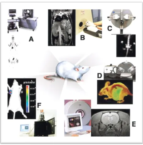
Examples of typical instruments available for small-animal molecular imaging are shown in Figure 1.2 and Table 1.1; they summarize the most relevant characteristics of imaging modalities mentioned above, including the major strengths and weaknesses. (13)
Figure 1.2 - Multiple imaging modalities are available for small-animal molecular imaging. There are views of
typical instruments available, and illustrative examples of the variety of images that can be obtained with these modalities.(A) microPET whole-body coronal image of a rat injected with 18F-FDG; (B) microCT coronal
image of a mouse abdomen after injection of intravenous iodinated contrast medium. (C) microSPECT coronal image of a mouse abdomen and pelvic regions after injection of 99mTc methylene diphosphonate, (D) Optical
reflectance fluorescence image of a mouse (E) microMRI coronal T2-weighted image of a mouse brain. (F) Optical bioluminescence image of a mouse. (*adapted from(13)).
5
Table 1.1 - Relevant characteristics of the imaging modalities used in clinical set.
Techniqu e resolution Spacial Amount of molecular probe used
Main use Advantages Disadvantages
Str u ctu ra l im ag ing MRI 25-100 m mg-g Morphological/ Metabolic
Higher spatial resolution, combines morphological and
functional imaging
Low sensitivity, amount of probe CT 50-200m applicable Not
Morphological
Bone and tumor imaging,
anatomical imaging Limited soft tissue resolution US
(ultrasons) 50-500 m mg-g Real time imaging, low cost
Limited spatial resolution Fu nc ti ona l Im ag ing SPECT 1-2 mm ng Metabolic
Highest sensitivity, quick, easy, low cost, relatively high
throughput
Low spatial resolution PET 1-2 mm substitute naturally occurring High sensitivity, isotopes can
atoms
Cyclotron or generator needed
From the nuclear imaging modalities, SPECT requires a radiopharmaceutical containing a radionuclide that emits gamma (γ radiation with energies between 80 and 300 KeV and a gamma camera for patient imaging). PET requires a radiopharmaceutical labeled with a positron-emitting radionuclide (+) and a PET camera. (15)
SPECT Imaging - The gamma rays are
emitted by the radionuclide present in the injected radiopharmaceutical. These rays are detected by a gamma camera and an image of localized radioactivity is produced. In SPECT, the gamma camera rotates around the patient's body and the image is reconstructed to produce image slices.
PET Imaging - The nucleus of the radionuclide decays by the
emission of a positron (Positive electron) which will then combine with an electron from the surroundings tissues and annihilate to produce two characteristic γ-rays of energy equivalent to the rest mass energy of an electron (511keV). These are emitted at approximately 180º degrees away from each other. The PET scanner, used to detect the two photons, consists of a ring of scintillation detectors of which opposing detectors are in coincidence so that the
Figure 1.3 – SPECT device from Philips.
Figure 1.4– PET principle of
radionuclide can be determined to be in the region between the detector pair. Reconstruction of events between these multiple detector pair leads to a functional image of the brain.
Although SPECT imaging has lower sensitivity in the detector and less quantitative accuracy than PET, it has broader clinical applications. In fact, the radioisotopes used in SPECT have longer half-life (t1/2) and therefore can be produced away from the site of
administration and transported whenever required.(16) Moreover, the detection period can be widened, allowing the observation of biological processes in vivo over several hours or days after the administration of the radioactive probe.(17)
The γ-emitting radionuclides with the most suited characteristics for SPECT imaging are 99mTc (t1/2 = 6.02 h, Eγ(max) = 140 keV), 123I (t1/2 = 13.20 h, Eγ(max) = 159
keV), 67Ga (t1/2 = 78.26 h, Eγ(max) = 296 keV), 201Tl (t1/2 = 72 h, Eγ(max) = 167 keV) and 111In (t1/2 = 67.9 h, Eγ(max) = 245 keV).
The β+-emitting radioisotopes used in PET imaging are in general short-lived
nonmetallic isotopes, such as 18F (t1/2 = 109.8 min, Eβ+ (max) = 202 keV), 11C (t1/2 = 20.4
min, Eβ+ (max) = 326 keV), 15O (t1/2 = 2.03 min, Eβ+ (max) = 650 keV), and 13N (t1/2 = 9.98
min, Eβ+ (max) = 432 keV). [18F]-2-fluoro-2-deoxy-D-glucose (18F-FDG) is the most widely
used positron-emitting radiopharmaceutical for PET imaging, making 18F the most used
radionuclide in this nuclear imaging technique. 18F-FDG has also shown clinical usefulness
in cardiology and neurology, but it is used mainly in oncology, in the diagnosis, staging and post-therapy evaluation. (18,19)
Most PET radionuclides have the disadvantage of requiring costly technology, namely cyclotrons for their production and sophisticated automated methods for the radiosynthesis of the radiopharmaceuticals. Owing to the short half-life of most β+
-emitting radioisotopes, the production and synthesis must occur close to the administration site. A few β+-emitting radiometals such as 64Cu (t1/2 = 12.7 h, Eβ+(max) =
660 keV) and 68Ga (t1/2 = 1.1 h, Eβ+(max) = 1899 keV) are also of great interest in
developing new PET radiopharmaceuticals. In particular, 68Ga is emerging as a PET
radionuclide with clinical relevance, due not only to its physical and chemical properties, but mostly to its availability from a long-lived 68Ge/68Ga generator system. (3,5,7,20–22)
7
1.1.5.
Nuclear TherapyAs mentioned before, radiopharmaceuticals for systemic radiotherapy have in their composition a radionuclide that emits ionizing radiation with a high LET to destroy selectively cells or tissues ( β- emitters, α- and Auger electron emitters). (7) These particles
have different ranges in the tissues and can be described by the amount of transferred kinetic energy as a function of distance.
The selection of a radionuclide for therapy depends not only on the type, energy, half-life and range of emitted particles, but also on the size of the tumor or tissue to irradiate and treat. Since the penetration depth range of β- particles in biological tissues is
relatively long (0.1 – 10 mm), radiopharmaceuticals containing β--emitting radionuclides
can be used for the treatment of large solid tumors. (9,23)
Among β--emitting radionuclides with potential application, 131I (t1/2 = 8.0 d, E
β-(max) = 0.81 MeV, Eγβ-(max) = 0.364 MeV), 90Y (t1/2 = 2.7 d, Eβ- (max) = 2.27 MeV), 186Re (t1/2
= 3.8 d, Eβ-(max) = 1.07 MeV, Eγ(max) = 0.137 MeV), and, more recently, 188Re (t1/2 = 0.7 d,
Eβ- (max) = 2.10 MeV, Eγ(max) = 0.155 MeV), are used in the clinical onset for the
treatment of different tumor types. Moreover, the radiolanthanides 153Sm (t1/2 = 1.9 d, E
β-(max) = 0.8 MeV, Eγβ-(max) = 0.103 MeV), 166Ho (t1/2 = 1.1 d Eβ- (max) = 1.86 MeV, Eγ(max)
= 0.081 MeV) and 177Lu (t1/2 = 6.7 d, Eβ- (max) = 0.497 MeV, Eγ(max) = 0.208 MeV) have
been fully explored both at the preclinical and clinical levels as bone palliative agents (153Sm, 166Ho) and for peptide receptor radionuclide therapy in the case of 177Lu-labeled
somatostatin analogs. (23–27)
The α particles are heavy and charged helium nuclei with high LET. These particles “travel” only short distances and cause the most ionizing damage over a small distance (30 – 80 μm). Hence, α-emitting radionuclides are appropriate for the treatment of small tumors and/or metastasis. (9) Most of the α-emitters have a half-life that is too long to be compatible with in vivo applications. As a result, only a few of these radioisotopes have received serious attention for radiotherapeutic applications, namely,
211At (t1/2 = 7.2 h, Eα(avg) = 6.8 MeV), 212Bi (t1/2 = 1 h, Eα(avg) = 7.8 MeV) and 223Ra (t1/2 = 11.4 d,
Eα(avg) = 5.65 MeV). (23,24,28,29) It is worth mentioning that 223RaCl2 (Alpharadin®) was
explored in the treatment of skeletal metastases and due to the promising results obtained this compound is currently under clinical evaluation for the treatment of bone metastases resulting from prostate cancer.
The Auger electrons are less energetic particles with a very short range of penetration (< 1 μm), having a LET similar to that of α particles, which makes them potentially interesting for therapy. Unlike α and β- particles, treatment based on Auger
electron-emitters requires the targeting of individual cells, specifically the DNA in the nucleus. Despite the multiple obstacles that have to be overpass, Auger electron therapy approaches remain very appealing. Among the available Auger electrons-emitting radionuclides, 125I (t1/2 = 57 d, 24 electrons/decay), 111In (t1/2 = 67 h, 14 electrons/decay), 67Ga (t1/2 = 78 h, 4 electrons/decay) and 99mTc (t1/2 = 6.02 h, 4 electrons s/decay) are the
most interesting for potential clinical applications. (9,24,30–32)
Perfusion and Specific Radiopharmaceuticals
The radiopharmaceuticals can be classified according to their biodistributions profile as perfusion or first generation radiopharmaceutical and specific or second generation radiopharmaceutical. Most of the radiopharmaceuticals in clinical use are perfusion agents, but nowadays most of the research efforts are directed to specific radiopharmaceuticals for diagnostic and/or therapy. The biodistribution of perfusion agents depends mainly on their lipophilicity, size and charge. These agents are transported in the blood and delivered to the target organ in the proportion of the blood flow. A wide variety of compounds were successfully developed for imaging organs, such as liver, kidney, heart or brain, giving information on a pathologic condition. (6,33,34)
In the last decade, due to the advances in molecular biology and the consequent identification and comprehension of molecular mechanisms that are the base of several diseases, the most recent radiopharmaceuticals in the market are specific radiopharmaceuticals. The targeting capability of these compounds depends on a biologically active biomolecule (BM), which can be a small organic BM, a small peptide, an antibody, a nanobody, sugars, inhibitors or substrates of enzymes, nucleotides or oligonucleotides. The capacity of the BM to recognize a certain molecular target (receptor, antigen, enzyme, DNA, mRNA, etc) will determine the radiopharmaceutical uptake in the target organ or tissue.
Since the end of 1980’s, an intensive research for developing specific radiopharmaceuticals is being carried out, but only some cases were successful in getting to clinical use. (4,6) In fact, this is a challenging task, since it is necessary to conjugate the radionuclide to a biomolecule without interfering with its biological properties. For the design of specific radiopharmaceuticals there are three general strategies: the integrated, the hybrid and the bifuncional approach (Figure 1.5). (6,23,34–36)
9
M
X Y
X Y
Figure 1.5– Representation of the integrated approach.
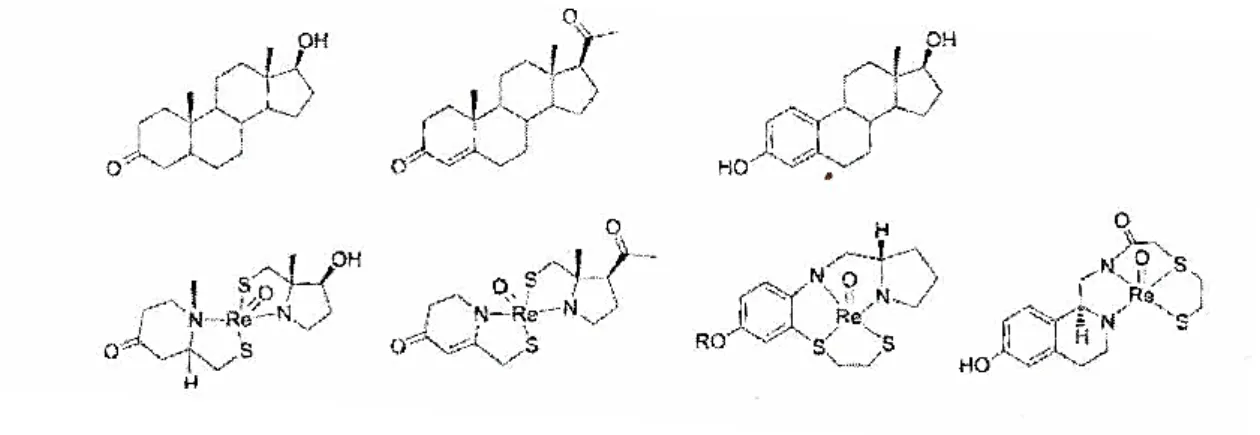
The integrated approach involves the substitution of a part of a biologically active molecule by a metal unit. This replacement must minimize changes in the structure, molecular size and conformation. Figure 1.5 presents the basis of the integrated approach and figure 1.6 shows examples of this approach. So far, integrated approach did not provide good results, as the resulting molecules presented low specificity and biological affinity for the receptors.(36,37)
Figure 1.6 – Exemples of the Integrated approach. Rhenium complexes which mimic the structure of
dihydrotestoterone, progesterone and estradiol. (36)
In the hybrid approach, the metal is stabilized by functional groups naturally occurring or synthetically introduced in the BM. These groups are usually tripeptides, such as GLY-GLY-GLY, CYs- GLY-GLY or CLYs-GLY-CYs. The small peptide sequence can be part of a linear or cyclic polypeptide. In some cases, the coordination of the radiometal to such amino acids can promote the cyclization of the polypeptide, improving the affinity for the receptors and increasing the in vivo stability. An example of such approach is the 99m
Tc(V)-Depreotide (NeoTect®) (Figure 1.7) a radiopharmaceutical for imaging of somatostatin
Figure 1.7 – Molecular configuration of NeoTect®. (38)
The bifunctional approach (Figure 1.8) has been the most exploited for the development of specific radiopharmaceuticals.
Figure 1.8 –Representation of the Bifuntional Approach (BFA); BM= biomolecule
This approach uses a bifunctional chelator (BFC) that strongly coordinates the metal ion, through appropriate coordinating groups, and is covalently attached to the biomolecule, which much interact specifically with the desired target. The nature of the BFC depends on the metal and its oxidation state. A spacer/linker between the metal and the biomolecule may exist, to modulate either the pharmacokinetics of the compound and/or its biological activity. This approach has been successfully used for the development of some radiopharmaceuticals, as it is for example the case of Octreoscan®, a
radiopharmaceutical based on 111In in clinical use for the imaging of neuroendocrine
11
Figure 1.9 – 111In-OctreoScan. In this case, the BFC used is
2-[Bis[2-[bis(carboxymethyl)amino]ethyl]amino]acetic acid (DTPA).
1.2. TECHNETIUM AND RHENIUM COORDINATION CHEMISTRY RELEVANT FOR NUCLEAR
MEDICINE
Technetium was discovered in 1937 by Perrier and Segrè in a sample of molybdenium, which was irradiated by deuterons. The new element received its name from the Greek word technetos, meaning artificial, because technetium was the first element previously unknown on earth to be made artificially. In 1939 Seaborg and Segrè observed that molybdenum-98 irradiated with slow neutrons gave rise to 99Tc through
decay of the metastable isomer, 99mTc. Eventually 21 isotopes of technetium were
discovered ranging from 90Tc to 110Tc, with technetium-110 having the shortest half life
(0.86 Sec) and 97Tc the longest (2.6 x 10⁶ y). All technetium isotopes are radioactive. In the
1950s, a purification work on the tellurium-132 / iodine-132 generator at Brookhaven National Laboratory (BNL) turned up a contaminant which was proved to be technetium.
The technetium contaminant was due to the presence of 99Mo in the chemical separation,
which was also present, because it had followed tellurium in the chemical separation process. This discovery eventually led to the production of the 99Mo/99mTc generator in
1957 at BNL. Final improvements were made by Powell Richards. (40)
Rhenium, first detected by Noddack, Tacke and Berg in 1925 in the X-ray spectra of certain mineral concentrate, was the last of the stable elements to be discovered. Rhenium occurs naturally as a mixture of two isotopes: 185Re(37.4%) and 187Re(62.6%) and has two
radioactive isotopes with an interest in nuclear medicine: 186Re and 188Re, two β- emitters
that can be used in therapy. Due to its availability from the generator (188W/188Re) and
favorable nuclear properties, 188Re is being intensively studied for the design of
therapeutics radiopharmaceuticals.
Technetium (Tc) and rhenium (Re) are group 7 transition metals, belonging respectively to the 2nd and 3rd transition series, with atomic numbers 43 and 75 and
electronic configurations [Kr]4d55s2 and [Xe]4f145d56s2, respectively. Tc complexes can
have the metal in the oxidation states -I (d8) to +VII (d0), whereas for Re complexes the
metal presents oxidation states -III (d10) to +VII (d0). These metals present very similar
atomic radius (Tc, 1.36 Å; Re, 1.37 Å), forming structurally analog complexes. For that reason, the characterization of 99mTc complexes (obtained at very low concentration, 10-10
-10-7M) is usually supported by the synthesis of analogs rhenium complexes. However,
there are differences between both metals that must be taken into consideration in the preparation of Tc and Re complexes. The most striking difference is related to the higher kinetic inertness and easier oxidation associated to Re complexes. (7,41)
1.2.1.
Radiochemistry of TechnetiumThe growth and wide application of diagnostic nuclear medicine have been mainly driven by the coordination chemistry and unique features of technetium. Among the 21 known artificial isotopes, 99mTc is the most useful radioisotope in nuclear medicine. The
importance of 99mTc in nuclear medicine is related to its almost ideal characteristics: (4,34)
- Half‐life of 6.02h is optimal for diagnostic, which is long enough to examine metabolic processes and yet short enough to minimize the radiation dose to the patient;
- γ-emitter (140 keV) with energy sufficiently high to penetrate easily the human body and to be detected externally by a gamma camera, and sufficiently low to minimize the dose to the patient;
- Available at reduced prices from a commercial 99Mo/99mTc generator, being one of
the greatest advantages of this radionuclide;
- Diverse coordination chemistry, which enables the preparation of a wide variety of complexes with different physicochemical and biological properties.
The prominence achieved by 99mTc as a useful radioisotope in nuclear medicine is
directly related to the design and development of a 99Mo/99mTc generator in the late
1950’s. (42–44)
Molybdenium-99 (99Mo), with a half-life of 66h, undergoes a radioactive decay,
emitting β‐ particles, in which 87% of the 99Mo atoms is converted directly into the
13
Figure 1.10 – Left: illustration of the contents of a 99Mo/99mTc generator;Right: Decay scheme of 99Mo. There is
a 2-keV isomeric transition from the 142-keV level to the 140-keV level, which occurs by internal conversion. Approximately 87% of the total 99Mo ultimately decays to 99mTc and the remaining 13% decays to 99Tc. (7)
1.2.2.
Coordination chemistry of 99mTcThe design of 99mTc-radiopharmaceuticals depends on the understanding of the
coordination chemistry of 99mTc. Factors such as stable oxidation states and core
structures are crucial in the design of effective target-specific 99mTc-radiopharmaceuticals.
The [99mTcO4]- anion, also known by pertechnetate, presents the metal in the
highest oxidation state possible (+VII) being an easily accessible precursor in the synthesis of all 99mTc-based complexes for imaging application. It is the most stable form of
technetium in aqueous solution, however, does not bind directly to any ligand and thus, for the synthesis of a 99mTc-radiopharmaceutical, reduction of 99mTc(VII) to a lower
oxidation state in the presence of a suitable ligand is a prerequisite. Several reducing agents such as tin salts (SnCl2, tin citrate, tin tartrate) or sodium borohydride, as well as
different reaction conditions (pH, temperature, concentration) are used to reduce [TcO4]
-in aqueous solution, yield-ing a technetium core -in a lower oxidation state that is stabilized by a suitable chelator. (5,9) When reduced in the presence of a ligand, the [99mTcO4]
-usually does not release all the oxygen atoms, leading to complexes with different 99mTc
cores.(45)
99mTc is usually used in the oxidation state +V as [99mTcO]3+ or [99mTcO2]+ cores.(6)
The coordination chemistry of the [99mTcO]3+ core has been explored for a long time but,
more recently, a lot of attention has been focused on the use of organometallic cores for the radiolabelling of BMs. In fact, the 99mTc(I)- and Re(I)-tricarbonyl core [M(H2O)3(CO)3]+
(M = 99mTc/188Re), or simply the [M(CO)3]+ core, has been adopted as an alternative
strategy to the 99mTc(V) complexes for the synthesis of 99mTc-radiopharmaceuticals.
Nowadays, the research is focused on the synthesis of new chelators that can coordinate strongly to the [M(CO)3]+ core. (6,20,33,45)
1.2.2.1. The [M(CO)3]+ core (M = 99mTc, Re)
The [M(CO)3]+ core (M = 99mTc, Re) has been adopted as an alternative strategy to
the 99mTc(V) complexes for the synthesis of 99mTc-radiopharmaceuticals. Nowadays, the
research is focused on the synthesis of new chelators that can coordinate strongly to the [M(CO)3]+ core. (6,20,33,45)
Alberto et al. were the first to report the one-step synthesis of an organometallic Tc(I) aqua-ion, [99mTc(H2O)3(CO)3]+, useful as a precursor for the radiolabelling of BMs for
diagnostic purposes. The Tc(I)-tricarbonyl precursor was synthesized by direct reduction of the [99mTcO4]- from the oxidation state +VII to +I, using sodium boranocarbonate
(Na2[H3BCO2]) as reductant and also as an in situ source of carbon monoxide. (46)
Nowadays, the precursor [99mTc(H2O)3(CO)3]+ can be obtained in quantitative yield by
directly adding [99mTcO4]- in 0.9% NaCl to a lyophilized commercial kit (IsoLink® kit,
Mallinckrodt-Covidien, Petten, The Netherlands), followed by incubation at 100 °C for 20 minutes. The ready availability of the precursor directly from the [99mTcO4]- constitutes
one advantage of the use of this 99mTc(I)-tricarbonyl precursor. The preparation of the Re
homolog [188Re(H2O)3(CO)3]+ was also accomplished, although using a procedure slightly
different from that envolved the preparation of the Tc(I)-tricarbonyl precursor, since Re is more difficult to reduce and reacts, in general, much slower, as previously mentioned. (45,47)
Figure 1.11 - Synthesis of the organometallic precursor [99mTc (H2O)3(CO)3]+.
The [M(CO)3]+ core is stable, even at high temperature and extended reaction
time.(48) Complexes of [M(CO)3]+ exhibit a d6 low-spin electronic configuration and, for
that reason, are kinetically inert. The core contains three tightly coordinated CO ligands and three water molecules, which are very labile and can be readily replaced by mono-, bi- or tridentate chelators.(49) The tridentate chelators form 99mTc(I)-tricarbonyl complexes
15 with the highest in vivo stability, which is essential for medical applications.(50) The [M(CO)3]+ core is very compact, being significantly smaller than the complexes in the
oxidation state (+V). This fact can be an advantage since it is thought that the smaller the complex, the higher the possibility that the biological activity will not be altered.(17,49)
The three water molecules in the precursor fac-[99mTc(CO)3(H2O)3]+ are labile with
respect to substitution, and the precursor can interact with potential coordination sites in proteins in human serum (e.g. histidine and cysteine residues). Therefore, the precursor itself is not suitable for diagnostic purposes, displaying a very unfavorable biological profile as demonstrated by biodistribution studies in mice. (51) Hence, to develop suitable radiopharmaceuticals based on this core, it is necessary to replace the water molecules by mono, bi, or tridentate ligands to form stable complexes. A wide variety of organometallic complexes have already been described, with those stabilized by tridentate chelating systems displaying the highest stability both in vitro and vivo. (52) In Figure 1.12 are represented selected complexes, stabilized by tridentate bifunctional chelators, bearing or not pendant targeting moieties. The diverse coordination chemistry of the [M(CO)3]+ core
offers an opportunity for the development of new chelators.
Figure 1.12 - Examples of M(CO)3-complexes stabilized by various types of chelating agents (BM =
biomolecule; M = Re/99mTc). 1 - Functionalized cysteine (53); 2 - Funcionalized 2,3-diamino propionic acid
derivatives (51); 3-Functionalized histidine derivatives (54,55); 4 – Cyclopentadienyl (56) ; 5, 6 and 7 - Functionalized picolinic acid derivatives (57,58); 8 – Triazacyclononane(59) ; 9 and 10 - Functionalized pyrazolyl-diamine containing ligands (60–64); 11 – Functionalized bis(mercaptoimidazolyl)borates.(65)
1.2.3.
99mTc RadiopharmaceuticalsThe 99mTc-based radiopharmaceuticals are roughly divided into two general
categories, as mentioned before. Examples of the perfusion agents, commercially available and respective clinical application are presented in Figure 1.13. (4,7,66)
Myocardial Perfusion imaging
A 99mTc-Sestamibi (cardiolite®) B 99mTc-tetrafosmin( Myoview®)
Bone Imaging Renal Imaging
C 99mTc-MDP(techneScan MDP®) D 99mTc-MAG3 (techneScan MAG3®)
Brain Imaging
E 99mTc-HMPAO (Ceretec®) F 99mTc-L,L, EDC (Neurolite®)
Figure 1.13 - 99mTc-based radiopharmaceuticals for diagnosis in clinical use. (MDP =
methylenediphosphonate, MAG3 = mercaptoacetyl-triglycine, HMPAO = hexamethylpropyleneamine oxime, EDC = cysteinate dimer).
The growing demand for more specific 99mTc-radiopharmaceuticals has prompted
the development of targeted radiopharmaceuticals mainly based on the BFC approach (Figure 1.8). As mentioned before, the targeting ability of specific radiopharmaceuticals relies on the capacity of the pendant biologically active molecule in the metal complex to recognize its target in vivo (e.g. membrane receptor, antigen or enzyme). (67–69) Besides the targeting moiety, also the spacer/linker between the metal and the biomolecule has a decisive influence in the overall biological properties of the radiopharmaceutical. (36) The modulation of the metal coordination environment with different chelators is a imperative parameter that opens great opportunities towards the design of innovative target specific radiopharmaceuticals with improved biological properties. Indeed, the design and