2017/2018
Cláudio André Melo Rodrigues
Síndrome
Cardiorrenal
Tipo 2 /
Cardiorenal
Syndrome
Type
2
Mestrado Integrado em Medicina
Área: Medicina Tipologia: Monografia
Trabalho efetuado sob a Orientação de:
Doutor Manuel Joaquim Lopes Vaz da Silva
Trabalho organizado de acordo com as normas da revista:
Revista Portuguesa de Cardiologia Cláudio André Melo Rodrigues
Síndrome
Cardiorrenal
Tipo
2
/
Cardiorenal Syndrome Type 2
Title: Cardiorenal Syndrome Type 2 Título: Síndrome Cardiorrenal Tipo 2
Nome: Cláudio André Melo Rodrigues
Orientador: Doutor Manuel Joaquim Lopes Vaz da Silva Faculdade de Medicina da Universidade do Porto
Al. Prof. Hernâni Monteiro, 4200-319 Porto
Index Abstract ………. 3 Resumo ……….. 4 List of abbreviations………... 6 Introduction ………... 8 Methods ………... 14
Chronic Heart Failure ……….…….. 15
Chronic Kidney Disease………... 18
Definition, Diagnosis and Biomarkers ………. 20
Pathophysiology of Cardiorenal Syndrome Type 2 ………...……... 27
1. Hemodynamic Factors ………...… 28
2. Neurohormonal Activation, Oxidative Stress and Inflammation ……...………… 29
3. Anemia ………... 35
4. Impact of Pharmacotherapy for the Management of Heart Failure …………..….. 36
Treatment Considerations in Cardiorenal Syndrome Type 2 ………... 38
Conclusion ………... 45
References ………...……… 46
Abstract
Cardiac and kidney disease are becoming progressively more prevalent in the population and may occur simultaneously. One of the most recognisable comorbidities in heart failure is renal dysfunction.
Cardiorenal Syndrome Type 2 (CRS2) is a term used to designate clinical conditions in which chronic abnormalities in cardiac function through a chronological and causal relationship leads to chronic kidney disease. This syndrome has recently become the subject of increasing discussion related to its diagnosis, pathogenesis and treatment since it is associated with a significant morbidity and mortality.
Data from clinical and experimental studies reveal that CRS2 is a condition characterized by a gradual deterioration of glomerular filtration rate, mild to moderate proteinuria and expression of high levels of renal injury biomarkers. Renal disease progression in the setting of CRS2 is elicited by triggers as maladaptive chronic neurohormonal stimulation, chronic increases in renal venous pressure as well as chronic oxidative and inflammatory state which leads and perpetuates structural renal injury, involving tubulointerstitial fibrosis and glomerulosclerosis.
Much remains to be clarified about CRS2. There are still significant questions concerning its pathophysiology and the existing therapy is actually a fragmented one, and therefore there is a need for interventional trials in this setting, regarding new criteria to optimize the management and improve outcomes in these patients.
The aim of this review is address the current knowledge about CRS2, including its definition, pathophysiology, diagnosis and treatment strategies.
Keywords: Cardiorenal syndrome type 2, Chronic heart failure, Chronic kidney disease,
Resumo
As doenças cardíaca e renal têm-se tornado cada vez mais prevalentes na população, sendo que podem ocorrer simultaneamente. Uma das comorbilidades mais reconhecidas na insuficiência cardíaca é a disfunção renal.
A Síndrome Cardiorrenal Tipo 2 (CRS2) é um termo usado para designar a condição em que as anomalias crónicas da função cardíaca, numa relação causal e cronológica, conduzem ao desenvolvimento de doença renal crónica. Esta síndrome tem sido recentemente alvo de um crescente debate em relação ao seu diagnóstico, patogénese e tratamento, uma vez que está associada com morbilidade e mortalidade significativas.
Dados de estudos clínicos e experimentais revelam que a CRS2 é uma condição caracterizada por uma deterioração gradual da taxa de filtração glomerular, proteinúria ligeira a moderada e expressão de níveis elevados de biomarcadores de lesão renal. A progressão da doença renal no contexto da CRS2 é despoletada por desencadeadores como a estimulação neurohormonal crónica e mal adaptativa, aumentos crónicos na pressão venosa renal assim como um estado oxidativo e inflamatório crónico que conduz e perpetua a lesão renal estrutural, envolvendo fibrose tubulo-intesticial e glomerulosclerose.
Muito permanece por ser esclarecer na CRS2. Há ainda muitas questões relevantes acerca da sua patofiosiologia, e a terapêutica atual é na verdade uma terapêutica fragmentada e, por isso, é necessário conduzir mais estudos intervencionais no sentido de se criar critérios que otimizem a abordagem e melhorem os outcomes nestes pacientes.
Palavras-Chave: Síndrome Cardiorrenal tipo 2, Insuficiência Cardíaca Crónica, Doença Renal
Crónica, Interação Coração-Rim, Lesão Renal, Inflamação, Stress Oxidativo, Ativação Neurohormonal
List of abbreviations
ACEI Angiotensin converting enzyme inhibitor ACR Albumin-to-creatinine ratio
ACS Acute coronary syndrome
ADH Antidiuretic hormone
AER Albumin excretion rate
AIM Antioxidant inflammation modulator
AKI Acute kidney injury
ANGII Angiotensin II
ARBs Angiotensin II receptor antagonist BNP Brain-Type Natriuretic Peptide CAD Coronary artery disease
CHF Chronic heart failure
CKD Chronic kidney disease
CO Cardiac output
CRS Cardiorenal syndrome
CRS2 Cardiorenal syndrome type 2
cTns Cardiac troponins
CRT Cardiac resynchronization therapy
CysC Cystatin C
EF Ejection fraction
FGF-23 Fibroblast growth factor 23 GFR Glomerular filtration rate GPCR G protein–coupled receptor
HF Heart failure
HFpEF Heart failure with preserved ejection fraction HFrEF Heart failure with reduced ejection fraction KIM-1 Kidney injury molecule-1
LAE Left atrial enlargement
LV Left ventricle
LVAD Left ventricular assist devices LVEF Left ventricular ejection fraction LVH Left ventricular hypertrophy
MI Myocardial infarction
NADPH oxidase Nicotinamide adenine dinucleotide phosphate-oxidase
NF-κB Nuclear factor kappa B
NAG N-acetyl-beta-D-glucosaminidase NT-proBNP NT-pro-brain natriuretic peptide NSAIDs Non-steroidal anti-inflammatory drugs
PD Peritoneal dialysis
RAAS Renin-angiotensin-aldosterone system
ROS Reactive oxigen species
sCr Serum creatinine
SNS Sympathetic nervous system SVR Systemic vascular resistance
Introduction
The organ systems are closely linked. In the normal state, this connection helps maintain optimal homeostasis and the functioning of the human body. The interaction between heart and kidney is a striking example. Both organs are regulators of vital functions, such as blood pressure, vascular tone, circulatory volume homeostasis, peripheral perfusion, diuresis, natriuresis, and tissue oxygenation and the two organs have endothelial functions as well as a
role in cellular and humoral signaling.1
It is well known that many hospitalized patients simultaneously exhibit varying degrees
of cardiac and renal dysfunction.2 The interaction is bidirectional so that the leading disease of
the heart or kidney frequently involves injury or dysfunction in the other organ.3 Founded on
this cross-talk, the term cardiorenal syndrome has arisen. The most widely used classification is that established by the Consensus Conference by the Acute Dialysis Quality Group, which divides the CRS fundamentally into two core groups, the cardiorenal and renocardiac syndromes, according to the origin of the disease (cardiac or renal); both CRS are then divided
into acute or chronic according to the onset of the disease.4 This classification was not proposed
to be unchangeable, as many patients may transit between different CRS subtypes (Table 1).5
Recently some authors such as Hatamizadeh et al. and Naranjo et al. have proposed propose a new classification of cardiorenal syndromes (Table 2), not only focusing in the timing and causality of the syndrome but also taking into account clinical manifestations and current
therapies and new treatment strategies,5,6 with Obi et al. additionally highlighting the related
Table 1. Cardiorenal syndrome (CRS) subtypes.
Cardiorenal
Subtype Description Examples/Etiology
CRS Type 1 (acute CRS)
Rapid worsening of cardiac function leading to acute kidney
injury
Acute MI with cardiogenic shock, acute decompensation of chronic heart failure, acute
valvular insufficiency
CRS Type 2 (chronic CRS)
Chronic abnormalities in cardiac function leading to chronic
kidney disease
CHF with chronic inflammation, long-term RAAS and SNS activation, chronic hypoperfusion
CRS Type 3 (acute renocardiac
syndrome)
Acute worsening of renal function leading to cardiac dysfunction (HF, arrhythmia, and
so forth)
Acute kidney injury with uremia causing impaired contractility, hyperkalemia causing arrhythmias,
volume overload causing pulmonary edema
CRS Type 4 (chronic renocardiac
syndrome)
Chronic worsening of renal function leading to worsening
cardiac function
CKD leading to LVH, coronary disease and calcification, diastolic dysfunction, and so forth
CRS Type 5
Acute or chronic systemic disease leading to both cardiac
and renal dysfunction
Diabetes mellitus, amyloidosis, sepsis, vasculitis, sarcoidosis, systemic lupus erythematosus
CHF: chronic heart failure; CKD: chronic kidney disease; HF: heart failure; LVH: left ventricular hypertrophy; MI: myocardial infarction; RAAS: renin-angiotensin-aldosterone system; SNS: sympathetic nervous system. Adapted from Ronco et al. (2008),3 Cole et al. (2012)8 and De Vecchis et al. (2014).9
Table 2. The new CRS classification proposed by Hatamizadeh et al.
CRS category and
definition Current strategies Potential strategies Potential harms
Hemodynamic
Hemodynamic compromise is the major clinical
manifestation Diuretics Ultrafiltration Vasodilators Inotropic agents Natriuretic peptides ACE inhibitors Digitalis Dopamine Mechanical circulatory assist devices Heart and/or kidney
transplantation Vasopressin V2-receptor antagonists ARBs Peritoneal dialysis Exercise training Calcium sensitizers Endothelin-receptor antagonists
Luso-inotropic agents (e.g. istaroxime) Cardiac myosin activators
Dual/triple RAAS blockade, antiarrhythmic
drugs (except for amiodarone and dofetilide), calcium-channel blockers (except for amlodipine), NSAIDs,
thiazolidinediones, long-term inotropic agents
Uremic
Uremic manifestations are the most prominent clinical
appearances
Conventional peritoneal dialysis
and hemodialysis therapies
Toxin removal by super-high-flux hemofiltration and/or novel
absorbents
High-protein diet
Vascular
Cardiovascular and/or renovascular manifestations
are the most prominent clinical findings Statins Atherosclerosis risk factor modification Antiplatelet agents Anticoagulants ACE inhibitors ARBs Endothelin-receptor antagonists Aldosterone-receptor antagonists Nitric oxide
Correction of pump failure and anemia (by increasing shear stress and improving endothelial function)
Exercise training
Smoking
Neurohumoral
Electrolyte disorders, acid– base disorders or dysautonomia is the most
prominent finding ACE inhibitors β-blockers Aldosterone-receptor antagonists ARBs FGF23-receptor blockers Adenosine A1-receptor antagonists
Direct renin inhibitors Exercise training Kidney sympathectomy
Long-term inotropic agents, ACE inhibitors or
ARBs, aldosterone receptor blockers
Anemia and/or iron metabolism
Dysregulation of iron metabolism and anemia
Iron ESAs Folic acid Cyanocobalamin
Red blood cell transfusion (in certain
circumstances)
Nutritional support Vitamin C
Carnitine Anti-hepcidin therapy
ACE inhibitors or ARBs, aldosterone receptor
blockers
Mineral metabolism
Dysregulation of calcium and phosphorus and their regulators including vitamin
D and FGF23 are the most
Vitamin D agents Phosphorus binders Calcimimetics FGF23-receptor blockers FGF23 antibodies Calcium, warfarin, overdosed active vitamin
It should be noted that the category of any patient is dependent on the current clinical evaluation and may vary over time. Attention must first be paid to the patient's most relevant clinical manifestation.
ACE: angiotensin-converting enzyme; AIM: antioxidant inflammation modulator; ARB: angiotensin-receptor blocker; CRS: cardiorenal syndrome; ESA: erythropoiesis-stimulating agent; FGF23: fibroblast growth factor 23; NSAIDs: non-steroidal anti- inflammatory drugs; RAAS: renin-angiotensin-aldosterone system.
Adapted from Naranjo et al. (2017),5 Hatamizadeh et al. (2013),6 e Obi et al. (2016).7
The prevalence of heart failure (HF) and chronic kidney disease (CKD) in Europe is solidly increasing, much at the expense of the growing incidence of acute and chronic cardiovascular disease, such as acute decompensated HF, arterial hypertension and cardiac
valve disease.10 However, this scenario seems to occur in other regions of the world, with data
from the United States that indicate a prevalence of symptomatic congestive HF estimated at 2% in those over 45 years of age with a lifetime risk of CHF estimated at 20%. CHF is the prominent cause of admission in persons over 65 years, accounting for more than 1 million
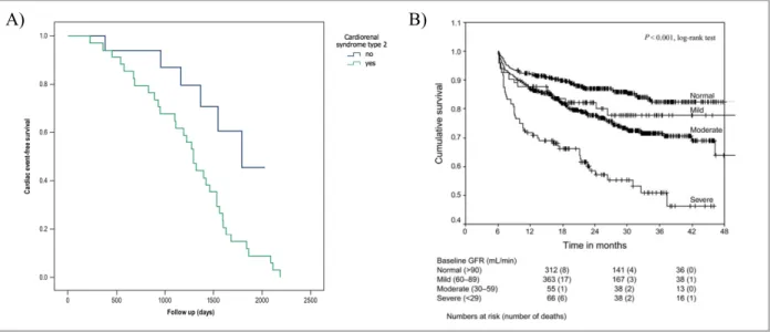
hospitalizations yearly.5 In the context of CRS2, the impact of worsening heart failure is once
again clear through higher rates of death and re-hospitalization compared to those without
CRS211 (Figure 1A).
Renal failure is frequent among patients with chronic HF, in both reduced (HFrEF) and
preserved ejection fraction (HFpEF) as well as symptomatic and asymptomatic patients,5 with
a variable prevalence ranging from 20% to 57% worldwide,12 but according to K/DOQI clinical
Malnutrition– inflammation–cachexia
Malnutrition, cachexia and inflammatory state is the
most prominent clinical manifestation Nutritional support Appetite stimulators Exercise training ACE inhibitors ARBs AIM β-blockers Ghrelin Growth hormone Anti-inflammatory and/or
anti-oxidative agents Volume overload correction (to
improve gut wall edema and nutrient absorption) Muscle enhancers (e.g.
practice guidelines for chronic kidney disease, nearly 63% of patients with HF meet the classification of stage 3–5 CKD with an estimated glomerular filtration rate < 60 ml/min/1.73
m2.13 Renal dysfunction takes a significant responsibility in the progression of cardiovascular
disease and performs as an independent risk factor for morbidity and mortality in patients with
heart failure5 (Figure 1B) and is correlated with elevated risk of readmission.12
It was shown even slight reductions in eGFR notably increase mortality risk.14
Figure 1 Kaplan-Meier curves.
A): Cardiac event-free survival analysis in patients with and without cardiorenal syndrome type 2. Patients with CRS2 had higher rates of cardiac death and re-hospitalization due to worsening heart failure than those without CRS2.
Taken and adapted from Salim et al. (2017)11 with permission.
B) Calculated GFR (using the simplified modification of diet in renal disease (MDRD) prediction equation (186 ´ sCr-1.154 (mg/dL) ´ Age-0.203 (years) ´ 0.742 if female, ´ 1,212 if black) and relationship to prognosis in patients
with chronic heart failure. Patients with worse renal function at baseline had a poorer prognosis. Taken and adapted from de Silva et al. (2006)12 with permission.
The aim of the present work is to review the current knowledge about chronic cardio-renal syndrome, also known as CRS type 2. First, key concepts in this syndrome such as "chronic heart failure" and "chronic kidney disease" will be described. Then, the definition, diagnosis, biomarkers and pathophysiology will be addressed. Finally, the current options for the treatment of CRS2 will be outlined.
Methods
A literature search using PubMed’s data base was performed. Articles up to March 2018, with no inferior date limitation, written in English or Portuguese were selected for inclusion. The most recent articles were selected whenever possible. The search was conducted based on MeSH terms using the following combinations: [cardiorenal syndrome type 2 AND (chronic heart failure OR chronic heart disease) AND chronic renal disease]. A preliminary assessment of eligibility was made through titles and abstracts. Potentially applicable articles were retrieved and reviewed independently for final decision on inclusion. Unavailable and irrelevant articles were excluded. Additional relevant articles found in the reference lists were also included.
Chronic Heart Failure
Chronic Heart Failure (CHF) is a condition in which the heart is incapable to perform its major pump function effectively (ie, failing to provide sufficient blood flow to meet the requirements of the various organs and apparatus of the organism or providing it at elevated
intracardiac pressures).9 It is a clinical syndrome in which patients have signs or symptoms of
HF for some time and objective evidence of an anomalous heart structure or function at rest,
generally confirmed by echocardiographic parameters quantification.15 In CHF there is
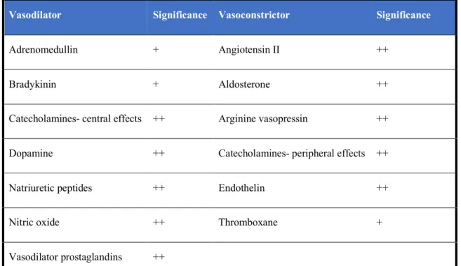
overexpression of biologically active substances, with cardiac myocytes contributing to that production, and whose effects may prove to be harmful both by autocrine action and by
paracrine activity, particularly at the renal level (Table 3).16
Table 3. Biologically active substances overexpressed in CHF.
Vasodilator Significance Vasoconstrictor Significance
Adrenomedullin + Angiotensin II ++
Bradykinin + Aldosterone ++
Catecholamines- central effects ++ Arginine vasopressin ++
Dopamine ++ Catecholamines- peripheral effects ++
Natriuretic peptides ++ Endothelin ++
Nitric oxide ++ Thromboxane +
Vasodilator prostaglandins ++
+ = minor effect; ++ = major effect Adapted from Preeti et al. (2016)16
HF is recognized as one of the 21st century epidemics and a public health problem,
affecting 1 to 2% of the adult population in industrialized countries,17 with its prevalence
growing in patients over 70 years of age (³10%).18 Hospitalization for HF is the flagrant cause
of admission in Western countries and accounts for a large percentage of the financial impact
of HF,17 estimated in $37.2 billion spent each year on HF in the United States.19 In hospital,
mortality is between 4-7% and within 12 weeks of discharge Mortality is around 13% in
European cohorts, and 5-year mortality reaching 60%.16 The leading cause of CHF, excluding
renal impairment, includes ischemic heart disease, diabetes mellitus, the metabolic syndrome
a Signs may not be present in the early stages of HF (especially in HFpEF) and in patients treated with diuretics;
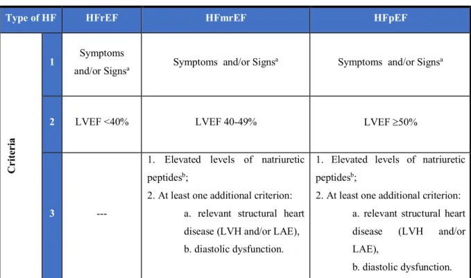
Table 4. Definition of HF with preserved, mid-range and reduced ejection fraction.
Type of HF HFrEF HFmrEF HFpEF
Cr
it
er
ia
1 Symptoms
and/or Signsa Symptoms and/or Signs
a Symptoms and/or Signsa
2 LVEF <40% LVEF 40-49% LVEF ³50%
3 ---
1. Elevated levels of natriuretic peptidesb;
2. At least one additional criterion: a. relevant structural heart disease (LVH and/or LAE), b. diastolic dysfunction.
1. Elevated levels of natriuretic peptidesb;
2. At least one additional criterion: a. relevant structural heart disease (LVH and/or LAE),
left atrial enlargement; LVEF: left ventricular ejection fraction; LVH: left ventricular hypertrophy; NT-proBNP: N-terminal pro-B type natriuretic peptide.
Adapted from Ponikowski et al. (2016).20
Patients with CHF have an increasingly longer life expectancy and are dying less
frequently of primary arrhythmias, so that CRS is expected to become more frequent.21
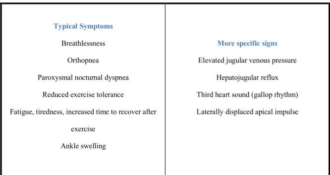
The signs and symptoms of HF and its definition (HT with preserved, mid-range and reduced ejection fraction) are presented in Tables 4 and 5.
Table 5. Symptoms and signs of heart failure.
Typical Symptoms
Breathlessness Orthopnea
Paroxysmal nocturnal dyspnea Reduced exercise tolerance
Fatigue, tiredness, increased time to recover after exercise
Ankle swelling
More specific signs
Elevated jugular venous pressure Hepatojugular reflux Third heart sound (gallop rhythm) Laterally displaced apical impulse
Chronic kidney disease
CKD is defined as the occurrence of anomalies of kidney function or structure or
decreased glomerular filtration rate (GFR<60 mL/min/1.73 m2) for at least three months.22
Kidney injury is defined as pathological alterations or markers of damage, including
abnormalities in blood or urine analyses or imaging techniques (Table 6)23 and overall CKD is
characterized by the gradual destruction of normal renal parenchyma due to a relentless process
of scarring.24 CKD can be categorizes according to GFR and albuminuria categories (Table
7).22
CKD: chronic kidney disease; GFR: glomerular filtration rate Adapted from KDIGO 2012.22
The global prevalence of CKD is 8-16% and is growing worldwide,25 and latest
epidemiological studies have showed CKD is a substantial risk for cardiovascular events Table 6. Definition of chronic kidney disease.
Criteria for CKD (either of the following for more three months)
Markers of kidney damage (one or more)
Albuminuria
Urine sediment abnormalities
Electrolyte and other abnormalities due to tubular disorders Abnormalities detected by histology
Structural abnormalities detected by imaging History of kidney transplantation
50% of all deaths in this circumstance.5 The majority of CKD patients might never reach the
circumstance of needing renal replacement therapy to support life, since they are more
susceptible to die prematurely because of faster cardiovascular disease evolution.24 However,
those with ESRD treated with dialysis are more prone to death by primary cardiac arrhythmias whilst in the general population coronary heart disease is the main cardiovascular cause of
death.27 Hence, CKD remains a critical health problem associated with high healthcare costs
and most important, high morbidity and mortality.25
In the absence of evidence of kidney damage, neither GFR category G1 nor G2 fulfill the criteria for CKD. ACR: albumin-to-creatinine ratio; AER: albumin excretion rate; CKD: chronic kidney disease; ESRD: end-stage renal disease; GFR: glomerular filtration rate.
Adapted from KDIGO 2012.22
Table 7. Glomerular filtration rate albuminuria categories in chronic kidney disease.
GFR category Terms GFR (ml/min/1.73m2)
G1 Normal or high ³90
G2 Mildly decreased 60-89
G3a Mildly to moderately decreased 45-59
G3b Moderately to severely decreased 30-44
G4 Severely decreased 15-29
G5 Kidney failure, including ESRD <15
Albuminuria category Terms AER (mg/24h) ACR (mg/g)
A1 Normal to mildy increased <30 <30 A2 Moderately increased 30-300 30-300 A3 Severely increased >300 >300
Definition, Diagnosis and Biomarkers
CRS2 is described by chronic anomalies in cardiac function that predispose to or cause progressive renal injury and / or dysfunction, a condition frequently observed in clinical
practice.9,28 The definition of chronic cardiac impairment includes many different heart
conditions (as constrictive pericarditis, congenital cyanotic heart disease and atrial fibrillation)
but chronic HF remains the main cause, and henceforth is the principal focus on this review.29
CRS2 is the most common CRS and has been reported in 63% of patients admitted with
congestive HF.30
Observational animal and human studies have shown evidence that CHF leads to variable levels of albuminuria / proteinuria, gradual reduction in GFR and expression of renal injury biomarkers (Figure 2). Although several mechanisms have been suggested, neurohormonal activation, hemodynamic factors as renal hypoperfusion and venous
congestion, inflammation and oxidative stress appear to be the principal ones.4,31
Figure 2 Cardiorenal Syndrome Type 2 and Renal Injury Biomarkers.
Serum creatinine
CRS2
Tubular injury/insterstitial fibrosis
Cardio-Renal Syndrome Type 1
LCOS and Congestion
Acute
Kidney
Injury
Acute
Heart
Disease
• Upon initial recognition, AKI induced by primary cardiac dysfunction
implies inadequate renal perfusion until proven otherwise. This should
prompt clinicians to consider the diagnosis of a low cardiac output state
Biomarkers Decreased GFR Glomerular injury Galectin-3, NGAL, KIM-1, L-FABP, IL-18, Cystatin C Urinary albumin-creatinie ratio
In addition to the more specific mechanisms, it is postulated that multiple episodes of
acute cardiac or renal decompensation may contribute to the progression of HF or CKD,28 as
suggested by data showing that prior hospitalizations for HF are predictors of mortality after adjusting for other HF risk factors. Recurrence of admissions for HF is independently related
with the development of CKD (eGFR <60mL/min).4
The diagnosis, prevention and treatment of this syndrome are generally fragmented on pathologies, focused on a single organ rather than a multidisciplinary approach. In 2010, for the first time the “Acute Dialysis Quality Initiative Consensus Group” presented the CRS definition, classification, and recommendations for the its diagnosis, prevention and management, with emphasis on the recommendation of a close collaboration between
cardiologists and nephrologists to optimize the outcome of these patients.32 Given the
complexity of CRS, there is no severity scale so far, and therefore it is recommended to use the
specific classifications for HF (NYHA) and CKD (KDIGO/KDOQI).32
It is essential to note that the simple co-occurrence of cardiovascular disease and CKD is not enough to establish a true diagnosis of CRS2. In the precise context of stable CHF, two prerequisites are required to establish the diagnosis of CRS2: (1) CHF and chronic renal failure coexist in the patient; (2) CHF is causally underlying the occurrence or progression of chronic renal failure (Figure 3). This latter aspect should be sustained by temporal connection (ie, the presumed or documented onset of chronic congestive HF temporally precede the occurrence or progression of chronic renal failure) and by pathophysiological plausibility (ie, the expression and grade of renal disease is reasonable justifiable by underlying heart disease) albeit available studies are often inconclusive in determining which of the two processes is primary versus secondary.9,28
Figure 3 Definition of Cardiorenal Syndrome Type 2. Adapted from Cruz et al. (2013)28 and KDIGO 2012.22
The findings revealing renal dysfunction in the context of CRS2 include increased serum creatinine (sCr) or, in individuals with lower muscle mass, a value of sCr in the
near-normal range, since it is associated with low eGFR values (<60mL/min/1.73m2), calculated
through the Modified Diet in Renal Diseases study or Cockcroft-Gault equations. Further laboratory findings may be useful, such as the coexistence of albuminuria or anemia, or both. The evaluation of renal dysfunction in HF clinical studies has been quite restricted to conventional biomarkers such as creatinine (or its derivative, eGFR), and urinary protein excretion. In CHF, both compromised renal function (denoted by elevated creatinine or
and albuminuria has been shown to have a prognostic value in relation to long-term renal
outcomes. However, this did not occur in the setting of CHF.10,28
Traditionally, eGFR stands as gold standard for the evaluation of renal function. However, its use in the context of acutely decompensated HF or CHF is difficult, since formulas that estimate eGFR are only validated when sCr is in a steady-state. SCr also presents important limitations. The sCr reflects only the eGFR and not directly the tubular lesion, and the tubular lesion can better predict and characterize acute kidney injury (AKI) and the progression of chronic renal damage. In addition, sCr shows a relatively late elevation during an episode of AKI, often when there is little to be done to prevent or counterbalance renal damage. In addition, sCr levels are influenced by age, ethnicity and muscle mass, which in hospitalized patients with HF, especially women and elderly individuals with low muscle mass, may lead to a lesser
recognition of renal failure,9 since eGFR could already have considerably decreased when sCr
levels surmount reference levels.15 Kervella et al. showed recently that the use of cystatin C for
GFR estimation in CRS2 may diagnoses with more accuracy reduced kidney function in patients with CHF, since these patients often have muscle wasting leading to a misclassification
of CKD stages with the use of creatinine for eGFR.33,34
Ultrasound imaging plays an relevant role in the diagnosis and management of CRS.35
Renal ultrasound may show the reduction of cortical thickness, corticomedullary ratio and hyperechogenicity of the parenchyma. The echocardiogram, in turn, may show high volumes or atrial areas such as volume overload, normal or reduced ejection fraction (EF), right chambers dilation and increased pulmonary artery pressure, pericardial effusion, and valvular
disease (calcific disease).4,10
If dysfunction in one organ can quickly lead to distant and harmful effects in the other, it seems natural that early acknowledgment provides the best chance for opportune intervention
that these patients may have ongoing kidney damage with constitutive release of biomarkers of renal injury as for example, KIM-1, NAG and Cystatin C, uncoupled from loss of renal
function.36 In this regard, new renal biomarkers such as NGAL and those mentioned above
(Table 8) were studied in patients with CHF and in many but not all studies, levels of these
biomarkers were increased even if at modest levels in CHF compared to control subjects.28
Tubular damage evaluated by urinary markers such as NAG, NGAL, and KIM-1 is frequently
present among patients with chronic HF and highly associated with mortality.37 Unfortunately,
most are investigational instruments being evaluated for clinical usefulness.38
Table. 8 Cardiac and renal biomarkers of cardiorenal syndrome.
Cardiac biomarkers
Natriuretic peptides
- The ability of BNP to predict the outcome of patients with CKD has been investigated in different studies, and in many of them there has been a positive association between high levels of BNP and mortality (total and cardiovascular) and progression of renal disease (HR of 1.38 95% CI 1.09 –1.76), and a even stronger association for NT-proBNP (HR of 2.28 CI 1.76 – 2.95).1,38,39 Moreover, there is evidence that increased levels of NT-proBNP are predictive of
subsequent death in pre-dialysis CKD patients (HR 9.6, P < 0.007).1,40
- Although valuable in the diagnosis of CHF, BNP and NT-proBNP are partially excreted by the kidneys thus decreasing its specificity as a marker of CHF.18
Cardiac troponins
- These markers can identify subclinical myocardial injury.41
- Current studies enrolled relatively small groups of patients and excluded patients with severe CKD, so the troponin clinical significance in patients with HF and severe CKD is not fully elucidated, but there are data to suggest that elevated levels of cTns may predict death among ESRD patients without symptoms of ACS.31
- The troponin elevation may be justified by glomerular filtration reduction.41,42
Galectin-3
- This b-galactoside-binding lectin plays an relevant regulatory role in cardiac fibrosis and remodeling.38,43
- It also has been implicated in the development of fibrosis in the liver, and together with TGF-b, these are the mediators prominently involved in kidney fibrosis. 38,43
Tabela 8. Continued.
Renal biomarkers
Cystatin C
- An early marker of impaired glomerular filtration instead of tubular lesion.42,44
- Serum levels of Cystatin C may represent a more accurate test of renal function than sCr levels.9
- An independent predictor of death, cardiac transplantation and hospitalization for HF (HR 2.27, 95% CI 1.12-4.63).28,45 It was also correlated with the levels of
NT-pro-BNP and measures of LV dysfunction.28,46
KIM-1 - Kidney injury molecule 1
- Quantified in the urine after an ischemic or nephrotoxic insult to the proximal tubular cells,9 and so it is a early marker of proximal tubular damage and useful in
early detection of acute kidney injury, especially in CHF.17
- Urine levels are elevated in symptomatic HF versus controls,28 and in CHF they
were the best predictors of worsening renal function.37
- CHF patients show higher levels even with normal kidney function.34 NAG-
N-acetyl-beta-D-glucosaminidase
- A sensitive marker of acute renal impairment or renal dysfunction worsening.17
- Increases significantly in congestive HF, with an important prognostic role independent from the glomerular filtration rate.28,42,47
- This protein is also raised in HF patients with preserved renal function.34
Neutrophil-associated lipocalin gelatinase
(NGAL)
- Involved in immune modulation, inflammation and neoplastic transformation.38 It
is expressed at reduced levels in various human tissues including kidney, lung, stomach and colon.9
- NGAL expression is prominently increased in epithelial cells that have suffered injury - for example, in the renal tubular epithelium after any type of insult or stress, in the myocardium in failure, in myocarditis and also in atherosclerotic plaques.9,28
- The increase in serum NGAL levels occurs prior to sCr, and still correlates with the degree of renal tubular injury.9
- Serum and urine NGAL may be sensitive early markers of renal dysfunction in patients with CHF and normal sCr but reduced eGFR.41
- In some clinical studies, NGAL blood and urine levels in general also correlated with clinical and biochemical markers of HF severity,28 and recently a study
demonstrates that plasma NGAL predicts mortality in patients with HF, both in patients with and without CKD.48
- Serum NGAL is not substantially correlated with EF or NYHA functional class or with other biomarkers like urinary KIM-1 and NAG.34
Albuminuria
- Micro- and macro-albuminuria are present in 20–30% and 5–10%, respectively, of the patients with HF.44
- Albuminuria, evaluated as the albumin-to-creatinine ratio in urine, is an recognized criterion for the diagnosis of CKD.44
- Correlations between reduced renal blood flow and albuminuria, elevated NT-proBNP levels and physical exam findings of volume overload have been proven, suggesting potential participation of arterial underfilling and venous congestion in the mechanism of albuminuria in HF.34
- It has been associated with an augmented risk of death that persists significant after adjustment for renal function or diabetes.44
Blood Urea Nitrogen to Creatinine Ratio
(BUN/Cr)
- BUN/Cr has been broadly used clinically to differentiate intrinsic kidney disease from pre-renal renal dysfunction, so that it offers supplementary diagnostic and prognostic value in CRS.34
- Renal clearance of urea is determined by the quantity filtered and the degree of tubular reabsorption. Decreased GFR resulting from any etiology can lead to reduced urea filtration. On the other hand, the tubular reabsorption of urea is mainly influenced by neurohormonal activation with increases in angiotensin II and vasopressin leading to higher urea concentration in the proximal tubule and increased urea transporters in the collecting duct respectively, thereby boosting absorption.34,49
- In HF, with increased neurohormonal activation, the BUN is elevated disproportionately to serum creatinine on the contrary of intrinsic kidney disease, where filtration may be decreased, but tubular reabsorption of urea is preserved producing a lower BUN/Cr.34
ACS: acute coronary syndrome; BNP: Brain-Type Natriuretic Peptide; CAD: coronary artery disease; CHF: chronic heart failure; CKD: chronic kidney disease; CRS: cardiorenal syndrome; cTns: cardiac troponins; EF: ejection fraction; ESRD: end-stage renal disease; eGFR: estimated glomerular filtration rate; HR: hazard ratio; LV: left ventricular; NT-proBNP: NT-pro-brain natriuretic peptide; sCr: serum creatinine, TGF-b: transforming growth factor beta.
Pathophysiology of Cardiorenal Syndrome Type 2
In CRS2, the pathogenic mechanisms by which cardiac dysfunction triggers renal dysfunction de novo or provoke the harmful progression of a pre-existing CKD is not so clear. Indeed, in this case, worsening of renal function occurs in patients who are unaffected by clinical signs and symptoms of hemodynamic imbalance (CRS2 patients do not have acute decompensated HF by definition). Potential mechanisms involved, namely hemodynamic factors, neurohormonal activation, oxidative stress, inflammation and anemia, which will be detailed below, but overall it is supposed the underlying mechanisms of HF, co-morbidities and/or their treatment affect renal function with subsequent development of renal failure (Figure 4).
Figure 4 Pathophysiology of chronic cardiorenal syndrome (type 2). CO: cardiac output; RAAS: renin-angiotensin-aldosterone system Adapted from Cruz et al. (2013)28 and KDIGO 2012.22
Cardio-Renal Syndrome Type 1
LCOS and Congestion
Acute Kidney Injury Acute Heart Disease
• Upon initial recognition, AKI induced by primary cardiac dysfunction implies inadequate renal perfusion until proven otherwise. This should prompt clinicians to consider the diagnosis of a low cardiac output state (LCOS) and/or marked increase in venous pressure leading to kidney congestion.
• It is important to remember that central venous pressure translated to the renal veins is a product of right heart function, blood volume, and venous capacitance which is largely regulated by neuro-hormonal systems.
• Specific regulatory and counter-regulatory mechanisms are activated with variable effects depending on the duration and the intensity of the insult.
• Inflammatory pathways • Arterial underfilling
• Decreased CO and effective circulating volume
• Chronic neurohormonal activation (RAAS and sympathetic tone) • Chronic venous congestion • Systemic inflammation • Chronic renal hypoperfusion • Tubular fibrosis • Tubular atrophy • Glomerulosclerosis Chronic worsening of renal function Chronic cardiac dysfunction Type 2 CRS
1) Hemodynamic Factors
Decreased glomerular filtration rate may be the result of reduced cardiac output in HFrEF, but in HFpEF, in addition to decreased cardiac output as a result of increased filling pressures, elevation of venous pressure is a relevant alternate cause of worsening renal function
in these patients.50-53 Thus, chronic HF is more likely to be illustrated by a long-standing renal
venous congestion and reduced renal perfusion, often predisposed by macro and microvascular
disease in the context of the same risk factors linked to cardiovascular disease.9,54
Although a large fraction of patients with a low eGFR have a more severe NYHA functional class, the association between LVEF and eGFR has not been consistently proven.
Hence, patients with HFpEF appear to have an eGFR similar to patients with HFrEF.3,55-57 But
not all patients with HF and decreased renal function have hypotension or reduction in cardiac output, and many patients with hypotension do not have reduced renal function, and even
increasing cardiac output does not necessarily improve renal outcomes.58
The prospective ESCAPE study did not demonstrate an association between cardiac index and baseline renal function. The only relationship found was with increased pressure in the right atrium, suggesting that renal congestion may be an important factor of renal
dysfunction that should be considered.59,60
Signs and symptoms of congestion, including increased jugular venous pressure, ascites, peripheral edema, and orthopnea are often found and associated with decreased renal function
in patients with CHF.61,62 The most plausible explanation for the impact of venous congestion
on renal function is that increased renal venous pressure (renal perfusion pressure is calculated by subtracting central venous pressure at mean arterial pressure) decreases the arterio-venous
tubules, and possibly inducing local hypoxia,63 but since intratubular pressure is one of the
driving forces of glomerular filtration, any increase in intratubular pressure may oppose
glomerular filtration and decrease eGFR.64
Right ventricular dilation and dysfunction may also adversely affect renal function through elevated venous pressure as well as compromising left ventricular filling, and
consequently, the forward output.5,58
Redirecting of blood from the medullary areas of the kidney in states of compromising renal blood flow may cause hypoxic lesion of the highly metabolic renal tubular cells. The hypoxic tubular lesion initiates a progressive loss of nephrons and gradual renal failure. Microalbuminuria in HF patients may reflect early tubular damage that compromises the
absorption of albumin filtered by the damaged tubular cells.15 Furthermore, the reduction in
renal perfusion, coupled with atherosclerotic changes underlying possible comorbidities such as diabetes mellitus and hypertension, may rapidly aggravate any pre-existing renal dysfunction
in this particular population.61
2) Neurohormonal Activation, Oxidative stress and Inflammation
In HF, reduction of left ventricular systolic or diastolic function results in decreased cardiac output, ejection volume, arterial bed underfilling, elevated atrial pressure and venous
congestion.5 The decrease in the effective intravascular blood volume verified in chronic HF is
sensed at the level of the high-pressure baroreceptors, which are found in the left ventricle,
carotid sinus and aortic arch.60,64,65 This results in an activation of the SNS, with increased heart
rate and peripheral and renal vascular resistance. Increased renal adrenergic tone stimulates RAAS. The activation of the SNS also stimulates the supra-optic and paraventricular nuclei of
the hypothalamus, resulting in the non-osmotic release of ADH66 and in this point it is known
that higher plasma ADH concentration is associated with higher cardiovascular mortality at 1 year.67
The SNS is activated to maintain the decreased cardiac output in chronic HF,31 therefore
increasing the activation of the RAAS, but at the expense of producing reactive oxygen species (ROS) and the activation of the immune system. Chronic stimulation of the SNS has a growth promoting effect at the level of the intrarenal blood vessel wall, which has recently been
identified as being mediated, at least in part, by the production of ROS.31 Moreover, it has long
been known that the kidneys of patients with HF release considerable amounts of renin into the
circulation68 due to increased activity of the SNS in addition to decreased renal artery pressure,
increased renal venous pressure, and decreased delivery of sodium to the distal nephron.69 SNS
activation indirectly leads to congestion because it causes arterial and venous tachycardia and vasoconstriction which rises afterload and decreases preload, leading to a reduction in LV
function and eventually to LV remodeling.65
The RAAS is also activated in CHF, as previously mentioned, and ANGII has multiple adverse effects on the cardiovascular system in HF patients, through a significant effect on peripheral and renal vascular resistance, and also having a contribution on the stimulation of the SNS, promoting the release of noradrenaline at pre-synaptic level. ANGII is an important mediator of myocardial hypertrophy and remodeling, and high plasma concentrations of this neurohormone further contribute to the deposition of extracellular matrix and fibrosis in the
kidney.8 ANGII has an important vasoconstrictive effect on the afferent and efferent arterioles
and shedding as intraglomerular pressure increases due to its preponderant effect on efferent
arterioles.70 In addition to its (de)regulation of extracellular volume and vasoconstriction, one
of the most deleterious effects of angiotensin on CRS2 results from the activation of NADPH oxidase in endothelial cells, renal tubules and cardiomyocytes, with consequent formation of
free radicals.31,57,71,72 As it potentiates modifications in the cellular redox state, ANGII is
implicated in vascular inflammation through the nuclear factor kappa B (NF-kB), which
induces the production of adhesion and chemotactic molecules.72,73 Hence, ANGII has been
shown to play an important role in the synthesis of proinflammatory cytokines in the kidney, regulation of cell proliferation, fibrosis and apoptosis as well as vascular and myocardial
hypertrophy and endothelial dysfunction.49,66 Besides participating in the same activation
processes mediated by ANGII, high levels of serum aldosterone stimulate overexpression of
TGF-b and fibronectin production, leading to renal fibrosis and glomerulosclerosis.4,56
Endothelin represents one of the most potent vasoconstrictors and its increase in HF contributes
to vasoconstriction, particularly in the renal circulation,65 and also for renal inflammation and
fibrosis.56
In experimental studies in HF, a reduction in glomerular plasma flow was observed along with eGFR (efferent arteriolar constriction); if these changes persist, focal and segmental glomerulosclerosis can ensue, often associated to local renal increase in SNS activity and RAAS activation.4,74
Recently, Kamal et al. (2017)75 have investigated the role of chronic stimulation of G
protein-coupled receptors (GPCRs), including adrenergic and endothelin receptors, and their work suggest that GPCR-Gbg inhibition can be a future promising therapeutic approach on preventing the heart and kidney damage in the setting of CRS2.
Natriuretic peptides are increased in HF and promote natriuresis, vasodilation, and inhibition of the SNS and the RAAS, effects that even being beneficial are not effective in HF
patients. There is evidence to suggest that there is resistance to the effects of natriuretic peptides
as CHF worsens.76 There are several explanations for this phenomenon: down-regulation of
renal natriuretic peptide receptors; secretion of inactive precursor peptides; increased endopeptidase activity, which restricts the delivery of natriuretic peptides to the distal nephron level; increase of sodium reabsorption at the proximal level, decreasing its concentration in the
distal nephron, the local of activity of natriuretic peptides.66
Nitric oxide (NO) is produced from L-arginine under the coordination of the enzyme nitric oxide synthase and has great importance in renal control of extracellular volume and blood pressure since it induces vasodilation, natriuresis and desensitisation of tubuloglomerular
feedback.5,31 Superoxide has an contrary influence on extracellular volume control conditioning
an increase in blood pressure, and it can inactivate NO, and consequently generating
peroxynitrite and decreasing NO activity.77 In CRS2, it has been proposed that the impairment
of the L-arginine-nitric oxide pathway secondary to anomalous neurohormonal activation, the physiological balance between NO and ROS is altered, with increased levels of ROS resulting
in low bioavailability of NO,31,60,69,77 which might have a significant responsibility in the
progression of CRS2 (Figure 5). Endothelial cells release NO in response to laminar shear stress. Accordingly, a plausible mechanism of endothelial dysfunction in HF may be the lower shear stress secondary to pump failure in HF. Shear stress also depends on blood viscosity and
Figure 5 L-arginine - NO pathway and its influence on Cardiorenal Syndrome Type 2.
L-arginine is the substrate for NO synthesis. Transmembrane transport of L-arginine in endothelial cells is mainly intermediated through CAT-1, located in the plasma membrane of endothelial cells. CAT-1 also seems to be the major L-arginine transporter expressed in the kidney and it is mostly concentrated in the inner medullary collecting duct. According to published evidence, renal NO bioavailability is dependent on L-arginine transport and its bioavailability is essential for regulation of normal renal vascular and tubular function. Chronic activation of the SNS and the RAAS present in CHF and CKD may be responsible for chronic elevations in oxidative stress which can impair L-arginine transport. Furthermore, additional factors for the low availability of NO are reduced eNOS expression and uncoupling and elevated levels of inhibitors, such as ADMA, which along with marked breakdown of NO due to high ROS levels create a vicious cycle of damage affecting concurrently the heart and kidney.
L-Intracellular L-Arginine
pools
Endothelial Cell
CHF
CKD
Chronic activation of the RAAS Chronic activation of the SNS
Endothelial dysfunction Oxidative stress CAT-1 eNOS L-Arg L-Arg NO ADMA Inhibits eNOS Extracellular space Reduces L-Arg transport and uncouples eNOS Buffers the
activation of the RAAS
arginine transport is a conceivable novel therapeutic target in CRS2 once enhancing L-arginine transport can mitigate the activation of the RAAS and decrease oxidative stress.
ADMA: asymmetric dimethylarginine; CAT-1: cationic amino acid transporter-1; eNOS: Endothelial nitric oxide synthase; L-Arg: L-arginine; NO: nitric oxide; RAAS: renin-angiotensin-aldosterone system; SNS: sympathetic nervous system.
Adapted from Rajapakse et al. (2016).71
Oxidative stress is a major enhancer of inflammation, with increased synthesis of proinflammatory cytokines namely IL-1, IL-6 and TNF-alpha, with the latter being also increased by the elevated venous congestion and cardiac myocytes under the stress of
mechanical stretch or ischemia.28,64,73,78 The venous stasis and arteriolar vasoconstriction can
result in intestinal ischemia due to altered intestinal permeability79 and release of endotoxins
from gut bacteria, activating the immune system.80 The chronic inflammation ensued might
cause progressive toxic injury and renal cell apoptosis, which causes permanent kidney damage and loss of kidney function. This inflammatory mechanism in the setting of high venous pressure is described as a prime path of kidney function impairment specially in patients with
chronic cardiac dysfunction and preserved left ventricular systolic function.74,81
Following the initial insult, the impaired kidney undertakes a series of events, such as fibroblast activation, in an effort to repair and recover from the injury. Accumulated renal collagen is cross-linked and resilient to breakdown, which inevitably ends in loss of function when normal tissue is supplanted with scar tissue. Consequently, renal fibrosis leads to renal changes as glomerulosclerosis, tubulointerstitial fibrosis and tubular atrophy. Glomerulosclerosis result in the obstruction of the capillary loops and eventually lead to loss of
35
3) Anemia
Anemia, CHF and CKD interrelate in a vicious circle to cause or aggravate each other.78
Overall, most studies in this matter estimate that the prevalence of anemia in the HF population
is greater than 20%82 and is associated with increased morbidity and mortality (Figure 6).71,82
Figure 6 Estimates of hazard ratio (with 95% CI) for all-cause mortality associated with a decrease in hemoglobin of 1g/dL. Decreased hemoglobin in patients with CHF has been recurrently shown to be independently associated with increased risk of hospitalization and all-cause mortality. Various studies have shown that a 1g/dL decline in hemoglobin was independently associated with appreciably augmented mortality risk.
Taken and adapted from Tang and Katz (2006).83
Multiple etiologies have been proposed, considering that it is not only the result of associated CKD, with increasing attention being given to the hostile effects of inflammatory
markers on erythropoiesis and iron metabolism.44 Hepcidin, which production increases with
inflammation, may contribute to the anemia observed in HF, due to the activation of
inflammatory pathways in this condition.5 However, the cause that may bring more insight to
the pathophysiology is resistance to erythropoietin. Indeed, patients with congestive heart failure typically have elevated EPO plasma levels but are disproportionately low for the degree of anemia compared to patients with ESRD who are functionally anephric and with absolute of severe anemia on blood viscosity, oxygen tension in the
microvasculature, and nitric oxide availability.44,45 Lesser
degrees of anemia may contribute to neurohormonal activa-tion and disease progression in patients with CHF.46 In
patients with !-thalassemia syndromes, heart failure is a common complication that is likely mediated by the hemo-dynamic and neurohormonal effects of severe chronic anemia and iron overload secondary to chronic transfusion require-ments.47 Aggressive iron chelation therapy greatly reduces
but does not eliminate the risk of heart failure in these patients.
Hemoglobin content in blood is an important determinant of oxygen delivery to skeletal muscle during exercise. Pa-tients with CHF lack normal physiological reserve to com-pensate for decreased hemoglobin and may manifest de-creased aerobic capacity in response to mild degrees of anemia.48 Several investigators have reported association
between reduced hemoglobin and greater functional impair-ment as defined by New York Heart Association classifica-tion.3,15,49 Kalra and colleagues50 reported a linear relation
between reduced hemoglobin values and peak oxygen con-sumption in anemic patients with CHF with hemoglobin !13.0 g/dL.
Cardiac mass increased by 25% in a rat model of chronic anemia.51 An inverse relation between hemoglobin value or
hematocrit value and left ventricular hypertrophy has also described in clinical studies of patients with dialysis-dependent and predialysis chronic kidney disease.52 In a
subgroup of patients with CHF enrolled in the Randomized Etanercept North American Strategy to Study Antagonism of Cytokines (RENAISSANCE) trial with available cardiac MRI data, a 1-g/dL increase in hemoglobin was associated with a 4.1-g/m2 decrease in left ventricular mass over 24
weeks.49 This observation was independent of study drug
treatment and does not provide evidence of a causal relation between changes in hemoglobin levels and changes in left ventricular mass. In two randomized trials of patients with chronic kidney disease, increased hemoglobin in response to erythropoietic agents was not associated with reduction in left ventricular hypertrophy.53,54
Anemia and Clinical Outcomes
Reduced hemoglobin in patients with CHF has been repeatedly shown to be independently associated with increased risk of hospitalization and all-cause mortality.2– 6,8,9,11–13,15,19,42,49These
findings in a diverse array of CHF populations are remarkably concordant and generally suggest a linear association between reduced hemoglobin and increased mortality risk. In studies that analyzed hemoglobin as a continuous variable, a 1-g/dL de-crease in hemoglobin was independently associated with signif-icantly increased mortality risk (Figure 2). The potential mech-anisms linking anemia to increased mortality risk in CHF have not been characterized but may be related to changes in ventric-ular loading conditions and cardiac structure, altered neurohor-monal activation, or reduced free radical scavenging capacity. It is also possible that anemia is a marker of more severe underlying myocardial disease.
Treatment Approaches
The clinical utility of blood transfusion in anemic cardiovas-cular disease populations is controversial. According to the guidelines from the American College of Physicians and the American Society of Anesthesiology, the “transfusion thresh-old” for patients without known risk factors for cardiac disease is a hemoglobin level in the range of 6 to 8 g/dL.55In
78 974 elderly patients hospitalized with acute myocardial infarction, blood transfusion was associated with a signifi-cantly lower 30-day mortality rate among patients with a hematocrit "30% on admission.56In 838 critically ill patients
(26% with cardiovascular disease), maintaining hemoglobin at 10 to 12 g/dL did not provide additional benefits on 30-day mortality compared with maintaining hemoglobin at 7 to 9 g/dL.57 Blood transfusion may be associated with other
adverse effects including immunosuppression with increased risk of infection, sensitization to HLA antigens, and iron overload.58,59 Given this profile of risks and benefits,
trans-fusion may be considered as an acute treatment for severe anemia on an individualized basis but does not appear to be a viable therapeutic strategy for the long-term management of chronic anemia in CHF.
Although erythropoietin levels are modestly elevated in patients with CHF, the increase is less than that observed in other anemic populations.27,38,60Accordingly, anemia in CHF
may be responsive to exogenous erythropoietin supplemen-tation. The primary mechanism by which erythropoietin stimulates red blood cell production is inhibition of apoptosis of bone marrow erythrocyte progenitors.23The erythropoietin
receptor is a member of the cytokine class I receptor superfamily.61 Ligand binding of erythropoietin to the
ho-modimeric erythropoietin receptor activates antiapoptotic sig-nal transduction pathways.23,62 Bone marrow erythroid
pro-genitor cells escape from apoptosis and proliferate to result in
Figure 2. Estimates of hazard ratio (with 95% CI) for all-cause
mortality associated with a decrease in hemoglobin of 1 g/dL. Estimates are derived from Cox proportional hazards models with adjustment for potential confounding variables. For studies with first author Al-Ahmad, Kosiborod and Mozaffarian, esti-mates of hazard ratio were derived from published data assum-ing a 1 g/dL decrease in hemoglobin is equivalent to a 3% decrease in hematocrit. The analysis by Mozaffarian included subjects with hematocrit "37.5%. Anand and colleagues did not report confidence intervals for their estimate of risk. Mag-gione and colleagues reported estimates of risk from 2 study populations enrolled in the IN-CHF registry and the Val-HeFT clinical trial.
2456 Circulation May 23, 2006
of severe anemia on blood viscosity, oxygen tension in the microvasculature, and nitric oxide availability.44,45 Lesser
degrees of anemia may contribute to neurohormonal activa-tion and disease progression in patients with CHF.46 In
patients with !-thalassemia syndromes, heart failure is a common complication that is likely mediated by the hemo-dynamic and neurohormonal effects of severe chronic anemia and iron overload secondary to chronic transfusion require-ments.47 Aggressive iron chelation therapy greatly reduces
but does not eliminate the risk of heart failure in these patients.
Hemoglobin content in blood is an important determinant of oxygen delivery to skeletal muscle during exercise. Pa-tients with CHF lack normal physiological reserve to com-pensate for decreased hemoglobin and may manifest de-creased aerobic capacity in response to mild degrees of anemia.48 Several investigators have reported association
between reduced hemoglobin and greater functional impair-ment as defined by New York Heart Association classifica-tion.3,15,49 Kalra and colleagues50 reported a linear relation
between reduced hemoglobin values and peak oxygen con-sumption in anemic patients with CHF with hemoglobin !13.0 g/dL.
Cardiac mass increased by 25% in a rat model of chronic anemia.51 An inverse relation between hemoglobin value or
hematocrit value and left ventricular hypertrophy has also described in clinical studies of patients with dialysis-dependent and predialysis chronic kidney disease.52 In a
subgroup of patients with CHF enrolled in the Randomized Etanercept North American Strategy to Study Antagonism of Cytokines (RENAISSANCE) trial with available cardiac MRI data, a 1-g/dL increase in hemoglobin was associated with a 4.1-g/m2 decrease in left ventricular mass over 24
weeks.49 This observation was independent of study drug
treatment and does not provide evidence of a causal relation between changes in hemoglobin levels and changes in left ventricular mass. In two randomized trials of patients with chronic kidney disease, increased hemoglobin in response to erythropoietic agents was not associated with reduction in left ventricular hypertrophy.53,54
Anemia and Clinical Outcomes
Reduced hemoglobin in patients with CHF has been repeatedly shown to be independently associated with increased risk of hospitalization and all-cause mortality.2– 6,8,9,11–13,15,19,42,49These
findings in a diverse array of CHF populations are remarkably concordant and generally suggest a linear association between reduced hemoglobin and increased mortality risk. In studies that analyzed hemoglobin as a continuous variable, a 1-g/dL de-crease in hemoglobin was independently associated with signif-icantly increased mortality risk (Figure 2). The potential mech-anisms linking anemia to increased mortality risk in CHF have not been characterized but may be related to changes in ventric-ular loading conditions and cardiac structure, altered neurohor-monal activation, or reduced free radical scavenging capacity. It is also possible that anemia is a marker of more severe
Treatment Approaches
The clinical utility of blood transfusion in anemic cardiovas-cular disease populations is controversial. According to the guidelines from the American College of Physicians and the American Society of Anesthesiology, the “transfusion thresh-old” for patients without known risk factors for cardiac disease is a hemoglobin level in the range of 6 to 8 g/dL.55In
78 974 elderly patients hospitalized with acute myocardial infarction, blood transfusion was associated with a signifi-cantly lower 30-day mortality rate among patients with a hematocrit "30% on admission.56In 838 critically ill patients
(26% with cardiovascular disease), maintaining hemoglobin at 10 to 12 g/dL did not provide additional benefits on 30-day mortality compared with maintaining hemoglobin at 7 to 9 g/dL.57 Blood transfusion may be associated with other
adverse effects including immunosuppression with increased risk of infection, sensitization to HLA antigens, and iron overload.58,59 Given this profile of risks and benefits,
trans-fusion may be considered as an acute treatment for severe anemia on an individualized basis but does not appear to be a viable therapeutic strategy for the long-term management of chronic anemia in CHF.
Although erythropoietin levels are modestly elevated in patients with CHF, the increase is less than that observed in other anemic populations.27,38,60Accordingly, anemia in CHF
may be responsive to exogenous erythropoietin supplemen-tation. The primary mechanism by which erythropoietin stimulates red blood cell production is inhibition of apoptosis of bone marrow erythrocyte progenitors.23The erythropoietin
receptor is a member of the cytokine class I receptor superfamily.61 Ligand binding of erythropoietin to the
ho-modimeric erythropoietin receptor activates antiapoptotic sig-nal transduction pathways.23,62 Bone marrow erythroid
pro-Figure 2. Estimates of hazard ratio (with 95% CI) for all-cause
mortality associated with a decrease in hemoglobin of 1 g/dL. Estimates are derived from Cox proportional hazards models with adjustment for potential confounding variables. For studies with first author Al-Ahmad, Kosiborod and Mozaffarian, esti-mates of hazard ratio were derived from published data assum-ing a 1 g/dL decrease in hemoglobin is equivalent to a 3% decrease in hematocrit. The analysis by Mozaffarian included subjects with hematocrit "37.5%. Anand and colleagues did not report confidence intervals for their estimate of risk. Mag-gione and colleagues reported estimates of risk from 2 study populations enrolled in the IN-CHF registry and the Val-HeFT clinical trial.
deficiency of EPO.82 The loss of sensitivity to EPO is thought to be mediated by chronic
inflammation.44,82 Hemodilution due to excessive venous congestion and gastrointestinal tract
edema that causes malabsorption and/or malnutrition are other possible etiologies of anemia.56
Anemia may affect oxygen supply to the tubular cells producing chronic ischemia,
activation of renal interstitial fibroblasts and, ultimately, nephron loss and kidney damage.56 On
the other hand, as previously explained, anemia may be a cooperative factor for endothelial dysfunction through reduction of shear stress, and contribute to worsening of cardiac and renal
function.5
4) Impact of Pharmacotherapy for the Management of Heart Failure
Pharmacological management of HF may have potential negative consequences on renal
function.67 Hypovolemia associated with diuretics, the early introduction of RAAS blockade
and iatrogenic hypotension have long been considered contributing factors to the genesis and
aggravation of CRS2.9
Patients with CHF often require higher dosages of loop diuretics to achieve comparable
sodium excretion given the attenuation of maximal response.9 This is the so-called "resistance
to diuretics", which eventually leads to harmful therapeutic approaches, due to excessive dose increases of diuretics, which induce several detrimental phenomena such as exaggerated stimulation of the tubule-glomerular feedback mechanism and activation of RAAS, a subsequent reactive vasoconstriction of renal afferent arterioles and eGFR decrease, and