Pelvic Congestion Syndrome case series: results of
endovascular treatment
Síndrome da Congestão Venosa Pélvica e resultados do tratamento
endovascular: série de casos
Gilberto do Nascimento Galego1
*
, Pierre Galvagni Silveira1,2, Cristiano Torres Bortoluzzi2,
Rafael Narciso Franklin2, hiago Mezadri Ronchi1
Abstract
Pelvic Congestion Syndrome (PCS) is a cause of chronic pelvic pain that primarily afects multiparous women of reproductive age. Embolization of pelvic varicose veins ofers excellent results for treatment of this syndrome. We describe an initial series of patients treated with embolization of pelvic varicose veins and their respective postoperative follow-up results. We provide clinical data, details of the procedures performed and results of follow-up and imaging exams for six patients. he technical success rate with these patients was 100% and there were no reports of serious intraoperative or postoperative complications. In all cases there was relief from symptoms and improvements in the results of imaging exams during short-term follow-up. he results of this small series of cases indicate that embolization is a safe and efective treatment for PCS.
Keywords: Pelvic Congestion Syndrome; pelvic venous incompetence; chronic pelvic pain; embolization; endovascular treatment; case series.
Resumo
A Síndrome da Congestão Venosa Pélvica (SCVP) é uma causa de dor pélvica crônica, que afeta principalmente mulheres multíparas em idade reprodutiva. Para o tratamento desta síndrome, a embolização de varizes pélvicas tem demonstrado excelentes resultados. Relatamos uma série inicial de pacientes submetidas a tratamento com embolização de varizes pélvicas e os respectivos resultados de acompanhamento pós-operatório. São apresentados dados clínicos, detalhes do procedimento e resultados do acompanhamento e de exames de imagem de seis pacientes. Dentre estas pacientes, o sucesso técnico foi de 100% e não houve relato de complicações trans ou pós-operatórias graves. Em todos os casos, pôde-se observar alívio dos sintomas e melhora nos resultados de exames de imagens no acompanhamento de curto prazo. Os resultados nesta pequena série de casos indicam que a embolização é um tratamento seguro e efetivo para a SCVP.
Palavras-chave: Síndrome da Congestão Venosa Pélvica; incompetência venosa pélvica; dor pélvica crônica; embolização; tratamento endovascular; série de casos.
1 Universidade Federal de Santa Catarina – UFSC, Florianópolis, SC, Brazil. 2 Clínica Coris Medicina Avançada, Florianópolis, SC, Brazil.
Financial support: None.
Conlicts of interest: No conlicts of interest declared concerning the publication of this article. Submitted: October 29, 2014. Accepted: May 05, 2015.
INTRODUCTION
Pelvic Congestion Syndrome (PCS) is a condition in which pelvic varicose veins cause dilation and venous stasis of the organs in the pelvic cavity and, as a consequence, chronic pelvic pain (CPP).1 The venous
dysfunction has its origins in a multifactorial process, in which an increase in abdominal pressure and the action of female hormones appear to be central factors and may explain the syndrome’s higher incidence among multiparous women of reproductive age and the disappearance of symptoms during menopause.1
A study by Asciutto et al.2 found that the left
gonadal veins and right internal iliac veins are most often affected (57.7% for both). In the majority of cases (53.5%) two or more veins will be incompetent.
Obstructive anatomic abnormalities of the pelvic venous system can also lead to secondary PCS.3 Extrinsic compression of the left renal vein,
impeding low to the inferior vena cava (nutcracker
phenomenon), is one possible cause of pelvic varicose veins and incompetence of the left gonadal vein that should be considered.4 Another possible cause of the
dysfunction, via a similar mechanism, is left common iliac vein compression (May-Thurner) syndrome.5
The most common clinical presentation of PCS is a
case of CPP with no evidence of inlammatory disease.
In general, the pain worsens during the perimenstrual period and with increased intra-abdominal pressure, generally accompanied by dyspareunia and postcoital discomfort, urinary symptoms (secondary to varicose veins in the bladder wall) and feelings of heaviness in the pelvis and legs.6 Physical examination may
reveal varicose veins involving the vulva, perineal
area and buttocks, and pain if the cervix of the uterus
is moved.3
Well-directed patient history and physical examination should lead to a diagnostic suspicion, which should
then be conirmed with supplementary examinations.7
These investigations should preferably begin with color Doppler ecography, which is a widely-available examination that offers the possibility of a dynamic
study of venous low, showing venous relux and stasis.6
Findings of gonadal veins with diameters larger than 5 mm on abdominal or transvaginal ultrasonography have a positive predictive value of 71.2%, increasing to 83.3% when diameters are greater than 6 mm.8
Investigation can also be accomplished using computed tomography (CT) angiography or magnetic
resonance (MRI) angiography. Diagnosis is conirmed
using criteria proposed by Coakley et al.,9 as follows:
four or more ipsilateral tortuous veins, with diameters > 4 mm, and a gonadal vein with a diameter > 8 mm.
Venography is the gold standard method for diagnosis.
The following indings should be present: gonadal vein with a diameter > 6 mm; retrograde venous low;
several collateral veins with tortuous paths, and slow drainage of the contrast after injection.3
Treatment of PCS by embolization of gonadal veins with minimally invasive endovascular procedures is becoming increasingly popular. It is conducted via the same catheterization used for diagnostic venography and, performed in this manner, the treatment has proved to be safe and effective for control of the condition, with low rates of relapse and complications.10
This article was approved by the local institutional Ethics Committee. The objective is to report an initial series of six cases of patients diagnosed with PCS who were treated using the endovascular technique at the Clínica Coris Medicina Avançada, in the city of Florianópolis, SC, Brazil, from 2011 to 2013, analyzing the results of clinical and imaging examinations conducted before treatment, intraoperatively and during the short-term postoperative period.
CASE REPORTS
Medical records were reviewed for six patients, all female, aged from 39 to 51 years. All were multiparous and their number of prior vaginal deliveries varied from 2 to 5. Three of these patients had varicose veins of the lower limbs, one had varicose veins
involving the left buttock. Five of them had a history
of non-cyclical CPP and one only reported long term feelings of pelvic ‘heaviness’. The majority of cases
(ive) had pelvic varicose veins identiied on color
Doppler echography. In three cases investigations were supplemented with another imaging exam: CT (one case) or MRI (two cases). One patient also exhibited ultrasonographic signs suggestive
of May-Thurner Syndrome, which were conirmed
by MRI. In one case pelvic varicose veins were an
incidental inding during CT. When questioned, this patient reported CPP of as yet unknown etiology.
Table 1 summarizes this information. In all cases,
venography conirmed the indings of the initial work-up examinations.
In all six cases an endovascular approach was used. Access was achieved by echo-guided puncture of the basilic or cephalic vein at the cubital fossa, with a proximal tourniquet. In all cases, a 5F sheath was used with a ‘roadrunner’ hydrophilic guidewire. Initially, an anatomic and hemodynamic investigation of the pelvic venous plexus was conducted, with
observation of the structure and the low of the
veins and internal iliac veins. In all patients, pelvic
varicose veins were identiiable, the diameter of at
least one of the gonadal veins was larger than normal (a dilated left gonadal vein was present in 100%) and
there were signs of relux in the pelvic venous plexus
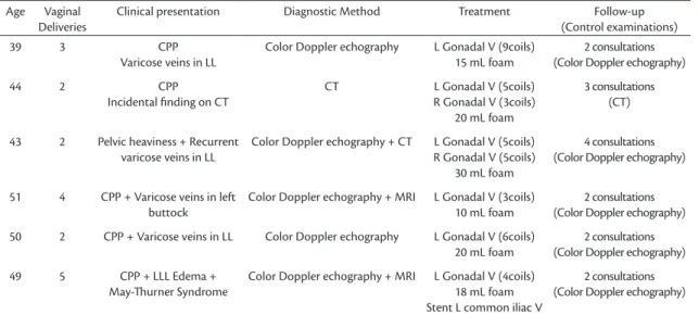
(Figure 1). After conirmation of venous dilation and
relux, selective catheterization of the vessel was
performed with a multipurpose, vertebral or mammary 5F catheter (the choice was made on the basis of each patients’ anatomy) followed by embolization of the pelvic varicose veins with dense polidocanol foam (10 to 20 mL at 1 to 3%). The polidocanol foam was diluted using room air at a proportion of 1 mL of
polidocanol to 4 mL of air. Making use of the same
selective catheterization, the vessel was embolized
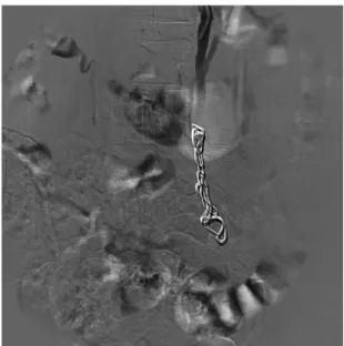
using an average of ive coils with ibers per treated
vein (Figure 2). Coils with diameters from 6 to 10 mm were used with uncontrolled release distally and controlled release in more proximal sites, with minimum oversizing of 20% and the sandwich technique in the distal portion. In just two patients the right gonadal vein was found to be dilated and was embolized. In all of the other patients, the diameter of the right gonadal vein was not large and catheterization was not performed. The patient who had been diagnosed with May-Thurner Syndrome was treated during the same intervention using a 22 × 60 mm Wallstent (R) self-expanding stent, placed in the left common iliac vein. None of the other patients had abnormalities of the iliac veins that required intervention. Vena cava
ilters were not used during the procedures. There
were no intraoperative or immediate postoperative
complications in any of the procedures. All patients were discharged from hospital less than 24 hours after the procedures.
During postoperative follow-up, all patients
reported signiicant improvements in symptoms at the irst follow-up consultation, an average of 7 days
after the procedures. All patients attended at least two postoperative follow-up consultations. Just one of them exhibited mild hematoma at the puncture site. At the time of writing, none had required reintervention or
Table 1. Summary of results of review of medical records for the patients studied.
Age Vaginal
Deliveries
Clinical presentation Diagnostic Method Treatment Follow-up
(Control examinations)
39 3 CPP
Varicose veins in LL
Color Doppler echography L Gonadal V (9coils)
15 mL foam
2 consultations (Color Doppler echography)
44 2 CPP
Incidental inding on CT
CT L Gonadal V (5coils)
R Gonadal V (3coils) 20 mL foam
3 consultations (CT)
43 2 Pelvic heaviness + Recurrent
varicose veins in LL
Color Doppler echography + CT L Gonadal V (5coils)
R Gonadal V (5coils) 30 mL foam
4 consultations (Color Doppler echography)
51 4 CPP + Varicose veins in left
buttock
Color Doppler echography + MRI L Gonadal V (3coils)
10 mL foam
2 consultations (Color Doppler echography)
50 2 CPP + Varicose veins in LL Color Doppler echography L Gonadal V (6coils)
20 mL foam
2 consultations (Color Doppler echography)
49 5 CPP + LLL Edema +
May-hurner Syndrome
Color Doppler echography + MRI L Gonadal V (4coils)
18 mL foam Stent L common iliac V
2 consultations (Color Doppler echography)
CPP= chronic pelvic pain; LL= lower limbs; LLL= left lower limb; CT= computed tomography angiography; MRI= magnetic resonance angiography; V= vein; L= left; R= right.
Figure 1. Digital subtraction venography image of selective
suffered relapse of their symptoms. Postoperative control examinations were conducted using color
Doppler echography in ive patients and CT in one
patient. None of the control examinations found pelvic
varicose veins, conirming successful embolization. All patients considered the inal result satisfactory.
DISCUSSION
The data compiled for this article revealed that all of the patients treated exhibited clinical improvement
and their imaging exams conirmed the eficacy of
embolization. Despite the study’s limitations – the
short follow-up period and the lack of standardization
of postoperative assessments – the results reported
are comparable with those of studies undertaken with
larger samples.
For example, Kies and Kim10 conducted a review of
studies published between 1993 and 2008. They analyzed
12 studies and the results showed signiicant relief
from symptoms, varying from 50 to 100.
Laborda et al.11 followed 202 patients for ive
years with repeated assessments and observed a reduction in visual pain scale scores from 7.34 ± 0.7 (preoperative) to 0.78 ± 1.2 (5 years later). An earlier study conducted by Kim et al.12 with 127 patients and
a similar design reported comparable preoperative visual pain scale scores (7.6 ± 1.8), but the results
observed during follow-up were less signiicant, with
a mean score of 2.9 ± 2.8.
The only randomized study of PCS comparing different types of intervention was published by
Chung and Huh13 and showed that embolization
was superior in several areas, such as reduction of symptoms and length of hospital stay. However, the scarcity of comparative studies means that the choice of the most appropriate technique for the procedure remains controversial. The choice of the most effective method is also dependent on the physician’s preference and experience, the methods available and the treatment setting.
Once embolization has been chosen, the most accepted approach recommends bilateral intervention in both gonadal veins, since this is associated with better results. Notwithstanding, the right gonadal
vein very often has a small diameter and is dificult to
see, which was the case in four out of the six patients studied here. In these circumstances treatment of the left gonadal vein only is acceptable, since with these
anatomic indings it is unlikely that the right gonadal
vein is responsible for the symptoms.6
None of the patients described here suffered serious complications. Mild intercurrent conditions observed were hematoma of the puncture site and postoperative abdominal pains, which did not affect
the inal results. The most worrying complication of
this procedure is migration of the coils into pulmonary
circulation and this did not take place in any of the
patients in this series.
Embolization is suggested as treatment of choice for PCS and has a 2B recommendation, according to the Society for Vascular Surgery and the American Venous Forum.14 However, more widespread use of
this method is primarily prevented by underdiagnosis
of PCS and the lack of more precise indications and of deinitions of symptoms considered suficiently
important to justify intervention.15
In this study, embolization proved to be a safe and effective method. Even so, we believe that technological advances and improvements to the
materials employed could improve the inal results still
further. Additionally, use of protocols standardizing diagnostic investigations, the treatment approach and
analysis of results could deine groups of patients
who would beneit even more from this method.16
REFERENCES
1. Liddle AD, Davies AH. Pelvic congestion syndrome: chronic pelvic pain causes by ovarian and internal iliac varices. Phlebology. 2007;22(3):100-4. http://dx.doi.org/10.1258/026835507780807248. PMid:18268860.
2. Asciutto G, Asciutto KC, Mumme A, Geier B. Pelvic venous incompetence: reflux patterns and treatment results. Eur J Vasc Endovasc Surg. 2009;38(3):381-6. http://dx.doi.org/10.1016/j. ejvs.2009.05.023. PMid:19574069.
Figure 2. Digital subtraction venography image after embolization
3. Ignacio EA, Dua R 4th, Sarin S, et al. Pelvic congestion syndrome: diagnosis and treatment. Semin Intervent Radiol. 2008;25(4):361-8. http://dx.doi.org/10.1055/s-0028-1102998. PMid:21326577. 4. Kurklinsky AK, Rooke TW. Nutcracker phenomenon and nutcracker
syndrome. Mayo Clin Proc. 2010;85(6):552-9. http://dx.doi. org/10.4065/mcp.2009.0586. PMid:20511485.
5. Lou WS, Gu JP, He X, et al. Endovascular treatment for iliac vein compression syndrome: a comparison between the presence and absence of secondary thrombosis. Korean J Radiol. 2009;10(2):135-43. http://dx.doi.org/10.3348/kjr.2009.10.2.135. PMid:19270859.
6. Freedman J, Ganeshan A, Crowe PM. Pelvic congestion syndrome: the role of interventional radiology in the treatment of chronic pelvic pain. Postgrad Med J. 2010;86(1022):704-10. http://dx.doi. org/10.1136/pgmj.2010.099473. PMid:21106807.
7. Rane N, Leyon JJ, Littlehales T, Ganeshan A, Crowe P, Uberoi R. Pelvic congestion syndrome. Curr Probl Diagn Radiol. 2013;42(4):135-40. http://dx.doi.org/10.1067/j.cpradiol.2012.11.002. PMid:23795992. 8. Park SJ, Lim JW, Ko YT, et al. Diagnosis of pelvic congestion
syndrome using transabdominal and transvaginalsonography. AJR Am J Roentgenol. 2004;182(3):683-8. http://dx.doi.org/10.2214/ ajr.182.3.1820683. PMid:14975970.
9. Coakley FV, Varghese SL, Hricak H. CT and MRI of pelvic varices in women. J Comput Assist Tomogr. 1999;23(3):429-34. http:// dx.doi.org/10.1097/00004728-199905000-00018. PMid:10348450.
10. Kies DD, Kim HS. Pelvic congestion syndrome: a review of current diagnostic and minimally invasive treatment modalities. Phlebology. 2012;27(Supl 1):52-7. http://dx.doi.org/10.1258/phleb.2012.012S27. PMid:22312068.
11. Laborda A, Medrano J, Blas I, Urtiaga I, Carnevale FC, de Gregorio MA. Endovascular treatment of pelvic congestion syndrome: visual analog scale (VAS) long-term follow-up clinical evaluation in 202 patients. Cardiovasc Intervent Radiol. 2013;36(4):1006-14. http:// dx.doi.org/10.1007/s00270-013-0586-2. PMid:23456353. 12. Kim HS, Malhotra AD, Rowe PC, Lee JM, Venbrux AC. Embolotherapy
for pelvic congestion syndrome: long-term results. J Vasc Interv Radiol. 2006;17(2 Pt 1):289-97. http://dx.doi.org/10.1097/01. RVI.0000194870.11980.F8. PMid:16517774.
13. Chung MH, Huh CY. Comparison of treatments for pelvic congestion syndrome. Tohoku J Exp Med. 2003;201(3):131-8. http://dx.doi. org/10.1620/tjem.201.131. PMid:14649734.
14. Gloviczki P, Comerota AJ, Dalsing MC, et al. The care of patients with varicose veins and associated chronic venous diseases: clinical practice guidelines of the Society for Vascular Surgery and the American Venous Forum. J Vasc Surg. 2011;53(5 Supl):2S-48S. http://dx.doi.org/10.1016/j.jvs.2011.01.079. PMid:21536172.
15. Smith PC. The outcome of treatment for pelvic congestion syndrome. Phlebology. 2012;27(Supl 1):74-7. http://dx.doi. org/10.1258/phleb.2011.012S01. PMid:22312071.
16. Nicholson T, Basile A. Pelvic congestion syndrome, who should we treat and how? Tech Vasc Interv Radiol. 2006;9(1):19-23. http:// dx.doi.org/10.1053/j.tvir.2006.08.005. PMid:17145481.
*
Correspondence
Gilberto do Nascimento Galego Rua Menino Deus, 63, Bloco A, sala 504 - Centro CEP 88020-210 - Florianópolis (SC), Brazil Tel.: +55 (48) 3322-1043 E-mail: [email protected]
Author information
GNG and PGS - Professors of the Department of Surgery, Universidade Federal de Santa Catarina (UFSC). CTB and RNF - Vascular Surgeons, Clínica Coris Medicina Avançada. TMR - Medical student, Universidade Federal de Santa Catarina (UFSC).
Author contributions
Conception and design: GNG Analysis and interpretation: GNG, TMR Data collection: TMR, GNG, RNF Writing the article: TMR, GNG Critical revision of the article: TMR, GNG, PGS, CTB, RNF Final approval of the article*: TMR, GNG, PGS, CTB, RNF Statistical analysis: TMR, GNG, PGS, CTB, RNF Overall responsibility: GNG