Faculdade de Ciências
Departamento de Química e Bioquímica
Study of the Nonsense-Mediated Decay (NMD)
Mechanism in the Human
α
α
α
α
-Globin mRNA
Francisco José Cabral Pereira
Doutoramento em Bioquímica
(Genética Molecular)
2009
Tese orientada por:
Doutora Luísa Romão Loison (Instituto Nacional de Saúde Dr. Ricardo Jorge) Professora Doutora Margarida D. Amaral (Faculdade de Ciências da
Prefácio
O trabalho de investigação descrito nesta tese de Doutoramento foi realizado na Unidade de Investigação e Desenvolvimento do Departamento de Genética do Instituto Nacional de Saúde Dr. Ricardo Jorge, sob a orientação da Doutora Luísa Romão Loison, investigadora principal neste Instituto, e co-orientação da Prof. Doutora Margarida Amaral, professora auxiliar com agregação na Faculdade de Ciências da Universidade de Lisboa.
Com este estudo pretendeu-se contribuir para o aumento dos conhecimentos adquiridos relativamente ao mecanismo de nonsense-mediated mRNA decay (NMD), usando o mRNA de
α
-globina humana como modelo experimental, e comparando o seu perfil de submissão ao NMD com o perfil conhecido para o mRNA deβ
-globina. Este trabalho permitiu fornecer novos elementos para a compreensão dos fundamentos bioquímicos e moleculares que determinam as diferenças observadas na susceptibilidade ao NMD entre transcritos equivalentes deα
- eβ
-globina.Em conformidade com o disposto no nº 5 do artigo 41º do Regulamento dos Estudos Pós-Graduados da Universidade de Lisboa, deliberação nº 93/2006, publicado em Diário da República, 2ª série – Nº 209 – 30 de Outubro de 2006, esta tese apresenta-se escrita na língua inglesa e inclui um resumo em português com mais de 1200 palavras (ver Resumo).
De acordo com o nº 1 do mesmo artigo do referido Regulamento, na elaboração desta tese foi aproveitado o resultado de trabalho já publicado e obtido em colaboração, estando, neste caso, a minha contribuição pessoal devidamente indicada. Efectivamente, o trabalho descrito possibilitou a publicação de dois artigos científicos e um comentário, em revistas internacionais especializadas:
Pereira FJC, Silva MC, Picanço I, Seixas MT, Ferrão A, Faustino P, Romão L. (2006) Human alpha2-globin nonsense-mediated mRNA decay induced by a novel alpha-thalassaemia frameshift mutation at codon 22. Br J Haematol 133:98-102.
Silva AL, Pereira FJC, Morgado A, Kong J, Martins R, Faustino P, Liebhaber SA, Romão L. (2006) The canonical UPF1-dependent nonsense-mediated mRNA decay is inhibited in transcripts carrying a short open reading frame independent of sequence context. RNA 12:2160-2170.
Inácio A, Silva AL, Morgado A, Pereira FJC, Lavinha J, Romão L. (2007) Comment on 'Nonsense-mediated mRNA decay modulates clinical outcome of genetic disease'. Eur J Hum Genet 15:533-534.
Este trabalho foi, em grande parte, financiado pela Fundação para a Ciência e a Tecnologia (FCT) no âmbito de dois projectos de investigação (POCI/BIA-BCM/59140/2004 e POCI/SAU-MMO/57573/2004), através do Programa de Financiamento Plurianual do Centro de Investigação em Genética Molecular Humana (CIGMH), e na forma de uma Bolsa de Doutoramento com a referência SFRH/BD/14273/2003.
A Doutora Luísa Romão Loison, orientadora deste trabalho, desempenhou um papel fundamental na boa prossecução do mesmo, acompanhando de perto toda a sua evolução. As suas frequentes ideias e sugestões profícuas, bem como o seu empenho e capacidade de motivação, favoreceram muito este estudo. Agradeço-lhe por me acolher no seu grupo e me permitir desenvolver um trabalho estimulante num ambiente aprazível. Agradeço ainda por me transmitir alguma da sua sabedoria, competência, espírito crítico e rigor científico, pelo constante incentivo, disponibilidade, e por todo o apoio e dedicação. Faço questão de salientar também as suas notáveis qualidades humanas: a sua generosidade, amizade e preocupação genuína que sempre demonstrou.
Agradeço à Professora Doutora Margarida Amaral por aceitar a co-orientação deste trabalho de Doutoramento, pelo seu apoio, disponibilidade, e pelo interesse com que seguiu o progresso do estudo.
O Professor Stephen Liebhaber, da Universidade da Pensilvânia, contribuiu consideravelmente para o desenvolvimento deste trabalho através das suas sábias e valiosas sugestões, e de estimulantes e proveitosas discussões científicas.
Ao Presidente do Conselho Directivo do Instituto Nacional de Saúde Dr. Ricardo Jorge, Prof. Doutor José Pereira Miguel, bem como ao Coordenador do Departamento de Genética deste Instituto, Prof. Doutor Luís Nunes, manifesto o meu apreço.
Agradeço à Doutora Maria Guida Boavida e ao Doutor João Lavinha por me terem recebido no então Centro de Genética Humana, permitindo a realização do meu trabalho, e por todo o interesse que demonstraram pelo mesmo.
Aos meus colegas de laboratório (também de laboratórios vizinhos e de alguns mais afastados), agradeço o importante apoio que muitos me deram no trabalho prático, as discussões científicas mais descontraídas, o agradável ambiente de trabalho, toda a paciência, a cumplicidade e a amizade.
Acknowledgements
The largest part of the present work was financed by Fundação para a Ciência e a Tecnologia (FCT) in the form of two grants (POCI/BIA-BCM/59140/2004 and POCI/SAU-MMO/57573/2004), through Programa de Financiamento Plurianual of Centro de Investigação em Genética Molecular Humana (CIGMH) and a doctoral fellowship with the reference SFRH/BD/14273/2003.
Dr. Luísa Romão Loison, supervisor of this work, performed an essential role in its good accomplishment, by closely following its whole evolution. Her frequent ideas and useful suggestions, as well as her interest and enthusiasm, greatly helped this study. I am grateful for being allowed to join her research group and developing a stimulating work in a pleasant environment. I am also grateful for being able to receive her excellent scientific guidance and motivation, and for all her support and dedication. Her generosity, friendship and genuine concern with people are among her outstanding human qualities.
I thank Prof. Margarida Amaral for having accepted the co-supervision of my PhD, for all her kind support and her interest in following the progress of this study.
Prof. Stephen Liebhaber, from the University of Pennsylvania, has considerably contributed to this work with his wise and valuable suggestions, and through inspiring and productive scientific discussions.
I also thank Dr. Maria Guida Boavida and Dr. João Lavinha for receiving me in the former Centro de Genética Humana, allowing me to develop this study, and for all the interest they have kindly shown about my work.
To my colleagues in the lab (also those from neighbor labs or from more distant ones), I thank their valuable help in my work, the relaxed scientific discussions, the cheerful working environment, all their patience, support and friendship.
Index
Prefácio iii Acknowledgements vii Index ix Resumo xiii Palavras-chave xviii Abstract xix Keywords xx Abbreviations xxiChapter I – General Introduction 23
I.1. Human globin genes 25
I.2. Eukaryotic mRNA translation processes 29
I.2.1. Initiation 29
I.2.2. Elongation 31
I.2.3. Termination and recycling 32
I.2.4. Reinitiation 33
I.3. Nonsense-mediated mRNA decay 34
I.3.1. PTC recognition 35
I.3.1.1. A rule for PTC recognition in mammals 36
I.3.1.1.1. Exceptions 37
I.3.1.2. The exon-junction complex 38 I.3.1.3. Main NMD trans-acting factors 42 I.3.1.4. The singular first round of translation 44
I.3.2. NMD activation 45
I.3.3. Degradation mechanisms for nonsense-mutated mRNA 47 I.3.4. Poly(A)-binding protein influence on NMD 49 I.3.5. Subcellular localization of NMD 51
I.3.6. Physiological gene expression regulation by NMD 53 I.3.7. Clinical perspective of NMD 54
Chapter II – Human
α
2-globin nonsense-mediated mRNA decay induced by a novelα
-thalassemia frameshift mutation at codon 22 59Remark 61
II.1. Summary 63
II.2. Introduction 63
II.3. Patient and Methods 64
II.3.1. Hematological studies 64 II.3.2. DNA sequence analysis 65 II.3.3. Construction of expression vectors 65 II.3.4. Cell culture and transfections 65
II.3.5. RNA isolation 66
II.3.6. Reverse transcriptase-PCR analysis 66 II.3.7. Ribonuclease protection assays (RPA) 66
II.4. Results and Discussion 67
II.5. Acknowledgements 71
Chapter III – The extension of NMD inhibition in human
α
- andβ
-globin transcripts bearing a short ORF is modulated by its secondarystructure 73
Remark 75
III.1. Summary 77
III.2. Introduction 77
III.3. Materials and Methods 79
III.3.1. Construction of expression vectors 79 III.3.2. Cell culture and transfections 85 III.3.3. Transfection of siRNA 85
III.3.5. Ribonuclease protection assay (RPA) 86 III.3.6. Western blot analysis 87 III.3.7. Semi-quantitative RT-PCR 87
III.3.8. Luminometry assay 88
III.4. Results and Discussion 88 III.4.1. The AUG-proximity effect occurs in human
α
-globin mRNA andextends further downstream when compared with the same effect
in
β
-globin 88III.4.2. Translation reinitiation plays a modest role concerning NMD
inhibition in human
α
-globin transcripts carrying a PTC in exon 1 93 III.4.3. Forα
-globin transcripts carrying a PTC at the 3’-part of exon 1,extended NMD inhibition depends on the upstream sequence 98 III.4.4. The secondary structure of the ORF influences NMD efficiency for
α
- andβ
-globin transcripts carrying a PTC at the 3’-part of exon 1 101III.5. Acknowledgements 107
Chapter IV – General Discussion 109
IV.1. General Discussion 111
IV.2. Concluding remarks 116
IV.3. Future perspectives 117
Resumo
Um controlo rigoroso da qualidade da expressão dos genes após a transcrição contribui para o funcionamento adequado das células eucarióticas. O mecanismo de decaimento do RNA mensageiro (mRNA) mediado pela existência de codões que determinam a terminação prematura da tradução (codões nonsense), denominado nonsense-mediated mRNA decay (NMD), é um exemplo de mecanismo que actua na verificação do mRNA após a transcrição. Ao eliminar transcritos portadores de mutações nonsense, este mecanismo impede a produção de proteínas truncadas, evitando os efeitos dominantes negativos, resultantes da acumulação de péptidos não funcionais (Maquat 2004; Chang et al. 2007; Neu-Yilik e Kulozik 2008).
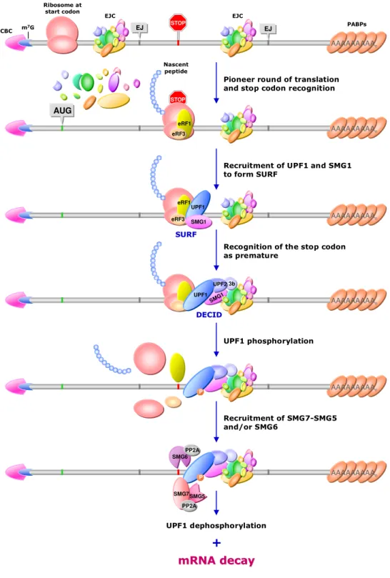
Nas células de mamíferos, o NMD é influenciado pela ocorrência de splicing (processo de remoção de sequências não codificantes – intrões) e depende da tradução. Aquando da remoção dos intrões, no núcleo da célula, ocorre a deposição de um complexo proteico no mRNA, cerca de 20-24 nucleótidos (nt) a montante de cada junção exão-exão, chamado exon-junction complex (EJC). Este complexo permanece associado ao mRNA durante o seu transporte para o citoplasma e durante pelo menos os primeiros passos da tradução. Considera-se que ao longo da primeira ronda de tradução, o ribossoma remove os EJCs que se encontram na porção codificante do transcrito. Assim, admite-se que se o ribossoma progredir suficientemente no mRNA, removerá todos os EJCs. Para que tal aconteça, é necessário que o codão de terminação da tradução se encontre a 3’ de um ponto situado 50-55 nt a montante da última junção exão-exão (Nagy e Maquat 1998). Quando o ribossoma não atinge esse ponto durante a tradução, pelo menos um EJC permanecerá associado ao transcrito, o que conduz à activação do NMD.
Os factores que promovem a correcta terminação da tradução (eRFs – eukaryotic release factors), eRF1 e eRF3, recrutam um factor essencial para a ocorrência de NMD, a proteína UPF1, para o complexo de terminação no final da primeira ronda de tradução, após o reconhecimento do codão de terminação (Czaplinski et al. 1998;
Wang et al. 2001; Hosoda et al. 2005; Behm-Ansmant e Izaurralde 2006; Kashima et al. 2006). Na maioria dos casos, quando existe um EJC ligado ao mRNA a jusante do codão de terminação, ocorre interacção entre a UPF1 e as proteínas UPF2 e/ou UPF3, componentes do EJC. Aparentemente como resultado desta interacção, a proteína cinase SMG1 promove a fosforilação da UPF1 (Kashima et al. 2006; Wittmann et al. 2006). No seu estado forforilado, a UPF1 consegue estimular a acção do complexo SMG5-SMG7 e/ou do factor SMG6, que se associam à fosfatase PP2A (Anders et al. 2003; Chiu et al. 2003; Ohnishi et al. 2003; Unterholzner e Izaurralde 2004; Fukuhara et al. 2005). A PP2A vai promover a posterior desfosforilação da UPF1 e pensa-se que os factores SMG5, 6 e 7 induzem a activação do mecanismo que conduz à degradação do mRNA (Unterholzner e Izaurralde 2004). Além disso, o SMG6 tem acção endonucleolítica e consegue mediar directamente a degradação de transcritos que contêm mutação nonsense (Huntzinger et al. 2008; Eberle et al. 2009), o que sugere que este factor pode ser responsável pela iniciação do processo de decaimento acelerado.
Por outro lado, estudos recentes efectuados com células humanas, têm vindo a atribuir um papel importante à PABPC1 [cytoplasmic poly(A)-binding protein 1 – proteína citoplasmática de ligação à cauda poli(A) 1] no impedimento da ocorrência de NMD e no aumento de eficiência da terminação da tradução (Eberle et al. 2008; Ivanov et al. 2008; Silva et al. 2008; Singh et al. 2008). De facto, a proximidade da PABPC1, no momento em que o ribossoma atinge o codão de terminação, permite a sua interacção com o eRF3, impedindo a ligação da UPF1 ao complexo de terminação (Uchida et al. 2002; Kashima et al. 2006). Desta forma o NMD não é activado e o transcrito é conservado, mesmo que exista um EJC situado a jusante. Esta actuação da proteína de ligação à cauda poli(A), na promoção de uma terminação correcta e na inibição do NMD, parece ocorrer em todos os eucariotas em que estes mecanismos têm sido estudados, pelo que se considera que esta função da PABPC1 terá sido conservada ao longo da evolução (Ivanov et al. 2008; Singh et al. 2008).
Estão descritos diversos exemplos de transcritos que não correspondem ao modelo, segundo o qual o codão de terminação será ou não reconhecido como prematuro,
de acordo com a sua posição relativamente à última junção exão-exão (Buzina e Shulman 1999; Romão et al. 2000; Asselta et al. 2001; Perrin-Vidoz et al. 2002; Wang et al. 2002; Bühler et al. 2004; Denecke et al. 2004; Inácio et al. 2004; Harries et al. 2005; Silva et al. 2006). No caso da
β
-globina humana, transcritos defeituosos que transportam mutações nonsense localizadas na parte 3’ do primeiro exão e em grande parte do segundo, conduzem o mRNA ao decaimento acelerado (Thermann et al. 1998; Zhang et al. 1998), encontrando-se de acordo com o referido modelo. Pelo contrário, mRNAs com mutações nonsense situadas na região 5’ do primeiro exão, apresentam um nível de acumulação nas células semelhante ao do mRNA deβ
-globina sem mutações (Romão et al. 2000; Inácio et al. 2004). Nestes casos demonstrou-se que a razão pela qual os transcritos estão a escapar ao NMD não está relacionada com uma alteração ao nível do splicing, com tradução deficiente ou com a ocorrência de reiniciação da tradução, mas sim com a proximidade do codão de terminação ao codão de iniciação da tradução, AUG (Inácio et al. 2004). Assim, a este resultado deu-se o nome de “efeito de proximidade ao AUG”.O gene de
α
-globina humana provém do mesmo ancestral que o deβ
-globina, e ambos partilham diversas características, como a sua organização geral, a ocorrência de permuta na expressão de formas embrionárias dos genes para formas fetais ou adultas, bem como a estrutura e função das proteínas que codificam (Choi e Engel 1988; Enver et al. 1990; Higgs et al. 1998; Zhang et al. 2002). Contudo, o mRNA deα
-globina contém um maior conteúdo de citidinas e guanosinas na sua sequência (Tuan e Forget 1980; Fischel-Ghodsian et al. 1987), e manifesta uma eficiência de tradução inferior à do mRNA deβ
-globina (Shakin e Liebhaber 1996; Chaisue et al. 2007). Estes factos indiciavam uma possível diferença no padrão de susceptibilidade ao NMD entre transcritos deα
- eβ
-globina com codões de terminação prematuros.Assim, com o estudo descrito na presente dissertação pretendeu-se verificar a ocorrência de NMD em transcritos de
α
-globina humana portadores de mutações nonsense; conhecer o nível de acumulação de mRNA deα
-globina com mutações nonsense em posições diferentes da sua região codificante; investigar apossibilida-de possibilida-de reiniciação da tradução após a terminação num codão prematuro; e averiguar a razão para possíveis diferenças no padrão de NMD entre
α
- eβ
-globina.O estudo iniciou-se com a análise do efeito de uma delecção pontual (–C) desco-nhecida, no codão 22 de um dos alelos do gene 2 de
α
-globina. Dados hematológi-cos indicavam a existência deα
-talassemia neste caso clínico, e a análise da sequência de nucleótidos no mRNA produzido revelou que a delecção conduzia ao aparecimento de um codão de terminação prematuro. Os níveis de acumulação do transcrito nestas condições indicavam que este era rapidamente degradado, pelo mecanismo de NMD, evitando-se a formação de uma proteína incompleta.A análise do nível de acumulação celular e da estabilidade do mRNA de
α
-globina, contendo mutações nonsense individuais em diferentes posições ao longo da região codificante, revelou uma inibição do NMD em transcritos mutados no primeiro exão. Esta inibição mostrou-se mais extensa que a encontrada emβ
-globina, verificando-se também para mutações situadas mais a jusante emα
-globina. Ao contrário do previamente observado no mRNA deβ
-globina (Inácio et al. 2004), demonstrou-se que ocorre reiniciação da tradução no codão 32 (AUG) deα
-globina, após a tradução de uma grelha de leitura curta. Por experiências com interferência de RNA, verificou-se que a inibição do NMD é atenuada pelo impedimento da reiniciação da tradução nos codões em que esta pode ocorrer. Contudo, este aumento da eficiência do NMD não suprime totalmente a inibição original, uma vez que não coloca os níveis de acumulação dos transcritos de acordo com os observados nos casos de ocorrência de NMD típico, pelo que a reiniciação da tradução não é suficiente para justificar a inibição do NMD. Estudos de luminescência, com genes híbridos deα
- ouβ
-globina e luciferase de pirilampo, demonstraram que a reiniciação da tradução ocorre eficientemente no mRNA deα
- mas não no deβ
-globina.Para investigar a origem da diferente extensão da inibição do NMD, em transcritos de
α
- eβ
-globina com mutações no exão 1, procedeu-se à substituição da região não traduzida a 5’ (5’-untranslated region – 5’-UTR) ou de toda a sequência amontante do codão nonsense, no mRNA de
α
-globina, pelas sequências equivalentes deβ
-globina; o procedimento recíproco foi aplicado em transcritos deβ
-globina. Os resultados obtidos indicam que a sequência a montante da mutação nonsense, mas não apenas a 5’-UTR, determina o nível de acumulação do mRNA.Adicionalmente, alterou-se a estrutura secundária da região codificante de transcri-tos de
α
- eβ
-globina com grelhas de leitura curtas, com o objectivo de alterar a duração da tradução. A introdução de uma sequência que induz a formação de uma estrutura secundária complexa, na grelha de leitura deβ
-globina, leva à redução da velocidade da tradução. Como consequência, o nível de acumulação de um mRNA, que apresentava elevada estabilidade, diminui devido à activação do NMD. Pelo contrário, a ausência de estrutura secundária na grelha de leitura leva a um aumento significativo da acumulação de um transcrito deβ
-globina, com mutação nonsense na parte 3’ do exão 1, que era originalmente degradado por NMD. Por sua vez, um codão de terminação prematuro na região 3’ do exão 1 deα
-globina não induz a activação eficiente do NMD, e esta inibição torna-se bem mais evidente quando a estrutura secundária é abolida. A aceleração da tradução de uma grelha de leitura pequena emα
-globina também potencia o aumento da taxa de reinicia-ção da tradureinicia-ção. Contudo, o aumento da inibireinicia-ção do NMD acontece independente-mente da reiniciação da tradução.Em síntese, no estudo descrito confirmou-se a ocorrência de NMD no mRNA de
α
-globina humana, verificou-se a inibição do NMD em transcritos portadores de codões de terminação no primeiro exão do transcrito, e mostrou-se que a tradução pode reiniciar-se no mRNA deα
-globina, após parar prematuramente. Em relação à diferente extensão observada para a inibição do NMD, entre transcritos deα
- eβ
-globina com grelhas de leitura curtas, verificou-se que esta diferença tem origem na natureza da sequência a montante do codão de terminação; em particular a estrutura secundária da grelha de leitura parece desempenhar um papel importante, provavelmente pelo seu efeito na velocidade da tradução.Sabendo-se que a PABPC1 interage com factores de iniciação da tradução (Kahvejian et al. 2001, 2005; Martineau et al. 2008) e que estes podem manter-se associados ao ribossoma nos primeiros momentos da elongação (Kozak 2001; Jackson 2005), os resultados aqui expostos sugerem que uma tradução muito breve pode permitir a proximidade da PABPC1 no momento da terminação, causando a supressão do NMD. Igualmente, a presença de determinados factores de iniciação da tradução no momento da sua terminação, aumenta a capacidade de reiniciação em
α
-globina.Este trabalho representa um importante progresso no seu domínio de investigação fundamental, inclui resultados já publicados em dois artigos científicos, e outros que serão submetidos para publicação em breve, promovendo a difusão da cultura científica. Contribui também com potenciais benefícios para a saúde, na medida em que as conclusões deste estudo poderão fundamentar a criação de estratégias terapêuticas aplicadas, por exemplo, em casos de NMD adverso, nos quais a produção de uma proteína incompleta é mais benéfica do que a sua eliminação. Nestes casos, uma terapia que favorecesse a aceleração da tradução, por abolição da estrutura secundária, ou o fortalecimento das interacções que permitem a permanência da PABPC1 associada ao ribossoma durante a elongação, poderiam ter um efeito benéfico para o doente.
Palavras-chave
mRNA de globinas humanas
tradução
codão de terminação prematuro
decaimento do mRNA mediado por codões nonsense
estrutura secundária
Abstract
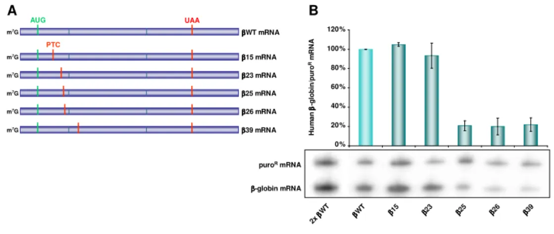
Eukaryotic cells have developed mechanisms to regulate many steps of gene expression, preventing defective protein production and ensuring cell survival. Nonsense-mediated mRNA decay (NMD) is a posttranscriptional surveillance pathway able to recognize and eliminate transcripts encoding truncated proteins. In mammalian cells, mRNAs containing premature translation termination codons (PTCs) placed more than 50-55 nucleotides upstream from the last exon-exon junction are generally committed to NMD. However, transcripts carrying a nonsense codon in this condition, located near the translation initiation codon, have been reported to escape decay.
The present work contains the description of a clinical case where a PTC, caused by a single nucleotide deletion in the human
α
-globin mRNA, induces accelerated mRNA degradation. On the other hand, data reveals that stop codons located in the first exon of theα
-globin transcript fail to induce NMD. This resistance to decay is further downstream-extended when compared with the equivalent effect observed in the humanβ
-globin mRNA, and this divergence is shown to be mainly determined by the sequence upstream to the PTC and the secondary structure of the open reading frame (ORF). Contrary toβ
-globin, theα
-globin mRNA allows translation reinitiation at a downstream AUG, after translation of a short ORF. However, NMD inhibition persists even when translation reinitiation is prevented. Results suggest that a less-structured short ORF is more rapidly translated and NMD-antagonistic factors, such as PABPC1, may remain associated with the ribosome when it reaches the stop codon.This work constitutes a basic research development with confirmed and potential scientific diffusion, and contributes with prospective benefits in public health, since its results may provide targets for new therapeutic strategies for genetic disorders caused by PTC-generating mutations.
Keywords
human globin mRNA
translation
premature termination codon
nonsense-mediated mRNA decay
secondary structure
Abbreviations
A adenosine
A-site aminoacyl-site
ATP adenosine triphosphate
ATPase adenosine triphosphatase
BMD Becker muscular dystrophy
bp base pair
C cytidine
CBC cap-binding complex
CBP cap-binding protein
cDNA mRNA-complementary DNA
C-terminal carboxy-terminal
DCP decapping protein
DECID decay-inducing complex
DMD Duchenne muscular dystrophy
DMEM Dulbecco’s modified Eagle’s medium
DNA deoxyribonucleic acid
DNase deoxiribonuclease
dNTP deoxinucleoside triphosphate
eEF eukaryotic translation elongation factor
eIF eukaryotic translation initiation factor
EJC exon-junction complex
eRF eukaryotic translation release factor
E-site exit-site
G guanosine
GAPDH glyceraldehyde-3-phosphate dehydrogenase
GDP guanosine diphosphate
GTP guanosine triphosphate
GTPase guanosine triphosphatase
Hb hemoglobin
HPLC high-performance liquid chromatography
HS hypersensitive site
IEF isoelectric focusing
Ig immunoglobulin
kb kilobase
LCR locus control region
Luc luciferase
MEL mouse erythroleukemia
MEM minimal essential medium
Met methionine
Met-tRNAi methionine-loaded initiator transfer RNA
mRNA messenger ribonucleic acid
mRNP messenger ribonucleoprotein
NMD nonsense-mediated mRNA decay
N-terminal amino-terminal
ORF open reading frame
PABP poly(A)-binding protein
PABPC1 cytoplasmic poly(A)-binding protein 1
PAGE polyacrilamide gel electrophoresis
Paip1 poly(A)-binding protein-interacting protein 1
PARN poly(A) ribonuclease
P-bodies processing bodies
PCR polymerase chain reaction
PIK phosphatidylinositol kinase
PIN PilT N-terminus
pk pseudoknot
poly(A) poly-adenilate
PP2A protein phosphatase 2A
pre-mRNA precursor messenger ribonucleic acid
P-site peptidyl-site
PTB polypyrimidine tract binding protein
PTC premature translation-termination codon
puroR puromycin-resistance
PVDF polyvinylidene difluoride
RNA ribonucleic acid
RNAi RNA interference
RNase ribonuclease
RNP ribonucleoprotein
RPA ribonuclease-protection assay
RT reverse transcription
RT-PCR reverse transcriptase-polymerase chain reaction
SKI superkiller
SDS sodium dodecyl sulphate
siRNA short interfering RNA
SMG suppressor with morphogenetic effects on genitalia
SSCP single-stranded conformation polymorphism
SURF SMG1-UPF1-eRFs complex
T thymidine
Tet tetracycline
TCR T-cell receptor
tRNA transfer ribonucleic acid
U uridine
uORF upstream open reading frame
UPF up-frameshift
UTR untranslated region
CHAPTER I
General Introduction
I.1. Human globin genes
The oxygen required for cell function is carried through the blood by hemoglobin, present in the erythrocytes – the red blood cells. Hemoglobin is a tetrameric protein composed by two identical
α
-like and twoβ
-like globin chains, each one linked to a heme group. These components of hemoglobin are encoded by two sets of genes located at two distinct gene clusters, where the genes are organized in tandem, ordered according to their developmental expression (Tanimoto et al. 1999; Tang et al. 2006). The ancestral ofα
- andβ
-globin genes diverged from their common ancestor probably 500 million years ago, having evolved and being modified by multiple genetic processes, including duplication events (Higgs et al. 1989, 1998).The human
α
-globin gene cluster, extended over nearly 80 kilobases (kb) in the telomeric region of the short arm of chromosome 16 (16p13.3), includes seven genes organized in the order telomere(5’)-ζ
-ψζ
-ψα
2-ψα
1-α
2-α
1-θ
-centromere(3’) (Figure I.1), whereψ
represents unexpressed pseudogenes (Higgs et al. 1998; Zhang et al. 2002). Their transcriptionally regulated expression results in a developmentally controlled switch in theα
-like globin gene being expressed:ζ
-globin in the embryonic stage andα
2- andα
1-globin throughout fetal and post-natal life. Theθ
-globin gene is also expressed in the adult stage although at a very low level and its function is still unknown (Marks et al. 1986; Cooper et al. 2005). Evidence of a very low erythroid-specific fetal expression ofψα
2 pseudogene was recently described and this newly discoveredα
-like globin gene was namedµ
-globin (Goh et al. 2005). Bothµ
- andθ
-globin genes exhibit minimal transcription levels when compared with the mainα
genes, and their deletion has no reported consequences on the clinical phenotype (Fei et al. 1988; Goh et al. 2005). Throughout fetal and adult life the twoα
-globin genes are highly expressed, producing an identical protein. However the levels of messenger RNA (mRNA) and protein synthesis from theα
2-globin locus are approximately 2.6 fold higher than those from theα
1-globin locus (Liebhaber et al. 1986; Albitar et al. 1992). Alpha-globin cluster genes specific erythroid transcription is controlled and strongly enhanced by a DNase I hypersensitive site(HS) located 40 kb upstream from
ζ
-globin gene, named HS-40 (Jarman et al. 1991; Bernet et al. 1995). HS-40 is actually placed in the intron of a ubiquitously expressed gene (Vyas et al. 1992).Figure I.1. Structural genes contained in the human αααα- and ββββ-globin clusters. Their positioning order and relative distances on chromosomes 16 and 11, respectively, are illustrated.
Green squares represent genes; orange squares represent pseudogenes. Adapted from Waggoner and Liebhaber (2003); chromosomes from Bank (2005).
The
β
-globin gene locus, spanning about 70 kb on the short arm of chromosome 11 (11p15.5), includes six genes arranged in the same orientation and in the order of their expression in erythroid development: 5’-ε
-Gγ
-Aγ
-ψβ
-δ
-β
-3’ (Figure I.1) (Tanimoto et al. 1999). Transcription of theε
gene in the embryonic yolk sac switches after 6–8 weeks of gestation to the transcription of the twoγ
genes in the fetal liver. A second switch occurs shortly before birth, to the transcription of theδ
(minor adult) and
β
(major adult) genes (Choi and Engel 1988; Enver et al. 1990).ψβ
is an unexpressed pseudogene. Upstream of theβ
-globin cluster is theβ
locus control region (β
LCR) which comprises five developmentally stable DNase I hypersensitive sites (designated HSs 1–5) located between 5 and 25 kb 5’ of theε
-globin gene0 kb 10 20 30 0 kb 10 20 30 40 β ββ β-globin cluster α αα α-globin cluster Chromosome 16 Chromosome 11 ζ ψζ ψα ζ ψζ ψα ζ ψζ ψα ζ ψζ ψα2 ψαψαψαψα1 ααα2 αα ααα1 θθθθ ε ε ε ε Gγ γ γ γ Aγ ψβ δ β γ ψβ δ β γ ψβ δ β γ ψβ δ β 5’ 5’ 3’ 3’
(Tuan et al. 1985). The
β
LCR plays a crucial role in gene expression by creating an active chromatin domain surrounding the cluster (Forrester et al. 1986) and acting as the enhancer for each one of theβ
-like globin genes.Hemoglobin represents up to 95% of the total soluble protein content of erythrocytes. Throughout the different stages of human development, the distinct globin genes expressed produce hemoglobin tetramers that assemble different subunits. During the embryonic stage, the erythropoiesis is performed in the yolk sac and hemoglobin is primarily composed of
ζ
2ε
2 (two zeta- and two epsilon-globinchains – Hb Grower 1) and later of
ζ
2γ
2 (Hb Portland) orα
2ε
2 (Hb Grower 2). Theerythrocytes generated in the fetal liver produce fetal hemoglobin (HbF) which consists of
α
2γ
2. Near birth time, erythropoiesis migrates to the bone marrow andthe hemoglobin produced includes
α
2β
2 (HbA), which represents 97%–98% of totaladult hemoglobin, and
α
2δ
2 (HbA2), composing the remainder 2%–3% (Ho and Thein2000; Waggoner and Liebhaber 2003).
Being two notably related gene clusters in evolution and function, human
α
- andβ
-globin genes are in very different chromosomal environments. Theα
-globin genes are situated in a gene-rich telomeric region with a constitutively open chromatin structure (characteristic of transcriptionally active genes) present in all cell types, and the genes have methylation-free CpG islands (Vyas et al. 1992). Theβ
-globin cluster is AT-rich, has no CpG islands and assumes an open chromatin structure only in erythroid cells (Arapinis et al. 1986). Furthermore, the two clusters have different control regions and different developmental switching mode: there is only one switching in theα
-globin gene cluster from the embryonic to the fetal stage of development, whereasβ
-globin cluster has one additional switching from the fetal to the adult stage. Despite great differences, expressions ofα
- andβ
-globin loci are temporally and quantitatively co-regulated to produce balanced amounts of protein to form stage-specific hemoglobin tetramers for oxygen transport.All globin genes are composed of three exons separated by two introns. Exons 1 and 3 are flanked by 5’ and 3’ non-coding sequences, respectively; the untranslated regions (UTR) (Huisman 1993). The human
α
-globin exon 1 comprises a 37-bp 5’-UTR and a 95-bp coding region, exon 2 includes 205 bp, and exon 3 contains 129 bp of coding region and 108 bp of 3’-UTR. Introns 1 and 2 have 117 bp and 142 bp, respectively. Together,α
-globin exons and introns constitute an 833-bp long gene. Concerning theα
-globin mRNA, it is composed of 574 nt with a coding sequence of 429 nt that encodes a chain of 141 amino-acids. Exon 1 contains the first 31 codons, then the second exon holds 68 codons, and the last one has 42 codons.The
β
-globin gene is 1610 bp long: exon 1 includes a 50-bp 5’-UTR and a 92-bp coding region, exon 2 has 223 bp, and exon 3 comprises a coding region of 129 bp and a 3’-UTR of 132 bp. Introns 1 and 2 contain 132 bp and 852 bp, respectively. In addition, theβ
-globin intron-free mRNA is composed of 626 nt, with a coding sequence of 444 nt that encodes a 146 amino-acid chain with a molecular weight of about 16 kDa. The first exon contains 30 codons, followed by 74 codons in the second exon, and 42 codons in exon 3 (Huisman 1993).Globin mRNAs present an extraordinary stability revealed by exceptionally long degradation half-lives, estimated between 16 and 48 hours, whereas the majority of mRNAs show half-lives that vary from minutes to a few hours. This outstanding stability allows very high accumulation levels of globin transcripts (more than 95% of all cellular mRNA) in terminally differentiated erythrocyte precursors (Russell and Liebhaber, 1996).
Reduced or absent synthesis of
α
- orβ
-globin chains of hemoglobin, due to mutations in the genes or in their regulatory sequences, results in thalassemia, the most common group of genetically inherited monogenic disorders (Gu and Zeng 2002; Mahajan et al. 2007). More important than the low hemoglobin production are the harmful effects caused by the imbalance of globin chains. Inα
-thalassemia, genetic defects that cause an insufficientα
-globin synthesis lead to the accumulation ofuncomplexed and unstable
γ
orβ
chains and a consequent hemolytic anemia (Liebhaber et al. 1986). Mostα
-thalassemia determinants are deletions involving one or both of the duplicatedα
-globin genes, although a growing number of point mutations have been described (Bernini and Harteveld 1998). Theβ
-thalassemias are characterized by reduced or absent production of hemoglobinβ
-chains. This deficit results in an excess of extremely unstableα
-globin chains; they precipitate in premature red blood cells causing hemolysis and ineffective erythropoiesis (Wheatherall 2001).I.2. Eukaryotic mRNA translation processes
I.2.1. Initiation
Protein synthesis starts by a complex process intended to properly position the ribosome at the translation start codon (AUG) of the transcript (Figure I.2). Eukaryotic mRNA translation begins with the formation of a ternary complex composed by the eukaryotic translation initiation factor (eIF) 2 coupled with GTP, and the methionine-loaded initiator transfer RNA (Met-tRNAi). This complex,
together with eIFs 1, 1A, 3 and 5, binds the small (40S) ribosomal subunit to generate the 43S pre-initiation complex. Meanwhile, eIF4F is thought to attach to the mRNA at the 5’-terminal cap structure (m7GpppN, where m denotes a methyl
group, p denotes a phosphate group and N denotes any nucleotide) and to trigger mRNA unwinding (Jackson 2005; Pestova and Hellen 2006). The eIF4F complex includes several proteins such as eIF4E, which directly binds to the cap structure; eIF4A, an RNA helicase that, stimulated by eIF4B, is thought to unwind secondary structures in the 5′-UTR; and eIF4G, a scaffold protein used as a platform for the assembly of other initiation factors, that interacts with eIF3, 4E and 4A (Gebauer and Hentze 2004).
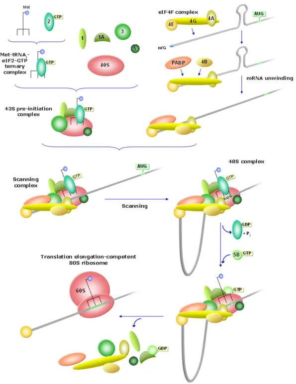
Figure I.2. Translation initiation. The methionine-loaded initiator transfer RNA (Met-tRNAi) binds
eIF2 coupled with GTP and forms a ternary complex, which, together with eIFs 1, 1A, 3 and 5, binds the small (40S) ribosomal subunit to create the 43S pre-initiation complex. Presumably, eIF4F complex binds the mRNA at the 5’-cap (m7
G) and recruits PABP and eIF4B. PABP may also bind the 3’-poly(A) tail and promote the formation of a closed-loop mRNP structure (not considered in the diagram). eIF4B induces eIF4A to trigger mRNA unwinding. The 43S complex joins the mRNA due to the interaction between eIF3 and the central domain of eIF4G, which promotes scanning. When the scanning complex reaches an AUG triplet, forms the 48S initiation complex. Then, eIF5 stimulates GTP hydrolysis and release of eIF2-GDP, and eIF1A recruits eIF5B-GTP to catalyze 60S subunit joining and the displacement of initiation factors, to form the translation elongation-competent 80S ribosome.
At least in mammals, the poly(A)-binding protein (PABP) also binds eIF4G and this interaction appears to be stabilized by the direct binding of PABP-interacting protein 1 (Paip1) to PABP and eIF3 (Kahvejian et al. 2001, 2005; Martineau et al. 2008). Besides, the simultaneous interaction of eIF4E and PABP with eIF4G is believed to circularize the mRNA, taking the 3′ UTR in close proximity to the 5′ end of the transcript (Wells et al. 1998; Sachs and Varani 2000; Amrani et al. 2008).
Moreover, the interaction between eIF3 and the central domain of eIF4G seems to promote loading of the 43S complex on the mRNA and the beginning of scanning. The 5’-most AUG triplet, influenced by the surrounding sequence, is usually chosen to be occupied by the anticodon of the Met-tRNAi (Pestova et al. 2001; Jackson 2005).
Start codon selection seems to be controlled by eIF1 and eIF1A, which stabilize a 43S conformation such that opens the mRNA binding channel during scanning and allow it to close upon start codon recognition (Fekete et al. 2007; Passmore et al. 2007; Mitchell and Lorsch 2008). Once at the start codon, the 43S complex forms a stable structure known as the 48S initiation complex, in which codon-anticodon base-pairing occurs (Pestova and Hellen 2006). Then, eIF5 stimulates GTP hydrolysis and release of eIF2–GDP, and eIF1A helps recruit eIF5B to the 48S complex to catalyze 60S subunit joining (Fringer et al. 2007), with the displacement of initiation factors, to form an active elongation-competent 80S ribosome, able to catalyze the formation of the first peptide bond (Pestova et al. 2000; Unbehaun et al. 2004).
I.2.2. Elongation
In peptide-chain elongation, amino-acids are added one by one and organized according to the nucleotide sequence of the mRNA. There are three tRNA-binding sites in the ribosome: the aminoacyl (A) site receives the new aminoacyl-tRNA specific for the newly encountered mRNA codon; the peptidyl (P) site harbors the tRNA with the peptide chain being formed; and the exit (E) site retains the deacylated tRNA prior to its release (Proud 1994). The eukaryotic translation
elongation factor (eEF) 1A recruits aminoacyl-tRNAs to the ribosomal A-site, in a GTP-dependent manner. The regeneration of active eEF1A-GTP complexes is promoted by eEF1B. The large ribosomal subunit catalyzes peptide bond formation connecting the aminoacyl-tRNA with the peptidyl-tRNA in the P-site, which causes repositioning of the peptide chain to the tRNA in the A-site. Then, in another GTP-dependent process, eEF2 catalyzes the translocation of the peptidyl-tRNA from the A- to the P-site and of the deacylated tRNA from the P- to the E-site, and also the movement of the ribosome relative to the mRNA by exactly three nucleotides, placing the following codon in the A-site prepared to receive the next aminoacyl-tRNA (Browne and Proud 2002; Abbott and Proud 2004).
I.2.3. Termination and recycling
When the ribosomal A-site reaches one of the possible three termination codons (UAA, UAG or UGA; also referred to as stop or nonsense codons) in-frame, hydrolysis of the peptidyl-tRNA ester bond occurs in the 60S ribosomal subunit, releasing the new polypeptide. This hydrolysis determines translation termination and depends on eukaryotic translation release factors (eRFs) 1 and 3 (Kisselev et al. 2003). The structure of eRF1 is similar to that of a tRNA molecule, which allows it to act like a tRNA and bind to the A-site of the ribosome (Frolova et al. 2000; Nakamura and Ito 2003). In the majority of eukaryotes, eRF1 recognizes all three stop codons (Frolova et al. 1999; Song et al. 2000) and interacts with a GTP-binding factor, eRF3, with GTPase activity (Chavatte et al. 2003). Mammalian eRF3 comprises two proteins, eRF3a and eRF3b (Kisselev and Buckingham 2000; Kisselev et al. 2003), but their function is not well understood. eRF1 and eRF3 form a stable complex through mutually binding their C-terminal domains, and cooperate in promoting fast hydrolysis of the ester bond between the polypeptide chain and the tRNA in the ribosomal P-site (Merkulova et al. 1999; Alkalaeva et al. 2006).
After completing translation of an open reading frame (ORF), post-termination complexes may be recycled subsequent to release of the mRNA and the deacylated tRNA, and to dissociation of the ribosome into subunits. These ribosomal subunits may become re-engaged in translation initiation on the same or another mRNA molecule (Rajkowitsch et al. 2004). Following release of the newly synthesized peptide, translation initiation factors 3, 1, 1A, and 3j, a subunit of eIF3, appear to promote recycling of eukaryotic post-termination complexes. eIF3 seems to be enhanced by eIFs 1, 1A and 3j in promoting post-termination ribosome-splitting into 60S subunits and tRNA- and mRNA-bound 40S subunits. eIF1 also catalyzes release of P-site deacylated tRNA, whereas eIF3j ascertains subsequent dissociation of the mRNA (Pizarev et al. 2007).
I.2.4. Reinitiation
Following translation-termination of a short upstream open reading frame (uORF), the 60S ribosomal subunit is supposedly released, whereas the 40S subunit remains bound to the mRNA, it may resume scanning and initiate another round of translation at a downstream AUG codon (Kozak 2002). For translation reinitiation, the 40S subunit must acquire a new Met-tRNAi, which may be easier when there is a
long intercistronic distance, providing more time for Met-tRNAi to bind (Kozak 1987;
Abastado et al. 1991). Reinitiation may occur after translation of a small ORF but not after translation of a long one. This may be due to the fact that some initiation factors dissociate from the ribosome only gradually during the course of elongation. Thus, if the elongation phase is brief, the factors required for reinitiation may still be present when the 40S subunit resumes scanning. It seems to be not so much the actual length of the ORF itself, but the time taken to translate it, what determines whether there is a resumption of scanning (Kozak 2002; Pöyry et al. 2004).
The induction of a pseudoknot structure, known to slow elongation (Somogyi et al. 1993), prevents reinitiation after translation of a short uORF that normally leads to
downstream reinitiation (Kozak 2001). This suggests that the initiation factors remain ribosome-associated for a few seconds during translation of the short ORF. The resumption of scanning and reinitiation may depend on the involvement of eIF4F in translation initiation of the short ORF and probably the upholding of the interaction between eIF4F (or at least the central one-third fragment of eIF4G) and the ribosome while the ORF is being translated (Pöyry et al. 2004). Also the retention of eIF3 on the ribosome during a uORF translation appears to be necessary for reinitiation. In fact, an interaction between the N-terminal domain of eIF3a and sequences 5’ of the uORF seem critically required to enhance reinitiation (Szamecz et al. 2008). If the mentioned interactions persist until termination of uORF translation, the associated factors can then promote further 40S subunit scanning and downstream reinitiation. Conversely, if the initiation factors detach prior to uORF translation ending, then both ribosomal subunits probably dissociate from the mRNA immediately after uORF translation termination (Jackson 2005).
I.3. Nonsense-mediated mRNA decay
Accurate regulation is crucial for proper gene expression, given the complexity of the biochemical processes involved. Eukaryotic gene expression includes a series of interrelated steps, starting with the mRNA precursor (pre-mRNA) being transcribed from DNA, followed by its processing with removal of introns (splicing) and addition of the 5’-cap structure and the 3’-poly(A) tail (polyadenylation); and the mature mRNA is then exported to the cytoplasm where it is translated to protein and finally degraded (Behm-Ansmant et al. 2007a; Chang et al. 2007). These steps are mutually integrated to increase their efficiency and fidelity (Orphanides and Reinberg 2002; Reed 2003), and cells have evolved mechanisms to control many of the steps, in order to prevent inappropriate gene expression and to ensure cell survival (Moore 2005).
Nonsense-mediated mRNA decay (NMD) is one of the best-studied quality control mechanisms of eukaryotic gene expression (Stalder and Mühlemann 2008).
This posttranscriptional surveillance mechanism exists in every eukaryotic cell that has been studied. It uses an elaborate network of nuclear and cytoplasmic processes to recognize and eliminate mRNAs which contain premature translation termination codons (PTCs or nonsense codons). Otherwise, the translation of PTC-containing transcripts would result in the production of C-terminally truncated nonfunctional proteins that might, instead, acquire deleterious gain-of-function or dominant–negative effects (Chang et al. 2007; Neu-Yilik and Kulozik 2008).
PTCs can arise from nonsense and frameshift point mutations (nucleotide insertions or deletions) in the DNA sequence, transcriptional errors and aberrant splicing events. Other sources are the existence of a uORF, the failure to incorporate selenocysteine at a UGA codon, or the insertion of a transposon or a retrovirus. Nonsense codons could also appear as a result of alternative splicing or missplicing, producing an intron-derived nonsense codon or a shift in the ORF that creates a downstream nonsense codon (Lejeune and Maquat 2005; Chang et al. 2007; Neu-Yilik and Kulozik 2008). Some genes, such as T-cell receptor (TCR) and immunoglobulin (Ig) genes, are subject to programmed DNA rearrangements to multiply their variety of antigen receptors. Two thirds of these rearrangements create a frameshift that may lead to a PTC at a downstream position (Li and Wilkinson 1998).
I.3.1. PTC recognition
To assure an appropriate mRNA surveillance function, the NMD machinery has to distinguish between premature and physiological termination codons. The mechanism used for this discrimination is still only partially understood (Chang et al. 2007). The ability of a nonsense codon to elicit rapid degradation of the mRNA depends on its location relative to downstream sequence elements and associated
proteins (Lejeune and Maquat 2005; Amrani et al. 2006; Eberle et al. 2008; Mühlemann 2008; Singh et al. 2008).
I.3.1.1. A rule for PTC recognition in mammals
The position effect for PTC recognition is evident in mammalian cells, where PTCs placed in the 3’-most exon or in the 3’-part of the penultimate exon, usually fail to elicit NMD, whereas more upstream-located PTCs direct the mutated transcript to rapid decay (Neu-Yilik and Kulozik 2008).
During pre-mRNA splicing (non-coding intron removal and exon-exon ligation), a multiprotein gathering named “exon-junction complex” (EJC) is deposited 20-24 nucleotides (nt) 5’ to the exon-exon junctions and remains associated to the mRNA throughout export to the cytoplasm and translation initiation (Kataoka et al. 2000; Le Hir et al. 2000a,b, 2001; Tange et al. 2004). Besides several other factors, EJCs recruit up-frameshift (UPF) proteins, which are required for NMD (Maquat 2004). If at least one EJC is located downstream to the translation stop codon, it functions as a spatial reference point for the discrimination between a premature and a physiological stop. Conversely, EJCs located within the ORF enhance translation (Nott et al. 2004; Wilkinson 2005; Kunz et al. 2006).
Generally, nonsense codons located more than 50-55 nt upstream of the 3’-most exon-exon junction elicit NMD (Figure I.3) (Nagy and Maquat 1998); downstream to this boundary or in an unspliced transcript, a stop codon is considered normal and the mRNA is preserved (Maquat and Li 2001; Neu-Yilik et al. 2001: Brocke et al. 2002). Consistent with this “50-55 nt boundary rule” is the fact that most natural termination codons lie in the final exons of the transcript (Nagy and Maquat 1998).
It is believed that ribosomes are capable of removing the EJCs that they encounter during translation elongation. As translation stops at a nonsense codon situated more than 50-55 nt upstream of an exon-exon junction, the translating ribosome
will not have progressed far enough along the mRNA to displace the EJC deposited near that exon-exon junction (Alkalaeva et al. 2006). On the other hand, it is thought that a ribosome that has reached a nonsense codon located either less than 50-55 nt upstream of an exon-exon junction or downstream from it will have removed the EJC (Dostie and Dreyfuss 2002; Lejeune et al. 2002).
Figure I.3. The rule for PTC recognition in mammalian mRNA establishes that only translation-termination codons located more than 50-55 nt upstream from the 3’-most exon-exon junction are considered as premature and elicit NMD.
The diagram represents a schematic mRNA with the 5’-cap structure (m7
G), exon-exon junctions (EJ) and the poly(A) tail (fading ‘A’s). The translation-initiation codon (AUG) is indicated in green and the boundary situated 50-55 nt upstream from the 3’-most exon-exon junction is indicated in yellow.
I.3.1.1.1. Exceptions
Despite the majority of the mammalian transcripts studied present an NMD behavior that follows the established rule, there are many reported examples of transcripts that disobey. PTCs within
β
-globin exon 1 fail to efficiently elicit NMD despite residing more than 50-55 nt upstream of an exon-exon junction (Romão et al. 2000; Danckwardt et al. 2002; Silva et al. 2006). Other expected NMD substrates have been reported to escape degradation, such as nonsense-mutated transcript of fibrinogen Aα
- orγ
-chains (Asselta et al. 2001; Neerman-Arbez et al. 2004), or BRCA1 (Perrin-Vidoz et al. 2002).NMD can be triggered by PTCs located very early in the ORF; a single peptide bond formation is sufficient to elicit NMD (Lew et al. 1998). However, there are many reports of nonsense-mutated transcripts that present a low NMD sensitivity when the PTC is located close to the 5’ end of the ORF (Buzina and Shulman 1999; Romão et al. 2000; Asselta et al. 2001; Perrin-Vidoz et al. 2002; Denecke et al. 2004; Inácio et al. 2004; Harries et al. 2005; Silva et al. 2006). In some cases, this results from translation reinitiation at a downstream AUG (Zhang and Maquat 1997; Lew et al. 1998; Buzina and Shulman 1999; Buisson et al. 2006; Paulsen et al. 2006), but in others, the mechanism appears to involve proximity of the PTC to the initiator AUG (Romão et al. 2000; Inácio et al. 2004; Silva et al. 2006).
Nonsense mutations located only 8-10 nt upstream of the 3’-most exon-exon junction of Ig
µ
and TCR-β
transcripts effectively trigger NMD, therefore breaking the rule (Wang et al. 2002; Bühler et al. 2004). In fact, these transcripts display a peculiar NMD behavior, since PTCs that reside closer to the 5’ end of the penultimate exon elicit NMD more efficiently than PTCs that reside near the 3’ end of the exon (Wang et al. 2002), being the only known mammalian mRNAs to exhibit a type of polar NMD. Another presumable case of EJC-independent NMD comes from studies withβ
-hexosaminidase (Rajavel and Neufeld 2001).I.3.1.2. The exon-junction complex
An exclusive feature of mammalian NMD is the involvement of the EJC. The nearly 350-kDa protein complex deposited about 20-24 nucleotides upstream of exon-exon junctions during pre-mRNA splicing, is composed of several factors (Le Hir et al. 2000a,b; Tange et al. 2004). The EJC is a dynamic structure that associates with the mRNA in the nucleus and remains bound, but modifies its protein composition, throughout mRNA processing, export to the cytoplasm, and translation (Dreyfuss et al. 2002; Jurica and Moore 2003; Reed 2003; Maquat 2004). The core of the EJC, composed by eIF4AIII, MAGOH, Y14 and MLN51 (Andersen et al. 2006; Bono et al. 2006), provides a
platform for the dynamic assembly (and disassembly) of many additional nuclear and cytoplasmic factors (Figure I.4) that perform functions in mRNA transport, subcellular localization, translation and turnover (Maquat 2004; Tange et al. 2004, 2005; Merz et al. 2007).
Figure I.4. EJC composition. Y14, MAGOH, eIF4AIII and MLN51 constitute the core of the complex and provide a platform for the binding of additional mRNA export factors, splicing activators and other associated proteins.
The eukaryotic translation initiation factor 4AIII (eIF4AIII) is an RNA helicase that binds directly to the mRNA and is thought to anchor the complex to the exon junction (Shibuya et al. 2004). MAGOH (a human homolog of Drosophila mago nashi protein) is an mRNA export factor (Shi and Xu 2003; Isken and Maquat 2007). Y14 is another mRNA export factor that interacts directly with the C-terminal domain of eIF4AIII (Ballut et al. 2005; Shibuya et al. 2004) and also interacts with UPF3a or UPF3b, necessary for NMD. Y14 forms a stable heterodimer with MAGOH and together inhibit the ATPase activity of eIF4AIII to stabilize its interaction with other EJC components and its binding to mRNA (Fribourg et al. 2003; Lau et al. 2003; Ballut et al. 2005). Even though Y14 contains an RNA-binding domain, its direct and very high
affinity interaction with MAGOH probably blocks its binding to the mRNA (Chang et al. 2007). MLN51 (also called Barentsz, BTZ) associates with and stimulates the RNA-helicase activity of eIF4AIII, and stabilizes its binding to RNA (Palacios et al. 2004; Ballut et al. 2005). Its ability to directly bind single-stranded RNA in the absence of eIF4AIII, suggests that MLN51 may function as an EJC-independent factor in some situations (Ballut et al. 2005).
RNPS1 (RNA-binding protein prevalent during S phase) is a splicing co-activator that also functions in mRNA export (Luo and Reed 1999; Mayeda et al. 1999; Zhou et al. 2000; Le Hir et al. 2001a) and it may recruit UPF3a or UPF3b to the EJC (Lykke-Andersen et al. 2001; Schwerk et al. 2003; Gehring et al. 2005). SRm160 (serine/arginine-related matrix protein of 160 kDa) also functions in splicing and enhances mRNA export (Luo and Reed 1999; Zhou et al. 2000; Le Hir et al. 2001a). UAP56 (U2 snRNP auxiliary factor-associated protein of 56 kDa) is an RNA helicase that functions in splicing and mRNA export (Gatfield et al. 2001; Luo et al. 2001). ACINUS forms a stable heterodimer with RNPS1 (Schwerk et al. 2003). SAP18 binds RNPS1 but its function as an EJC factor is unknown (Zhou et al. 2000; Schwerk et al. 2003). PININ also binds RNPS1 and is involved in mRNA export (Li et al. 2003). REF (RNA export factor; also known as ALY) is an mRNA-binding protein which interacts with UAP56 and Y14 (Zhou et al. 2000; Conti and Izaurralde 2001). TAP (tyrosine-kinase-interacting protein-associated protein; also called NXF1 – nuclear export factor 1) is also an mRNA export factor, loosely associated with the EJC, is recruited by REF and interacts with MAGOH and components of the nuclear pore complex (Stutz et al. 2000; Le Hir et al. 2001a; Stutz and Izaurralde 2003; Custódio et al. 2004). PYM is an RNA-binding protein which forms a trimeric complex with Y14-MAGOH (Bono et al. 2004). The NMD factors (characterized in I.3.1.3. Main NMD trans-acting factors, below) UPF2, UPF3a and UPF3b are also EJC components (Ishigaki et al. 2001; Kim et al. 2001a; Lejeune et al. 2002).
EJC factors are recruited to and released from a spliced mRNA at different times. In fact, some EJC proteins associate to the pre-mRNA even before splicing: REF can bind pre-mRNA before splice-site cleavage (Reichert et al. 2002). Others, such as
RNPS1, SRm160 and UPF3b, associate before exon-exon ligation (Reichert et al. 2002; Kataoka and Dreyfuss 2004). The EJC core factors eIF4AIII and Y14-MAGOH are recruited during or immediately after exon-exon ligation (Jurica et al. 2002; Reichert et al. 2002; Jurica and Moore 2003). UPF3b is assumed to remain bound to the EJC and to recruit UPF2 during or shortly after export on the cytoplasmic side of the nuclear pore. Some EJC proteins, such as RNPS1, dissociate during or shortly after mRNA export (Kim et al. 2001b; Le Hir et al. 2001a,b). Others, such as Y14-MAGOH, probably remain associated with the mRNA until the ribosome displaces them during the first round of translation (Dostie and Dreyfuss 2002). Whether the ribosome just physically removes the EJC or also biochemically transforms it to facilitate dissociation remains unclear. However, Y14 phosphorylation appears to be required for the dissociation of some EJC factors (Hsu et al. 2005).
Several lines of evidence indicate that translation in mammalian cells is stimulated by the EJC factors UPF2, UPF3a, UPF3b, RNPS1, Y14 and MAGOH (Wiegand et al. 2003; Nott et al. 2004; Wilkinson 2005). This translational enhancement appears to require efficient splicing (Gudikote et al. 2005; Le Hir and Séraphin 2008; Ma et al. 2008). Therefore, it has been suggested that the EJC is not required for PTC identification in mammals (Bühler et al. 2006), but merely functions as an NMD enhancer (Mühlemann 2008; Mühlemann et al. 2008). Other theories even postulate that the influence of EJCs on NMD is reduced to the enhancement of translation, which consequently leads to an improved turnover rate of nonsense-mutated transcripts (Hilleren and Parker 1999; Culbertson and Neeno-Eckwall 2005; Ford et al. 2006). In fact, although EJC factors UPF2, UPF3, RNPS1, eIF4AIII, MLN51, Y14 and MAGOH have been shown to play a role in mammalian NMD (Kim et al. 2001a; Le Hir et al. 2001a; Lykke-Andersen et al. 2001; Fribourg et al. 2003; Gehring et al. 2003, 2005; Ferraiuolo et al. 2004; Palacios et al. 2004; Shibuya et al. 2004), the activation of this pathway can occur independently of some of these EJC components, since UPF2-, UPF3-, RNPS1- or MLN51-independent NMD evidences have been reported (Gehring et al. 2003, 2005; Chan et al. 2007).
I.3.1.3. Main NMD trans-acting factors
Several proteins have been shown to be essential for NMD. In addition to some of the above-mentioned EJC components, the UPF (up-frameshift) proteins comprise the conserved core of the NMD system. They were originally discovered in Saccharomyces cerevisiae and more recently identified in higher eukaryotes (Leeds et al. 1992; Cui et al. 1995; Lee and Culbertson 1995). The SMG (suppressor with morphogenetic effects on genitalia) proteins are also fundamental NMD factors, by regulating the activity of UPF1.
The complex phosphoprotein UPF1 harbors multiple domains and is recruited to mRNAs following translation termination (Wang et al. 2001; Kobayashi et al. 2004; Kashima et al. 2006), where it interacts with eRF3 and eRF1 (Czaplinski et al. 1998). UPF1 also interacts with UPF2 and the cap-binding protein, CBP80 (Hosoda et al. 2005). It holds RNA/DNA-dependent ATPase activity and ATP-dependent 5’-to-3’ helicase activity, which are poorly understood but appear to be required for NMD (Weng et al. 1996; Bhattacharya et al. 2000). As expected for a protein involved in translation termination, human UPF1 is mainly cytoplasmic, although it shuttles between the nucleus and the cytoplasm (Mendell et al. 2002).
UPF2 is also a phosphoprotein (Wang et al. 2006) which bridges UPF1 with the EJC, and is capable of interacting simultaneously with both UPF1 and UPF3, through distinct binding domains (He et al. 1997; Lykke-Andersen et al. 2000; Mendell et al. 2000; Serin et al. 2001). Moreover, human UPF2 has one UPF1-binding domain in each of its termini (N and C), and both are required for NMD (He et al. 1996; Kadlec et al. 2004). UPF2 is a cytoplasmic protein that accumulates in the perinuclear region (Lykke-Andersen et al. 2000; Mendell et al. 2000).
Mammals own two UPF3 genes; human UPF3a (named also UPF3) is located on chromosome 13, and UPF3b (also known as UPF3x) is on the X chromosome (Lykke-Andersen et al. 2000; Serin et al. 2001). UPF3 proteins are mainly nuclear but shuttle to the cytoplasm. They both interact with Y14 to recruit UPF2 to the EJC (Gehring et al.
2003), however UPF3b interacts more strongly than does UPF3a. This difference in UPF3 proteins behavior is provided by the presence of an arginine residue in the C terminus of UPF3b, but not in UPF3a, which renders UPF3b the legitimate NMD effector, when judge against UPF3a (Kunz et al. 2006).
The human SMG proteins (SMG1, 5, 6 and 7) preserve characteristics identical to those attributed to their ancestors, initially identified in Caenorhabditis elegans. They determine the phosphorylation condition of UPF1 (Wilkinson 2003) and all are required for NMD (Pulak and Anderson 1993).
SMG1 is a PIK (phosphatidylinositol kinase)-related protein kinase that catalyzes the phosphorylation of UPF1, at specific serine residues, which is essential for NMD (Denning et al. 2001; Yamashita et al. 2001). This phosphorylation requires UPF2 or UPF3b – both factors can function independently (Gehring et al. 2005; Chan et al. 2007) – and may stimulate the dissociation of eRF3 from UPF1 (Pal et al. 2001; Grimson et al. 2004; Kashima et al. 2006; Wittmann et al. 2006). Assuming the opposite function of SMG1, the other three SMG factors are non-redundant proteins that instigate the dephosphorylation of UPF1 (Page et al. 1999; Ohnishi et al. 2003). SMG5, 6 and 7 bind phosphorylated UPF1 through a binding site for phosphoserine residues (14-3-3-like domain), present in each one of the three proteins (Fukuhara et al. 2005). Since none of these factors are phosphatases themselves, they appear to recruit the protein phosphatase 2A (PP2A) to mediate UPF1 dephosphorylation (Anders et al. 2003; Chiu et al. 2003; Unterholzner and Izaurralde 2004). SMG5 and SMG7 seem to work together; interact through their N-terminal domains to form a stable heterodimer and establish a larger complex with PP2A and phosphorylated UPF1 (Anders et al. 2003; Chiu et al. 2003; Ohnishi et al. 2003). In contrast, SMG6 appears to act more independently, yet also forms a complex with PP2A and phosphorylated UPF1 (Ohnishi et al. 2003; Unterholzner and Izaurralde 2004). Both SMG5 and SMG6 contain a C-terminal PIN (PilT N-terminus) domain, which is associated with ribonuclease activity. The PIN domain of SMG5 lacks essential catalytic residues and is inactive, but SMG6 presents effective RNA degradation activity (Glavan et al. 2006).