UNIVERSIDADE DE TRÁS-OS-MONTES E ALTO DOURO
In vitro and in vivo evaluation of the biological
effects of Celuvein
®and its components
Dissertação de Mestrado em Biotecnologia para as Ciências da Saúde
Sara Judite Pais Sario
Orientadora: Professora Doutora Isabel O'Neill de Mascarenhas Gaivão
Coorientadora: Professora Doutora Amélia Maria Lopes Dias da Silva
I
UNIVERSIDADE DE TRÁS-OS-MONTES E ALTO DOURO
In vitro and in vivo evaluation of the biological
effects of Celuvein
®and its components
Dissertação de Mestrado em Biotecnologia para as Ciências da Saúde
Sara Judite Pais Sario
Orientadora: Professora Doutora Isabel O'Neill de Mascarenhas Gaivão
Coorientadora: Professora Doutora Amélia Maria Lopes Dias da Silva
Composição do Júri:
Professora Doutora Estela Maria Bastos Martins de Almeida
Professora Doutora Isabel O’Neill de Mascarenhas Gaivão
Professora Doutora Paula Filomena Martins Lopes
Professora Doutora Cristina Maria Silveira Silva Pereira Wilson
II The ideas here exposed are the author’s full responsibility.
III
Acknowledgements
Ao Magnífico Reitor, Professor Doutor António Fontainhas Fernandes, da Universidade de Trás-os-Montes e Alto Douro pelas facilidades concedidas.
À Professora Doutora Isabel Gaivão e à Professora Doutora Amélia Silva pela orientação, ensinamentos e disponibilidade que sempre demonstraram, pelas palavras de encorajamento e apoio e por acreditarem em mim e no meu trabalho.
Ao Bruno Ponte, por permitir que eu fizesse este estudo com o Celuvein® e pela
disponibilidade que sempre demonstrou para ajudar no que fosse necessário.
À Lisete, da Unidade de Microscopia Eletrónica, pela disponibilidade desde logo mostrada para realizar a caracterização das partículas de dióxido de titânio.
A todos os que ao longo deste trabalho partilharam os laboratórios comigo e, de certa forma, também contribuíram para a realização deste trabalho, não só ajudando quando necessário, mas também por terem tornado o trabalho mais fácil com a boa disposição e companhia.
À Sofia e à Yana, pelos bons momentos, pelas brincadeiras e pelas gargalhadas. Obrigada pela vossa amizade.
Ao Nuno, obrigada pelos projetos, pelos bons momentos, pelo bom humor, por acreditares em mim e por estares sempre disposto a ajudar, seja no que for. Amigos como tu não se encontram facilmente.
À Carolina, obrigada pelas conversas, pelas saídas, pelas boas memórias ao longo destes anos e obrigada por, mesmo não estando em Vila Real estes últimos dois anos, continuares presente.
À Anicia, por estar sempre disposta a ouvir, mesmo que isso significasse sacrificar horas de sono tão preciosas para ela. Obrigada por confiares em mim desde os primeiros dias, pela enorme amizade, pelos bons e maus momentos, por seres como uma irmã mais nova. Obrigada por tudo.
À Bruna, por todo o carinho, palavras de incentivo, por me ensinares a encarar a vida e os problemas com um sorriso na cara. Foram 6 anos cheios de boas memórias, e tenho a certeza que as boas memórias não ficarão por aqui. A tua amizade é, sem dúvida, uma das melhores coisas que levo de Vila Real.
Ao António, obrigada por seres um exemplo, por todas as gargalhadas, todas as saídas, todos os bons momentos (que não foram assim tão poucos) ao longo destes 6 anos de amizade. És a melhor pessoa que alguma vez conheci. Obrigada por seres como um irmão para mim.
IV À Lúcia, que tanto fez por mim. Obrigada por me acolher quando não tinha de o fazer, por me dar uma segunda casa, por cuidar de mim, pelas palavras de incentivo, pela preocupação e pela boa disposição com que sempre me recebeu. Obrigada, acima de tudo, pela amizade, e por nunca me deixar desistir.
Ao Rafael. Acho que não há palavras suficientes para agradecer. Obrigada por me levantares quando eu estava em baixo, por olhares através de mim e saberes exatamente aquilo que penso, aquilo que sinto. Obrigada por me compreenderes, por esperares o tempo que fosse necessário até que eu conseguisse superar os meus medos e inseguranças. Obrigada por nunca me deixares cair e bater no fundo, por estares sempre ao meu lado, por me ajudares em tudo e mais alguma coisa, por fazeres o possível e o impossível para me fazeres feliz. Sei que, sem ti na minha vida, nada disto seria possível. Obrigada por seres a minha pessoa.
Por fim, mas definitivamente não menos importante, aos meus pais. Obrigada por nunca duvidarem que eu era capaz. Sei que nem sempre foi fácil, mas mesmo quando as coisas complicavam, nunca deixaram de acreditar em mim, de fazer de tudo para que eu pudesse continuar, seguir os meus sonhos e cumprir os meus objetivos. Obrigada por me deixarem ser quem eu quero ser, e por sempre apoiarem as minhas decisões, mesmo quando isso significava mover montanhas para tal. Obrigada por serem os melhores pais que alguém pode ter.
V
Resumo
A indústria cosmética está em constante crescimento, com produtos aliados a propriedades farmacêuticas, que surgem rapidamente. O Celuvein® é uma loção cosmética
com propriedades farmacêuticas (cosmecêutico) para o tratamento e melhoramento da celulite e varizes, dois dos problemas mais preocupantes principalmente para mulheres. Os seus ingredientes incluem o Liporeductyl® (uma formulação com cafeína), Algisium® (silanol,
sílica orgânica) e dióxido de titânio (TiO2), um pigmento branco com efeito protetor de raios
ultravioleta (UV), sendo um dos mais usados.
O objetivo deste estudo foi avaliar os efeitos destes três componentes do Celuvein®,
tanto in vivo como in vitro, principalmente o seu potencial citotóxico e genotóxico. Para os estudos in vivo foi usado como modelo animal a Drosophila melanogaster, uma vez que tem várias vantagens para estudos de toxicidade. Foram testados os efeitos na longevidade e prolificidade da Drosophila, bem como o potencial genotóxico, com o teste de mutação e recombinação somática (SMART). Para os estudos in vitro, os efeitos foram estudados em quatro linhas celulares: Caco-2, HaCaT, HepG2 e Raw264.7. A citotoxicidade foi avaliada com base no teste de viabilidade com Alamar Blue, e as células foram expostas durante 24 e 48 h aos compostos. Para determinar a genotoxicidade, foi realizado o ensaio de cometa em ambos os estudos, com neuroblastos de Drosophila nos estudos in vivo, e nas células HepG2 (com e sem FPG) para os estudos in vitro.
Os resultados in vivo mostraram que o Liporeductyl® (acima de 1%) é tóxico para as Drosophila, diminuindo o seu tempo de vida e prolificidade, e os resultados do SMART
também sugerem que este composto tem efeitos mutagénicos neste modelo animal. No entanto, não foram observados danos no DNA. No geral, o Algisium® e o TiO
2 parecem
melhorar a longevidade e prolificidade da Drosophila, contudo os resultados da mutagenicidade foram inconclusivos.
Os resultados in vitro, semelhante ao observado in vivo, sugerem que o Liporeductyl®
é citotóxico em todas as linhas celulares estudadas de um modo dependente da dose, no entanto, nas concentrações testadas no ensaio do cometa em HepG2, não houve qualquer aumento significativo de danos no DNA ou danos oxidativos. O Algisium® foi citotóxico nas
células HaCaT, HepG2 e Raw264.7, com recuperação da viabilidade celular quando a exposição por 24 h foi comparada com as 48 h. Os resultados do ensaio do cometa sugerem que a diminuição de viabilidade às 24 h de exposição está relacionada com danos de DNA e danos oxidativos, uma vez que houve um aumento significativo de ambos. O TiO2
VI qualquer dano oxidativo, mas houve um aumento de dano no DNA na concentração mais elevada.
Estes resultados demonstram a importância dos testes de cosméticos e cosmecêuticos, não só para comprovar a sua eficácia, mas para o seu potencial citotóxico e genotóxico. É importante considerar que as concentrações testadas não refletem as concentrações dos componentes no Celuvein®, logo não se devem fazer extrapolações
diretas. São necessários mais testes para determinar os potenciais efeitos destes componentes no corpo humano.
Palavras-chave: Genotoxicidade, cultura de células, toxicidade, Liporeductyl®, Algisium®,
VII
Abstract
The cosmetic industry is a growing field, with cosmetics starting to emerge with pharmaceutical properties at a fast rate. Celuvein® is a cosmetic lotion with pharmaceutical
properties (cosmeceutical) for the treatment and improvement of cellulite and varicose veins, two of the most concerning problems especially for women. Its ingredients include Liporeductyl® (a formulation with caffeine), Algisium® (an organic silicium) and titanium
dioxide (TiO2), one of the most vastly used white pigments and UV protectors.
The aim of this study was to evaluate the in vivo and in vitro effects of these three components of Celuvein®, especially their cytotoxic and genotoxic potential. For the in vivo
studies the Drosophila melanogaster animal model was used, as it has a lot of advantages for toxicity studies. Effects on longevity and prolificacy of Drosophila were tested, as well as the mutagenic potential, with the Somatic Mutation and Recombination Test (SMART). For the in vitro studies, the effects were studied in four different cell lines: Caco-2, HaCaT, HepG2 and Raw264.7. Cytotoxicity was evaluated based on the Alamar Blue viability assay, and cells were exposed to the components for 24 and 48 h. To assess the genotoxic potential of the components, the comet assay was performed in both studies, with Drosophila neuroblasts for the in vivo effects and with HepG2 cells (with and without FPG) for the in vitro effects.
The in vivo results showed that Liporeductyl® was toxic for Drosophila, decreasing its
lifespan and prolificacy (concentrations above 1%), and the SMART results also suggest that this component has mutagenic effects in this animal model. However, no DNA damage was observed. Algisium® and TiO
2, generally, seemed to improve the longevity and prolificacy of Drosophila, and the results of the mutagenicity tested were mainly inconclusive.
The in vitro results, similarly to what was observed in vivo, suggest that Liporeductyl® is
cytotoxic in all tested cell lines in a dose-dependent mode, though, in the tested concentrations in the HepG2 comet assay, there wasn’t a statistically significant increase in DNA or oxidative damage. Algisium® was cytotoxic in HaCaT, HepG2 and Raw264.7 cells,
with a recovery in cell viability when the 24 h of exposure were compared with the 48 h. The comet assay results suggest that the decrease in cell viability at the 24 h of exposure are related to DNA and oxidative damage, being that there was a statistically significant increase in both. TiO2 was cytotoxic to HaCaT and Raw264.7 cells in a dose-dependent way. No
significant oxidative damage was present, but there was an increase in DNA damage at the highest tested concentration.
These results show the importance of testing cosmetics and cosmeceuticals, not only for their efficacy but also for their cytotoxic and genotoxic potential. It’s important to consider
VIII that the tested concentrations don’t reflect the concentrations of the components in Celuvein®, so no direct extrapolation should be done. More tests are necessary to fully
assess the potential effects of these components on the human body.
Keywords: Genotoxicity, cell culture, toxicity, Liporeductyl®, Algisium®, titanium dioxide, Drosophila melanogaster,
IX General Index Acknowledgements ... 3 Resumo ... 5 Abstract ... 7 Figure Index ...11 Table Index ...14 Abbreviation’s list ...15 Conference Presentations ...18 1. Introduction ... 1
1.1. Venous insufficiency and cellulite ... 1
1.2. Celuvein® and its components ... 2
1.2.1. Liporeductyl® ... 3
1.2.2. Titanium Dioxide ... 6
1.2.3. Algisium C® ... 8
1.3. Skin structure ... 9
1.4. Cell Culture ...11
1.4.1. HaCaT Cell Line ...11
1.4.2. Caco-2 Cell Line ...11
1.4.3. HepG2 Cell Line ...12
1.4.4. Raw264.7 Cell Line ...13
1.5. Drosophila melanogaster ...13
1.6. Cytotoxicity and Genotoxicity ...14
1.7. Somatic Mutation and Recombination Test ...16
1.8. Comet assay ...17
1.9. Objectives ...19
2. Materials and Methods ...20
X
2.2. In vivo studies ...20
2.2.1. Biological material ...20
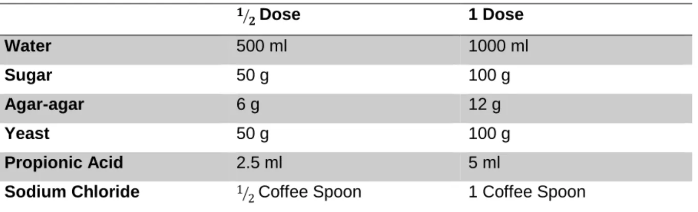
2.2.2. Growth Mediums ...20
2.2.3. Longevity and prolificacy ...22
2.2.4. SMART ...23
2.2.5. Comet assay ...25
2.3. In vitro studies ...27
2.3.1. Cell culture ...27
2.3.2. Culture mediums and solutions ...28
2.3.3. Cell culture maintenance ...29
2.3.4. Subculture and dissociation of adherent cells ...30
2.3.5. Cellular Viability: The Alamar Blue (AB) Viability Test ...32
2.3.6. Comet assay ...34
2.4. Statistical analysis ...35
3. Results...37
3.1. Longevity and Prolificacy ...37
3.2. w/w+ SMART ...39
3.3. Drosophila neuroblasts Comet Assay ...40
3.4. AB Viability Test ...41
3.5. Cell Comet Assay ...45
4. Discussion ...47 4.1. In vivo studies ...47 4.2. In vitro studies ...49 5. Conclusion ...53 6. References ...55 7. Annexes ...65
XI
Figure Index
Figure 1. Types of varicose veins. (A) Spider veins. (B) Reticular veins. (C) True varicose
veins. (Piazza, 2014) ... 1
Figure 2. Dimpling of the skin characteristic of cellulite as a result of the organization of the
adipocytes between fibrous septae (Draelos, 2010). ... 2
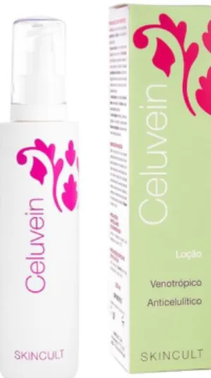
Figure 3. Celuvein® package (Skincult, 2017). ... 3
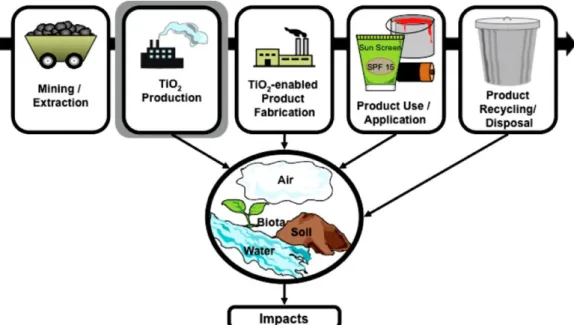
Figure 4. Life cycle of TiO2 and possible environmental impact (Robichaud et al., 2009). ... 8
Figure 5. Steps of the dermal/percutaneous absorption process: Penetration, Permeation
and Resorption (adapted from SCCS, 2016). ... 9
Figure 6. Schematic representation of the skin layers and the vascular network in human
skin, and factors that may influence the penetration of chemicals into and through the skin (Dancik et al., 2015). ...10
Figure 7. Sexual dimorphism in Drosophila melanogaster (Chyb and Gompel, 2013) ...14
Figure 8. DNA damage, repair mechanisms and consequences. a, Common DNA damaging
agents, examples of DNA lesions induced and most relevant DNA repair mechanisms. b, Acute effects of DNA damage (cell cycle and DNA metabolism) and long-term consequences of DNA damage and their biological effects. Abbreviations: cis-Pt (cisplatin), MMC (mitomycin C), PP (6-4 photoproduct), CPD (cyclobutene pyrimidine dimer, HR (homologous recombination), EJ (end joining) (Hoeijmakers, 2001). ...16
Figure 9. Schematic representation of the different genetic processes by which LOH could
occur (adapted from Marcos et al., 2014). ...17
Figure 10. Image obtained in the comet assay (Singh, 2016) ...18
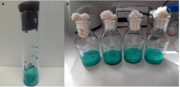
Figure 11. Vials used for the in vivo assays. a, Small tube used for the longevity and
prolificacy assay. b, 200 ml vial used for the SMART and comet assay. ...21
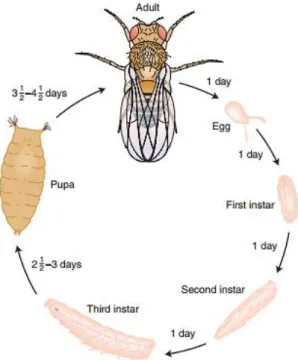
Figure 12. Life cycle of Drosophila melanogaster with different stages and development time
XII
Figure 13. Characteristics of virgin Drosophila melanogaster females (♀, top) and males (♂,
bottom) (Chyb and Gompel, 2013). ...24
Figure 14. Binocular magnifier and optic fiber (only one foci shown) used to observe the
eyes of Drosophila melanogaster. ...25
Figure 15. D. melanogaster brain ganglia and imaginal discs. ...26
Figure 16. Electrophoresis tank used in the comet assay. ...27
Figure 17. Cells used in the in vitro assays. a, Caco-2 cells; b, HaCaT cells; c, HepG2 cells;
d, Raw264.7 cells (ATCC, 2017; CLS, 2017). ...28
Figure 18. Characteristic growth pattern of cultured cells (Thermofisher Scientific Inc., 2016).
...31
Figure 19. Schematic representation of the subculture process. ...32
Figure 20. Conversion of resazurin (oxidized form) to resorufin (reduced form) that leads to
color change in the AB assay (Invitrogen, 2008)...33
Figure 21. Schematic layout of the 96-well plates used for the viability assay. Cells in the
wells from A-D were exposed for 24 h to the components and cells in the wells from E-D were exposed for 48 h. ...34
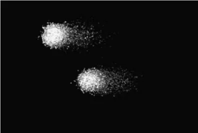
Figure 22. TEM photography of the TiO2 NPs agglomerates. Bars indicate 250 nm. ...37
Figure 23. Percentage of survival per day of Drosophila melanogaster adults exposed to the
indicated components for 72 h. ...38
Figure 24. Number of hatched adults chronically exposed to each concentration of the tested
components. * p<0.05, ** p<0.01. ...39
Figure 25. D. melanogaster eye with mutant spot. ...39
Figure 26. DNA damage expressed as arbitrary units, obtained from the visual scoring of
100 cells per gel (at least 2 gels observed in each concentration). ...41
Figure 27. Alamar blue assay for cell viability in Caco-2 cells after treatment with: (A)
Algisium®, (B) Liporeductyl® and (C) TiO
2 for 24 and 48 h. Cell viability is expressed as
XIII
Figure 28. Alamar blue assay for cell viability in HaCaT cells after treatment with: (A)
Algisium®, (B) Liporeductyl® and (C) TiO
2 for 24 and 48 h. Cell viability is expressed as
percentage of the control (untreated cells). *p<0.05, **p<0.01. ...43
Figure 29. Alamar blue assay for cell viability in HepG2 cells after treatment with: (A)
Algisium®, (B) Liporeductyl® and (C) TiO
2 for 24 and 48 h. Cell viability is expressed as
percentage of the control (untreated cells). *p<0.05, **p<0.01. ...44
Figure 30. Alamar blue assay for cell viability in Raw264.7 cells after treatment with: (A)
Algisium®, (B) Liporeductyl® and (C) TiO
2 for 24 and 48 h. Cell viability is expressed as
percentage of the control (untreated cells). *p<0.05, **p<0.01. ...45
Figure 31. HepG2 cells exposed to 0.1% of Algisium©, with FPG (A) and without (B), stained
with DAPI (400x). ...46
Figure 32. DNA damage expressed as arbitrary units (a.u.) in HepG2 cells. (A) HepG2 cells
XIV
Table Index
Table 1. Ingredients and quantities used for the Drosophila growth medium. ...20
Table 2. Tested concentrations, for the in vivo assays, for each component. ...21
Table 3. Reagents and solutions necessary to perform the comet assay. ...25
Table 4. Tested concentrations for the viability assay for each component. ...30
Table 5. Tested concentrations in the comet assay performed with HepG2 cells. ...35
Table 6. Results of the double-decision X2 based on the acceptance or rejection of H 0 and HA. ...35
Table 7. Percentage of eyes with spots observed in each concentration of the three tested components. (+) Positive; (-) Negative; (i) Inconclusive. ...40
XV
Abbreviation’s list
%AB reduction percentage of Alamar Blue reduction
(v/v) volume per volume
(w/v) weight per volume
786-O human renal cancer cell line
8-oxoGua 8-oxo-7,8-dihydroguanine
A549 human lung adenocarcinoma cell line
AB Alamar blue
AGEs advanced glycation end products
A-MuLV Abelson leukemia virus
AP apurinic/apyrimidinic sites
BSA bovine serum albumin
C6 rat glioma cells
Caco-2 human epithelial colorectal adenocarcinoma cell line
Caki-7 human renal cancer cell line
cAMP cyclic adenosine mono-phosphate
CEM acute T-lymphoblastic leukemia cell line
CRL5985 lung adenocarcinoma cell line
CYP cytochrome P450
DMEM Dulbeco’s modified Eagle’s medium DNA deoxyribonucleic acid
DU-145 castration-resistant prostate cancer cells
ECM extracellular matrix
EDTA ethylenediamine tetraacetic acid
EndoIII endonuclease III
FADH flavin adenine dinucleotide
Fapy formamidopyrimidines
FBS fetal bovine serum
FDA Food and Drug Administration
FPG formamidopyrimidine DNA glycosylase
GHK Glycil-hystidil-lysine
GHK-Cu Glycil-hystidil-lysine Copper
GST glutathione S-transferase
H0 null hypothesis
XVI
HaCaT human keratinocyte cell line
HBSS Hanks balanced salt solution
HeLa cervical carcinoma cells
HEPES 4-(2-hydroxyethyl)-1-piperazineethanesulfonic acid
HepG2 human hepatocellular carcinoma cell line
HL-60 promyelocytic human leukemia cell line
HLF human liver cancer cell line
HTB182 lung squamous carcinoma cell line
HuH-7 human hepatocellular carcinoma cell line
IARC International Agency for Research on Cancer
IL-6 interleucine-6
JB6 C1 41 mouse epidermal cell line
Jurkat human acute T-lymphoblastic leukemia cell line
Li-7 human hepatocellular carcinoma cell line
LMP low melting point
LOH loss of heterozygosity
LoVo human colon adenocarcinoma cell line
MCF-7 human breast cancer cell line
MDA-MB-231 human breast cancer cell line
MTT 3-(4,5-dimethylthiazol-2-yl)-2,5-diphenyltetrazolium bromide
NADH nicotinamide adenine dinucleotide
NADPH nicotinamide adenine dinucleotide phosphate
NF-кB nuclear factor кB
NMP normal melting point
NPs nanoparticles
OECD Organisation for Economic Co-operation and Development
Ok Oregon K
PBS phosphate buffer solution
PC-3 castration-resistant prostate cancer cells
PNT-2 castration-resistant prostate cancer cells
Raw264.7 mouse macrophage cell line
RNA ribonucleic acid
ROS reactive oxygen species
RPMI Roswell Park Memorial Institute
XVII
SH-SY5Y neuroblastoma cell line
SMART somatic mutation and recombination test
T47D human breast cancer cell line
TEM transmission electron microscopy
TNF-α tumor necrosis factor α
UV ultraviolet
UVA ultraviolet A
UVB ultraviolet B
X2 qui-square
XVIII
Conference Presentations
The data of the research work here exposed lead to the following presentations and workshops:
Sario, S.,Silva, A.M.,Ponte, B., Baptista, A.L., Sá Couto, J., Gaivão, I. (2016). Evaluation in
Drosophila melanogaster of the effects of titanium dioxide incorporated in cosmetic
products. Oral presentation at VIII Jornadas de Genética e Biotecnologia at University of
Trás-os-Montes and Alto Douro, March 10th-12th, pg. 37 (Annex I).
Sara Sario, Isabel Gaivão (2016). A Drosophila melanogaster como modelo in vivo no estudo da genotoxicidade. Workshop at I Fórum de Ciências Biológicas at University of
Trás-os-Montes and Alto Douro, May 6th-7th (Annex II).
Sara J. Sario, Isabel O. Gaivão, Amélia M. Silva (2016). Does titanium dioxide, incorporated in cosmetics, affect cell viability? Oral presentation at 19th Symposium of
Biology Students in Europe (Symbiose) at Lusofuna University (ULHT) and University of Trás-os-Montes and Alto Douro (UTAD), July 27th – August 5th, pg. 49 (Annex III).
Sara J. Sario, Isabel O. Gaivão (2016). How to assess the genotoxicity of compounds in
Drosophila melanogaster? Workshop at 19th Symposium of Biology Students in Europe
(Symbiose) at Lusofuna University (ULHT) and University of Trás-os-Montes and Alto Douro (UTAD), July 27th – August 5th, pg. 15 (Annex IV).
1
1. Introduction
1.1. Venous insufficiency and cellulite
Venous insufficiency and cellulitis are two health problems frequently associated with each other (Skincult, 2015).
Venous insufficiency is characterized by superficial veins that became abnormally large and cause symptoms that are cosmetically stressful to patients (Piazza, 2014). Varicose veins have an estimated prevalence of 5% to 30% in adult population, and their occurrence is higher in women than men (Eberhardt and Raffeto, 2014). There are various risk factors for the appearance of varicose veins, including sex (feminine), long standing periods, existence of a congenital
valvular dysfunction, venous hypertension due to obesity and multiple pregnancies. In cases where both parents have varicose veins, the hypothesis of their sons having the same problem is 90% (Hamdam, 2012). Different types of varicose veins include line type thin veins with a yellowish-blue color (figure 1A); reticular veins with a blue appearance (figure 1B); and true varicose veins, rope-like large veins with a sponge-like appearance to the touch that form protuberances in the skin surface (figure 1C) (Piazza, 2014). The majority of the patients with venous diseases only have a superficial venous reflux. Venous insufficiency alone doesn’t bring any immediate threats to patients’ life, however, it has been demonstrated that varicose veins treatment not only improves life quality as eases depression symptoms (Moore et al., 2013).
Cellulite is defined as a metabolic disturb located in the subcutaneous tissue that leads to body type alteration (Khan et al., 2010). It is a topographic skin alteration that occurs mainly in women in the pelvic region, inferior limbs and abdomen, being characterized by an orange peel or pillow appearance of the skin (Rossi and Verganini, 2000). Cellulite formation is related to a failure in microcirculation that can be caused by hereditary factors, lack of exercise, hormonal dysfunction, circulation issues, connective tissue weakness, bad nutrition, excessive alcohol consumption, among others (Dweck, 1995). The term cellulite was used for the first time in the 20’s to describe an esthetic modification in the cutaneous surface. Since then, more descriptive terms have been suggested, the most correct being gynoid lipodystrophy. Many authors mistake gynoid lipodystrophy with obesity, however that
Figure 1. Types of varicose veins. (A) Spider veins. (B) Reticular veins. (C) True varicose veins. (Piazza, 2014)
2 is incorrect because in cellulite there’s various structural
modifications in dermis, microcirculation and inside adipocytes (figure 2). These modifications can be associated with morphologic, histochemical, structural and biochemical additional modifications (Rossi and Vergnanini, 2000).
Both venous insufficiency, in the form of varicose veins, and cellulite affect significantly the patient’s self-esteem. If the symptoms are visible, the discomfort and psycho-emotional effects are frequently more serious than the physical changes caused by the disease itself (Hexsel et al., 2010a).
1.2. Celuvein® and its components
Generally, the preferential treatments for venous insufficiency are surgery, sclerotherapy and mechanical compression, still, pharmacological treatments are frequently used as they are easier to apply (Martinez-Zapata et al., 2005). There’s no drug that can cure varicose veins, but some drugs are capable of improving venous edema and venous ulceration (Smith, 2009). Some of these drugs include phlebotonics, most of them natural flavonoids extracted from plants, associated with circulation effects as the increase of the venous tonus and effects in microcirculation like capillary hipper permeability (Martinez-Zapata et al., 2005).
Similarly to phlebotonics, some anti-cellulite drugs also act at the microcirculation level, besides adipose and connective tissue. Both drugs for the treatment of venous insufficiency and anti-cellulite can be administered topically and systematically (Rossi and Vergnanini, 2000). Anti-cellulite products of topical administration can be divided into different groups according to its proposed action mechanism. These groups include agents that increase the microvascular blood flux, agents that reduce lipogenesis and promote lipolysis, agents that restore dermal structure and connective tissue that surrounds adipocytes and agents that prevent modifications in the fibrillar connective tissue (Barel and Clarys, 2014). The concentration and pharmacokinetics of the active compounds must be considered in the use of topical treatments to reduce the appearance of cellulite and to treat venous insufficiency, as well as the nature of the vehicle. The administration vehicles can be in gel form, pomade, foam, cream and lotion, whose objective is the efficient delivery of the active product to the skin layers and subcutaneous tissue (Hexsel et al., 2010b).
Figure 2. Dimpling of the skin characteristic of cellulite as a result of the organization of the adipocytes between fibrous septae (Draelos, 2010).
3 Celuvein® (figure 3)is a cosmeceutical product with scientifically proved efficacy in the
treatment of venous insufficiency and cellulite. Cosmeceuticals are a unique field with great expansion in the dermatologic and skincare industries. Usually, formulations with cosmetic purposes are used for aesthetic reasons, whereas pharmaceutical formulations are those that cure, treat, mitigate/prevent disease or affect the structure or function of the human body to treat or cure some pathology. Though an exact definition of cosmeceutical is not yet established, these are typically considered cosmetic products with components that can have medical benefits, so the term cosmeceutical is derived from the words cosmetic and pharmaceutical. However, the Federal Food, Drug, and Cosmetic Act by the Food and Drug Administration (FDA) does not recognize the term (Sivamani and Maibach, 2014; Severino et
al, 2016; FDA, 2017).
The main components of Celuvein® are caffeine, Ruscus aculeatus, milk thistle and
organic silicium (Skincult, 2017), however, there are many other components included in Celuvein® that have characteristics that allow us to include them in the groups previously mentioned.
1.2.1. Liporeductyl®
Liporeductyl® is a product developed by Lipotec that has lipolytic activity and venotonic
effects, capable of reducing and preventing cellulite formation and activate microcirculation. It consists of a combination of classical anti-cellulite extracts with the action of a tripeptide Glycil-hystidil-lysine (GHK). Liporeductyl® can be incorporated in body care formulas,
especially in anti-cellulite and sliming products. The components of Liporeductyl® include
caffeine, Ruscus aculeatus extract, Hedera helix extract, carnitine, escin and GHK (Lipotec).
4
Caffeine is an alkaloid metilxantine that can be consumed as a drink, administered as
a drug and applied for cosmetic purposes (Luo and Lane, 2015). Metilxantines are classified as β-agonists and are the main category with documented action in the treatment of cellulite, with caffeine as one of the principals metilxantines obtained from botanical extracts (Hexsel
et al., 2005). The biggest advantages reported for the use of caffeine in topical administration
cosmetic products are that caffeine prevents the excessive accumulation of fat in the skin, promote lymphatic drainage and protects skin from solar damage (Luo and Lane, 2015). Caffeine is capable of affecting the intracellular pathways involved in lipolysis by blocking the α-adrenergic receptors, preventing an excessive accumulation of fat and accelerating the lipolysis process. This compound also stimulates lipolysis via phosphodiesterase activity inhibition and by elevating the levels of cyclic adenosine mono-phosphate (cAMP) in adipocytes. Besides adipocyte action, caffeine also stimulates lymphatic drainage in adipose tissue by removing stored fat, toxins and unnecessary substances from lipolysis, that together can block microcirculation and lead to the occurrence of cellulite. Frequently, caffeine is used in concentrations of 1-2% and offers good skin penetration, so it’s rapidly absorbed and has a quick action (Hexsel et al, 2010b; Herman and Herman, 2013; Barel and Clarys, 2014).
Ruscus aculeatus, known as butcher’s-broom, is a hard shrub, green and erect with
cladodes that grow in the West, South and East of Europe, Asia Minor and North Africa (Hadzifejzovic et al., 2013). This plant is introduced as a garden and home plant in Great-Britain and North America, and its cladodes are valued as ornamental greens. Its rhizomes have steroid saponins (ruscogenisns) used in drugs and cosmetics due to their anti-inflammatory, venotonic and anti-hemorrhoid activity (Ivanova et al., 2015). It’s been a long time that the underground parts of the plant are used traditionally. In Palestinian popular medicine, the rhizome extract was used externally against skin diseases; in Turkey it was boiled and used internally against eczemas, kidney stones and nephrites; in some parts of Italy it was used to treat colitis and diarrhea, as well as a local treatment for inflammation and arthritis (Hadzifejzovic et al., 2013). Ruscus aculeatus is included in the group of agents that increase microvascular flux as it is a strong venous vasoconstrictor, with the capacity to decrease edema. It acts as a vein smooth muscle α-adrenergic receptors agonist and therefore reduces vascular permeability (Hexsel et al., 2010b) so it is considered a compound for the treatment of both cellulite and venous insufficiency (Smith, 2009; Barel and Clarys, 2014). The action of ruscogenin in the inhibition of pro-inflammatory factors, including effects of tumor necrosis factor α (TNF-α) and the activation of the nuclear factor кB (NF-кB), was also already proved (Vieira, 2010).
5
Common ivy (Hedera helix) is a plant that grows naturally in the West, Centre and
South of Europe, but is also introduced in North America and Asia, and is a popular ornamental plant in various countries (Lutsenko et al., 2010). The traditional use of common ivy goes back to the 19th century, and the dry extract of its leaves is nowadays used as an
anti-inflammatory, anti-bacteria, mucolytic and anti-spams (Mendel et al., 2011). Common ivy extract is efficient in cellulite treatment and itch relieving preparations and emollients with this component, including creams, lotions and shampoos are used in cosmetics and the treatment of skin disorders (Lutsenko et al., 2010). The most used parts of common ivy are the leaves and dry stems, the leaves have flavonoids as rutoside and rutinoside, and saponins as hederin, hederacoside and hederaginin. All saponins, especially hederin, improve venous and lymphatic drainage and reduce edema. Besides, hederin also has analgesic and anti-inflammatory effects. Common ivy extract also has vasoconstrictor and anti-exudative properties and have the capacity to reduce capillary permeability, improving circulation and reducing inflammation (Hexsel et al., 2010b).
Carnitine is a small component involved in various physiologic processes (Ingoglia et
al., 2016). Carnitine is obtained from endogenous biosynthesis from diet and isn’t
metabolized but is expelled as free carnitine in urine (El-Hattab and Scaglia, 2015). In mammals, it is mainly synthetized in the liver, testicles and kidney but also exists in the organism as acetyl-L-carnitine and other carnitine esters. Approximately 98% of the carnitine existent in our body is located in the skeletal muscle and heart (Wang et al., 2015b). The main function of carnitine in cellular metabolism is to facilitate the transport of long-chain fatty-acids to mitochondria; however, it also acts in the maintenance of non-esterified intra-mitochondrial coenzyme A availability or in the transference of shot-chain fatty-acids from peroxisomes to mitochondria (Rebouche, 2012). It has also been demonstrated that L-carnitine has an effective activity in the capture of reactive oxygen species (ROS) in vitro (Wang et al., 2015b). Carnitine and its esters have various interactions and physiological and pharmacological effects that were already demonstrated in animals, cellular lines and in vitro experiments. These interactions and effects can be physiological by nature but can be increased by the pharmacological use of these components (Rebouche, 2012). In anti-cellulite preparations, carnitine is used as stimulator of the transference of free fatty-acids to the mitochondria in order to produce energy (Roure et al., 2011).
The extract of horse-chestnut (Aesculus hippocastanum) has essentially escin, a triterpenoid saponin complex biologically active traditionally used in chronic venous insufficiency therapy, swelling, exudation and inflammation. Recently, escin attracted attentions due to its anticancer activity (Reuter et al., 2010; Piao et al., 2014). The horse-chestnut is distributed around the world as it has great resistance to environmental
6 conditions. This plant grows in Iran, Northern India, Asia Minor, and European Southeast and also in the United States of America (USA). The most used parts in medicine are the seeds and young trunks crust (Sirtori, 2001). The efficacy of this extract was already proved in various clinical studies. The efficiency is, presumably, based in the inhibition of proteoglycan catalytic degradation in capillary walls (Reuter et al., 2010). Escin also has the capacity to reduce the activity of lysosomatic enzyme by 30%, probably by stabilizing the content of cholesterol in lysosome membranes, reducing the releasing of the enzyme and capillary permeability. In venotonic and anti-cellulite preparations, the recommended concentration of this extract is 1-3% (Hexsel et al., 2010b).
GHK is a tripeptide with glycil-hystidil-lysine amino acid sequence (Pickart et al.,
2015a). It was isolated in 1973 from human plasma, and was identified as an activity that stimulated CO2 production and lipid synthesis in liver sections and stimulated protein,
ribonucleic acid (RNA) and deoxyribonucleic acid (DNA) production in cellular culture of normal neoplastic liver. It also increased cellular growth of hepatoma cells and promoted normal liver cell survival (Pickart et al., 2015b). Since then, much attention has been given to tripeptides, as GHK and its copper complex (GHK-Cu) that presents high activity and good skin tolerance. Recent studies suggest that its physiological role is related to cicatrization process, tissue repair and skin inflammation (Gruchlik et al., 2014). Studies to prove GHK action in skin quality improvement demonstrated that creams with GHK-Cu complex compressed loose skin and improved skin elasticity; improved skin density and firmness; reduced fine lines and profound wrinkles; improve skin clarity; reduce solar damage and mask hyperpigmentation locals; and strongly increased keratinocytes proliferation (Pickart et
al., 2015a). The antioxidant actions of GHK have been demonstrated in vitro in animal
wounds cicatrization studies. These actions include the inhibition of reactive carbonyl species formation, desintoxicating toxic products from lipid peroxidation, protecting keratinocytes from ultraviolet B (UVB) lethal reaction and preventing hepatic damage by dichloromethane radicals (Pickart et al., 2015b).
1.2.2. Titanium Dioxide
Titanium dioxide (TiO2) is a natural, highly insoluble, thermally stable, non-inflammable
and non-silicate mineral oxide found principally in the form of the minerals rutile, anatase, brookite and as the iron-containing mineral ilmenite (Iavicoli et al., 2011). The major source of TiO2 is ilmenite, while rutile and anatase pigments are mainly manufactured commercially
(Duan et al., 2010). TiO2 is a chemically inert, semiconducting material that also exhibits
catalytic activity in the presence of light with the same or higher energy than its band-gap energy (Skocaj et al., 2011). It has excellent physicochemical properties such as fatigue
7 strength, resistance to corrosion, machinability, biocompatibility, whitening and photocatalysis as well as great optical performance and electrical properties (Iavicoli et al., 2011). The TiO2 pigments are also excellent at resisting to chemical attacks and have
resistance to ultraviolet (UV) degradation (IARC, 2010).
TiO2 is used in a large quantity in various applications, both in its fine form (>100 nm)
and ultra-fine (<100 nm), and it’s also one of the most used nanoscale materials to date (Robichaud et al., 2009; Shi et al., 2015). This is the most common titanium compound and represents almost 95% of all titanium consumed (Jin and Berlin, 2015). During the last decades, TiO2 dusts have started to appear in several applications, essentially due to its
capacity to provide whiteness and opacity to numerous products like paints, papers and cosmetics (Skocaj et al., 2011). Other applications include the use of TiO2 as an antimicrobial
agent, air and water purification catalyzer, medical applications (for example in the pharmaceutical industry) and energy storage (Weir et al., 2012). Being and efficient solar protector, due to its ultraviolet A (UVA) light absorption (catalyzing ROS), TiO2 is often
included in a great variety of cosmetics, especially sunscreens (Gurr et al., 2005).
TiO2 has been classified as biologically inert in humans and animals and is considered
as a “natural” material, which in part contributes to its relatively positive acceptance by the public (Skocaj et al., 2011). Its particles are thought to have low toxicity, however, according to the International Agency for Research on Cancer (IARC), TiO2 is classified as a possible
group 2B carcinogenic for humans (IARC, 2010). Considering the impact of dermal exposure of TiO2 is especially relevant due to its inclusion in sunscreens and cosmetics that are
directly applied to the skin (Johnston et al., 2009). Even if titanium compounds are, generally, of low absorption by ingestion and inhalation, titanium can be detected in blood, brain and parenquimatose organs of general population individuals, with the higher concentrations in hilar lymphatic nodes and lungs (Jin and Berlin, 2015). There’s also an increased concern that the general population is at risk not only from direct usage of products containing TiO2
but also from indirect exposure through food, garbage, water supply and of course airborne means (Wright et al., 2016).
The potential environmental impact of TiO2, in either the bulk-scale or nanoscale,
occurs in multiple stages of the material’s life cycle. From the raw product until the final product there’s the potential release of TiO2 to the air, water, soil and biosphere, which will
8
1.2.3. Algisium C®
Algisium C® is a semi-natural silanol developed by Exsimol. Silanols are organic
derivatives of silicium, with various hydroxyl functions, and are synthetized when different radicals are present that give stability and specificity to the compound. All silanols have biological activity, and some properties are amplified by the nature of their radicals. In Algisium C®, the radical is mannuronic acid, extracted from a brown algae, Laminaria
(Exsymol; Velasco et al., 2008). Generally, brown and red marine algae are the algae types present in cosmetic products. The Laminaria saccharina extract has proteins, vitamins, minerals and polysaccharides that regulate the sebaceous glands activity and have anti-inflammatory and antiseptic functions. The algae extracts are rich in cosmeceutical ingredients like florotanines, polysaccharides, carotenoid pigments and fucosterol (Kim, 2014).
The organic silicium, present in Algisium C®, acts like a catalyzer in different enzymatic
phenomena, regulates cellular division and metabolism, inhibits free radical formation and has lipolytic activity by lipase enzyme activation (Co et al., 2007). Studies suggest that silicium may have a role against skin deterioration. In fact, silicium is one of the skins’ constituents (present in the dermis extracellular matrix (ECM), near the glycoproteins and glycosaminoglycan’s networks and very frequently near cellular membranes). The silicium levels decrease with age. This way, all silanols are known for preventing advanced glycation final products formation, stabilizing the ECM structure and inducing collagen production (Robin et al., 2012).
9
1.3. Skin structure
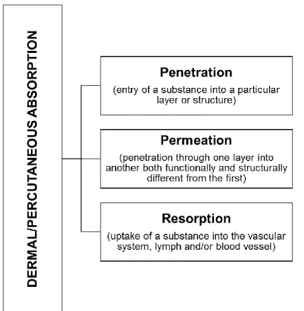
The skin, being the largest and outermost organ of the human body, is the only organ interfacing with the environment and thus has direct and important regulatory functions such as permeation, metabolism and thermoregulation and actively contributes to the sensorial function (Couturaud, 2014). It acts as an envelope to the whole body and is closely integrated to the underlying fascial endoskeleton through retinacular ligaments, blood vessels, nerves and lymphatics (Wong et al., 2015). The dermal/percutaneous absorption process is a global term which describes the passage of compounds across the skin and can be divided into three steps, as seen in figure 5. Systemic exposure and distribution to other organs further depend on dermal blood flow rate, itself dependent on neural and local mechanisms (as for example during thermoregulation) and on the inflammation status of the skin (Dancik et al., 2015).
Human skin is composed of three distinct layers: epidermis, dermis and hypodermis, with varying degrees of specialization within each layer (figure 6) (Wong et al., 2015). With cosmetics (and cosmeceuticals), epidermis is the most important layer of the skin, consisting of diverse cells. Their shape, size and structure depend on the layer in which they are located (Zielinska and Nowak, 2016).
Figure 5. Steps of the dermal/percutaneous absorption process: Penetration, Permeation and Resorption (adapted from SCCS, 2016).
10 The epidermis is a very thin protective epithelium whose more superficial layer constitutes the stratum corneum, composed of dead cells and keratin scales that are continuously shed and replaced by underlying layers. It serves as protection of the organism against external aggressions, ensured through the cohesion of keratinocytes, the epithelial cells (Burlando et al., 2010; Couturaud, 2014). This is the most biologically active layer, as the basal layer of the epithelium is constantly renewing (Wong et al., 2015). For a compound to be an irritant and to reach the systemic circulation following skin exposure, it must first be absorbed into and permeate through the stratum corneum. Besides this ability to penetrate the stratum corneum, the strength of an irritant is also dependent on its cytotoxic effect on living cells (Gibbs, 2009; Dancik et al., 2015).
The dermis is a connective tissue developing beneath the epidermis. Its main cell type are fibroblasts, and this layer is characterized by an extensive ECM containing high levels of collagen and elastin, matrix proteins conferring strength, firmness, and elasticity to the tissue (Burlando et al., 2010). The complexity of this layer and its strong blood supply and innervation allows the dermis to perform important physiological functions, such as nourishing other skin structures, absorption and storage of valuable nutritional components, among others (Zielinska and Nowak, 2016). The nature of the dermis impacts the access of compounds to the deeper blood vessels (Dancik et al., 2015).
The hypodermis is the most internal layer of the skin and consists of loose, conjunctive tissue, linked to the lower part of the dermis by expansions of collagen fibers and elastic
Figure 6. Schematic representation of the skin layers and the vascular network in human skin, and factors that may influence the penetration of chemicals into and through the skin (Dancik et al., 2015).
11 fibers (Couturaud, 2014). This tissue extends deeply to contact the connective sheaths of muscles and skeletal system, and acts as a thermal insulator, a reserve of fuel for the body’s metabolism, and a cushion that soothes the potentially damaging effects of bumps and other traumatic events (Burlando et al., 2010).
The skin appendages, in particular the hair follicle, constitute transport pathways through which chemicals may access the network of blood vessels surrounding the appendages as well as penetrate to the viable tissues bypassing the stratum corneum (Dancik et al., 2015).
1.4. Cell Culture
1.4.1. HaCaT Cell Line
There are four categories of models to study the effects of compounds in the skin, with increasing complexity: single-cell assays, epidermal equivalents, skin equivalents and excised skin (Gibbs, 2009). The HaCaT cell line is a human keratinocyte line derived from spontaneously immortalized human keratinocytes (Boukamp et al., 1988) and exhibits high differentiation potential that allows its use as a substitute for normal human keratinocytes (Schoop et al., 1999). This cell line belongs to the single-assay models and is widely used as a first-round screening to evaluate the toxicity of several compounds at the skin level (Gibbs, 2009; Pelin et al., 2017).
HaCaT cells are spontaneously immortalized cells that remain non-tumorigenic for an extended number of passages in culture (approximately 300) (Shrürer et al., 1993; Sawan et
al., 2017). They can be grown in traditional media and can be maintained for long periods of
time. While not entirely “normal”, these cells show basal cell properties and respond to different inducers of differentiation such as Ca2+ and high cell density. In fact, HaCaT cells in
culture can revert back and forth between a differentiated and a basal state upon changes in Ca2+ in the medium (Deyrieux and Wilson, 2007; Wilson, 2013). Even though HaCaT cells, as
an in vitro skin model, determine whether a compound is capable of causing cell damage, it does not determine if the compound can penetrate the stratum corneum of the epidermis and therefore reach systemic circulation (Gibbs, 2009).
1.4.2. Caco-2 Cell Line
The Caco-2 cell line, derived from human epithelial colorectal adenocarcinoma, is a very simplified model of the gastrointestinal tract and is considered the most common in vitro model used for investigation and prediction of intestinal drug absorption (Sambuy et al., 2005; Awortwe et al., 2014; Kaiser et al., 2017). The small intestine is the primary organ responsible for the absorption of nutrients, and serves as a physical and biological barrier to
12 digestive enzymes and ingested foreign substances. Even though a cosmeceutical is applied to the skin, other biological barriers can also be exposed to these compounds and be affected (Breeman and Li, 2005; Fisichella et al., 2012).
Although cancerous in origin, Caco-2 cells are unique in their ability to initiate spontaneous differentiation on reaching confluence under normal culture conditions (in the presence of glucose and serum) (Smetanová et al., 2011). With this differentiation, these cells become polarized cells expressing apical and basolateral surfaces with well-established tight junctions (Awortwe et al., 2014). The advantages of using the Caco-2 cell monolayer model system of intestinal absorption are that it models transcellular and paracellular permeability, vesicular transport, active transport, facilitated transport and efflux systems, however, it presents some limitations as its low throughput and low expression of intestinal cytochrome P450 isozymes (Li et al., 2003).
1.4.3. HepG2 Cell Line
The liver is responsible for a wide range of functions, including xenobiotic detoxification, protein synthesis, synthesis and storage of glucose, production of the bile necessary for digestion, and regulation of blood cholesterol and triglycerides (Vernetti et al., 2017). Being the main site of xenobiotic biotransformation due to the ability of this organ to express a lot of enzymes, both quantitatively and qualitatively, facilitates the generation of reactive metabolites that can cause liver damage. Therefore, cell lines of liver origin are widely used in biomedical research involving xenobiotic metabolism, including cytotoxicity and genotoxicity studies (Mersch-Sundermann et al., 2004; Vernetti et al., 2017). The toxicity of substances on hepatocytes can be measured by a variety of parameters designed to detect structural or functional perturbations that impair cellular capabilities and produce cell injury. Most of the biochemical indicators to determine these perturbations fall into categories of, as an example, measuring viability, membrane integrity or metabolic function (Dehn et al., 2004). The human hepatocellular carcinoma, the HepG2 cell line, has been extensively used in toxicity studies for the screening of classic toxicants as it preserves most of the phenotypic characteristics of normal hepatocytes (Sahu et al., 2016; Miranda et al., 2017).
The HepG2 cell line has a major advantage for mutagenicity/antimutagenicity studies as it expresses phase I and phase II drug metabolizing enzymes (Knasmüller et al., 1998). Phase I reactions such as oxidation, reduction, hydrolysis, among others, introduce functional groups into the molecule and phase II reactions render the xenobiotic substance or its metabolites more hydrophilic and excretable via bile or urine (SCCS, 2016). HepG2 cells also produce most of the plasma proteins, have biosynthetic capacities similar to those of normal hepatocytes, retain cell surface receptors and in serum-free culture medium are
13 capable of secreting liver-specific serum proteins (Dehn et al., 2004). However, these cells have the disadvantage of having cell functions lower than primary hepatocytes, low urea and albumin production that can be limiting in some studies (Vernetti et al., 2017).
1.4.4. Raw264.7 Cell Line
Macrophages represent the primary line of defense against foreign materials (Nishanth
et al., 2011), playing multiple roles in immune function such as replenish resident
macrophages and dendritic cells under normal states; and move quickly to sites of infection in the tissues and divide/differentiate into macrophages and dendritic cells to elicit an immune response (Zhou et al., 2015).
The macrophage-like Raw264.7 cell line was established in 1978 from mouse tumors that had the appearance of typical lymphocytic lymphomas induced by Abelson leukemia virus (A-MuLV) (Raschke et al., 1978). These cells have been widely used in inflammatory response studies because once activated they express inflammatory mediators [such as nitric oxide (NO)] and pro-inflammatory cytokines [such as interleucine-6 (IL-6)] (Makene and Pool, 2015). They are also used as a representative of a phagocytic cell line to study the toxicity of, for example, nanomaterials (Ninshanth et al., 2011; Zhou et al., 2015)
1.5. Drosophila melanogaster
Drosophila has been used productively has a model organism for more than a century
to study a diverse extension of biological processes, including genetics and genetic heritage, embryonic development, knowledge, behavior, and aging. Even if humans and flies don’t seem too similar, it has become established that most of the biological mechanisms and pathways that control development and survival are conserved through evolution between both species (Jennings, 2011). In fact, approximately 75% of the genes connected to human diseases have related sequences in Drosophila, suggesting that flies are an efficient model to study the function of a great variety of genes involved in human diseases (Kaun et al., 2012).
There are various advantages to Drosophila use, including factors like the cost, size, fecundity, and time scale. The relatively small lifetime of flies means that the genetic experiments that would take months or even years in vertebrate models like the mouse or zebrafish can be completed in a few weeks. Besides its lifetime, maintaining the flies in the lab is easy and cheap, and due to its small size, it’s possible to have different Drosophila genotypes in a typical lab (Jennings, 2011; Kaun et al., 2012).
The fly is considered to be multiple model organisms, each with its specific advantages, defined by their development stage: embryo, larvae, pupae and adult (Pandey
14 and Nichols, 2011). Once fertilized, the embryo develops in the egg for about one day (at 25ºC) before hatching in a larva. The larvae feeds and grows (passing through three changes) for five days until it becomes a pupa and suffers metamorphosis in an adult fly for four days. During metamorphosis, most of the embryonic and larvae tissue is destroyed, and the adult tissues (like the wings, legs or eyes) develop from a group of cells named imaginal discs (Jennings, 2011).
Drosophila melanogaster is a sexually dimorphic species, and the males and females
are easily distinguished based on various morphologic differences (figure 7). In terms of size, generally, females are bigger than males. The
color also differs with sex, as in males the posterior abdomen segments are completely dark and shiny and in the females these segments vary from pale to completely dark. In terms of external morphology, females have an abdomen with a pointy extremity and the males have a round abdomen that tends to curve to the inside. The male external genitals are bigger, more complex and darker than female external genitals. Other characteristic, easy to observe and trustworthy is the presence of sex combs in the male anterior legs. These combs consist in dark and thick hairs in the tarso first segment (Chyb and Gompel, 2013).
1.6. Cytotoxicity and Genotoxicity
Predictive testing of the mutagenic/genotoxic potential of compounds is an essential part of human risk assessment for pharmaceuticals, chemicals, food additives, plant protection products, biocides and cosmetic ingredients (Ates et al., 2014). The assessment of the toxicological potential lays on distinct toxicity studies, specific for different toxicological endpoints, representing the primary step in the hazard evaluation of a cosmetic ingredient (Almeida et al., 2017).
The safety requirements for the evaluation of cosmetic ingredients and products listed in the “The Scientific Committee on Consumer Safety (SCCS) Notes of Guidance for the Testing of Cosmetic Ingredients and their Safety Evaluation”, revised in April 2016, include the following parameters:
Figure 7. Sexual dimorphism in Drosophila melanogaster (Chyb and Gompel, 2013)
15 acute toxicity (adverse effects that may result from a single exposure to a
substance);
corrosion and irritation (irreversible damage to the skin and production of reversible damage of the skin, respectively);
skin sensitization (induction of specific immunological reactivity after contact with the skin and penetration into the epidermis);
repeated dose toxicity (adverse general toxicological effects, including reproductive, genotoxic and carcinogenic effects, occurring as a result of repeated daily dosing with, or exposure to, a certain substance);
reproductive toxicity (adverse effects induced on any aspect of mammalian reproduction);
mutagenicity/genotoxicity (induction of permanent transmissible changes in the amount or structure of the genetic material of cells or organisms and processes which alter the structure, information content or segregation of DNA, respectively);
carcinogenicity (induction of tumors or increasing their incidence, malignancy or shortening the time before tumor occurrence);
photo-induced toxicity; Human data (SCCS, 2016).
The term cytotoxicity is defined by the Organisation for Economic Co-operation and Development (OECD) as “the adverse effects resulting from interference with structures and/or processes essential for cell survival, proliferation, and/or function”. For most chemicals/substances, toxicity is a consequence of non-specific modifications in basal cell functions that may lead to effects on organ-specific functions and/or death of the organism. These effects may involve the integrity of membranes and the cytoskeleton, cellular metabolism, the synthesis and degradation or release of cellular constituents or products, ion regulation and cell division (OECD, 2010). Generally, in vitro cell-based cytotoxicity assays measure a signal correlated to a cell physiological event, for example, cell number, proliferation, membrane integrity or metabolic state, at increasing compound concentrations. These signals serve as a proxy to quantify cell viability and/or proliferation (Cortés-Ciriano and Bender, 2016).
As mentioned earlier, the term genotoxicity refers to processes which alter the structure, information content or segregation of DNA and are not associated with mutagenicity (SCCS, 2016). When a toxic agent interacts with DNA, this interaction may lead to chromosomal aberrations and/or changes in DNA structure that may affect the fidelity of
16 the message and lead to irreversible changes in the cell (Sponchiado et al., 2016). The mechanisms responsible for inducing genotoxicity can be divided into primary and secondary mechanisms, with the first being related to cellular uptake and the second to chronic inflammatory response and consequent immune cell activation (Doak et al., 2012). Tests for genotoxicity are considered short-term in nature and are an integral part of product safety assessment and include tests which provide an indication of induced damage to DNA via for example sister chromatid exchange, DNA strand breaks, DNA adduct formation or mitotic recombination (Baumstark-Khan et al., 2010; SCCS, 2016). The outcome of DNA damage is diverse and generally adverse, as for example cell cycle arrest or even cell death, and long-term effects result in irreversible mutations which may contribute to oncogenesis (Hoeijmakers, 2001). DNA damage, repair mechanisms and consequences are summarized in figure 8.
1.7. Somatic Mutation and Recombination Test
Mutagens can induce mutations by replacing a base in the DNA, alter a base so that it mispairs with another base, or damage a base so it can no longer pair with other bases under normal conditions (Griffiths et al., 2015a). The somatic mutation and recombination test (SMART) is an in vivo assay with Drosophila melanogaster used to study the genotoxic
Figure 8. DNA damage, repair mechanisms and consequences. a, Common DNA damaging agents, examples of DNA lesions induced and most relevant DNA repair mechanisms. b, Acute effects of DNA damage (cell cycle and DNA metabolism) and long-term consequences of DNA damage and their biological effects. Abbreviations: cis-Pt (cisplatin), MMC (mitomycin C), PP (6-4 photoproduct), CPD (cyclobutene pyrimidine dimer, HR (homologous recombination), EJ (end joining) (Hoeijmakers, 2001).
17 activity of chemical compounds (Graf et al., 1984). This is a sensitive, rapid and cheap test that assesses a wide variety of genetic alterations, including somatic recombination, and is based on the loss of heterozygosity (LOH) resulting from genotoxicity (Ávalos et al., 2015).
In individuals, heterozygous for “visible marker mutations”, certain mutagens can lead to the loss of the dominant wild-type allele opposite to the marker mutation, resulting in the expression of the recessive marker allele in a clone of subsequent mutant cells (Graf et al., 1984). If a genetic alteration occurs in one cell of the imaginal discs of Drosophila
melanogaster during the mitotic proliferation (figure 9), the result will be a clone of mutant
cells expressing the phenotype regulated by specific genetic markers (Marcos and Carmona, 2013).
There are two different SMART assays, the wing-spot test (mwh/flr) and the eye-spot test (w/w+). Although these assays have the same genetic base, the targeted adult tissues are different: the wings in the wing-spot test and the eyes in the eye-spot test (Marcos et al., 2014). In the wing-spot test, flies carrying heterozygous recessive mutations for the multiple wing hair (mwh) and flare (flr) phenotypes are exposed to a test compound and the wing hair phenotypes in adult flies are observed (Lombardot et al., 2015). The eye-spot test uses the X-chromosome white (w) gene as a recessive
marker to monitor the presence of white clones on wild-type eyes (Marcos et al., 2014). Any increase in the clone frequency, when compared to control, reflects genotoxic activity, which is characterized by the frequency of the clone induction: the more it induces, the bigger the extension of the genotoxic activity (Ávalos et al., 2015). For both assays, the results can be controlled by the capacity of the used strain to properly metabolize xenobiotic compounds (Marcos et al., 2014).
1.8. Comet assay
Measuring DNA damage is an important step in a lot of biomedical research studies as many environmental agents can damage the hereditary material of living organisms and lead to a variety of disorders in experimental animals and humans (Osipov et al., 2014; Pu et al.,
Figure 9. Schematic representation of the different genetic processes by which LOH could occur (adapted from Marcos et al., 2014).