HIV-1 Myristoylated Nef Treatment of
Murine Microglial Cells Activates Inducible
Nitric Oxide Synthase, NO
2
Production and
Neurotoxic Activity
Giorgio Mangino1,2☯, Marylinda Famiglietti1☯, Caterina Capone1, Caterina Veroni3, Zulema Antonia Percario1, Stefano Leone1, Gianna Fiorucci4, Sebastian Lülf5, Giovanna Romeo2,4, Cristina Agresti3, Tiziana Persichini1, Matthias Geyer5, Elisabetta Affabris1*
1Department of Science, University Roma Tre, Rome, Italy,2Department of Medico-Surgical Sciences and Biotechnologies, Sapienza University of Rome, Latina, Italy,3Department of Cell Biology and
Neuroscience, Istituto Superiore di Sanità, Rome, Italy,4Institute of Molecular Biology and Pathology, CNR, Rome, Italy,5Center of Advanced European Studies and Research, Group of Physical Biochemistry, Bonn, Germany
☯These authors contributed equally to this work.
*elisabetta.affabris@uniroma3.it
Abstract
Background
The potential role of the human immunodeficiency virus-1 (HIV-1) accessory protein Nef in the pathogenesis of neuroAIDS is still poorly understood. Nef is a molecular adapter that influences several cellular signal transduction events and membrane trafficking. In human macrophages, Nef expression induces the production of extracellular factors (e.g.
pro-in-flammatory chemokines and cytokines) and the recruitment of T cells, thus favoring their infection and its own transfer to uninfected cells via exosomes, cellular protrusions or cell-to-cell contacts. Murine cells are normally not permissive for HIV-1 but, in transgenic mice, Nef is a major disease determinant. Both in human and murine macrophages, myristoylated Nef (myr+Nef) treatment has been shown to activate NF-κB, MAP kinases and interferon re-sponsive factor 3 (IRF-3), thereby inducing tyrosine phosphorylation of signal transducers and activator of transcription (STAT)-1, STAT-2 and STAT-3 through the production of proinflammatory factors.
Methodology/Principal Findings
We report that treatment of BV-2 murine microglial cells with myr+Nef leads to STAT-1, -2
and -3 tyrosine phosphorylation and upregulates the expression of inducible nitric oxide synthase (iNOS) with production of nitric oxide. We provide evidence that extracellular Nef regulates iNOS expression through NF-κB activation and, at least in part, interferon-β
(IFNβ) release that acts in concert with Nef. All of these effects require both myristoylation OPEN ACCESS
Citation:Mangino G, Famiglietti M, Capone C, Veroni C, Percario ZA, Leone S, et al. (2015) HIV-1 Myristoylated Nef Treatment of Murine Microglial Cells Activates Inducible Nitric Oxide Synthase, NO2 Production and Neurotoxic Activity. PLoS ONE 10(6): e0130189. doi:10.1371/journal.pone.0130189
Academic Editor:Bruno Verhasselt, Ghent University, BELGIUM
Received:July 15, 2013
Accepted:May 17, 2015
Published:June 11, 2015
Copyright:© 2015 Mangino et al. This is an open access article distributed under the terms of the
Creative Commons Attribution License, which permits unrestricted use, distribution, and reproduction in any medium, provided the original author and source are credited.
Funding:This work was supported by grants from the Italian National Research Program on AIDS of the Istituto Superiore di Sanità (Affabris N.40.G1), Rome, Italy, and from PRIN-MIUR (Progetti Rilevante Interesse Nazionale-Ministero dell’Università e della Ricerca Scientifica) 2007 (Affabris
prot.20072J9RWM_002), Italy. The funders had no role in study design, data collection and analysis, decision to publish, or preparation of the manuscript.
and a highly conserved acidic cluster in the viral protein. Finally, we report that Nef induces the release of neurotoxic factors in the supernatants of microglial cells.
Conclusions
These results suggest a potential role of extracellular Nef in promoting neuronal injury in the murine model. They also indicate a possible interplay between Nef and host factors in the pathogenesis of neuroAIDS through the production of reactive nitrogen species in microglial cells.
Introduction
The term“neuroAIDS”encompasses different clinical syndromes including sensory neuropa-thy, myelopaneuropa-thy, HIV-associated dementia (HAD), HIV-associated encephalitis (HIVE) and cognitive/motor disorders. Despite their heterogeneity, these neurologic disorders are all char-acterized by neuronal loss, due to damage of central and peripheral nervous system by HIV (for review see [1]). Prior to highly active antiretroviral therapy (HAART) introduction, neuro-logical disorders were the first manifestation of symptomatic HIV-1 infection, affecting rough-ly 10/20% of patients and up to 60% of patients in the advanced stages of HIV-1/AIDS [2]. HAART has reduced the incidence of severe forms of AIDS-associated neurologic disorders such as HIV-1-associated dementia (HAD) but, with longer life span, the prevalence of milder forms of neurologic manifestations such as HIV-1-associated neurocognitive disorder (HAND) appears to be increasing [3,4].
Although substantial advances in deciphering the pathophysiological role of viral products in the development of neuroAIDS have been achieved, two main aspects of HIV-associated neuronal degeneration remain puzzling. First, neurological diseases are generally a later mani-festation of HIV-1 infection and tend to progress in parallel with the degree of immunosup-pression, while invasion of the nervous system may occur early upon viral infection, probably concomitant with initial systemic infection [5]. Second, since the virus is unable to infect neu-rons, the neuronal loss associated with neuroAIDS manifestations is not thought to be a direct cytopathic effect of HIV-1 itself [6,7]. Accumulating evidence suggests that over-activation of the immune system, particularly of the macrophage subset, is the pathophysiological event common to all of the neuroAIDS-associated syndromes [8]. Macrophage activation produces pro-inflammatory cytokines, platelet activating factor (PAF), nitric oxide, and free oxygen radi-cals that appear to contribute to the neuronal damage and dysfunction underlying the clinical syndromes [7,9–11]. Thus, neuroAIDS shares some similarities with other neurodegenerative syndromes such as Alzheimer’s disease, Parkinson's disease and multiple sclerosis, in which ac-tivated macrophages and microglial cells (i.e. the nervous system’s resident macrophages) act as crucial disease determinants by sustaining an inflammatory microenvironment that ulti-mately leads to nervous system damage.
specific target sequences located in theinospromoter region, thus inducing the synthesis of the enzyme [13–15]. In contrast to neuronal and endothelial NOS (nNOS and eNOS, respectively), iNOS is constitutively active once synthesized and acts in a Ca2+-independent manner [16, 17].
HIV-1 Nef is a small (MW 27–34 kDa) myristoylated, cytoplasmic, multifunctional viru-lence factor acting as an adaptor molecule inside the cell. It is partially associated with the cell membrane and plays multiple roles during HIV-1 replication [18–20]. Nef-defective viruses lead to an attenuated clinical phenotype with reduced viral load in mouse models, monkeys, and humans [21–25]. More recently, it has been shown that this viral protein can be transferred to uninfected cells via cellular nanotubes, cell-to-cell contacts and release of exosomes. These findings lead to the idea that Nef is able to regulate both the endocytotic and exocytotic cell pathways thereby inducing specific effects also in non-infected cells [26].
In human monocyte-derived macrophages (MDMs), both Nef expression within the cell and cell treatment with the recombinant protein induce a pro-inflammatory response charac-terized by synthesis and release of specific cytokines and chemokines [27–32]. Nef-induced pro-inflammatory state in macrophages is largely due to NF-κB activation [28,32–34]. In addi-tion, we reported that Nef treatment of MDMs activates IRF-3, the main transcriptional regula-tor leading to the synthesis of IFNβ[32] and, ultimately, to the induction of IRF-1. Based on these two premises, we hypothesized that Nef promotes synthesis and activation of iNOS in microglial cells as a result of its pro-inflammatory properties. Consequently, iNOS-derived ni-trogen reactive species might play a role in neuronal loss in a Nef-dependent manner. Due to the lack of an availablein vitrosystem based on human-derived microglial cells, we resorted to a well characterized murine microglial cell line (i.e. BV-2, see [35]) and tested the above hy-potheses by treating cells with myristoylated (myr+) Nef and by analyzing iNOS and RNS in-duction, as well as the ability of RNS to induce neuronal death. Murine macrophages respond to Nef treatment as human MDMs by activating IKKαand IKKβ, JNK, and p38 MAP kinases [36]. Activation of the NF-κB pathway is mandatory for the tyrosine phosphorylation of signal transducer and activator of transcription (STAT)-1, STAT-2, and STAT-3, which is induced within 2 h in an autocrine and paracrine manner. These data confirmed that murine and human macrophages respond similarly to myr+Nef treatment [36].
Although murine cells are not permissive for HIV-1 infection, several investigators have en-gineered transgenic (Tg) mice to model HIV-1-induced diseases and overcome such restriction [37–39]. The generation of Tg mice expressing selected HIV-1 genes revealed that Nef repre-sents a major disease determinant and that the murine system is a suitable model to investigate the mechanisms of Nef activity [37]. Nevertheless, in these studies the functions affected by Nef in monocytes/macrophages have not been extensively investigated. To the best of our knowledge, only two studies dealt with Nef expression in the brain of Tg mice: in the first one Nef was expressed in oligodendrocytes [40], whereas in the second Nef was expressed in mac-rophages/microglial cells but pro-inflammatory responses were not investigated [41].
Results
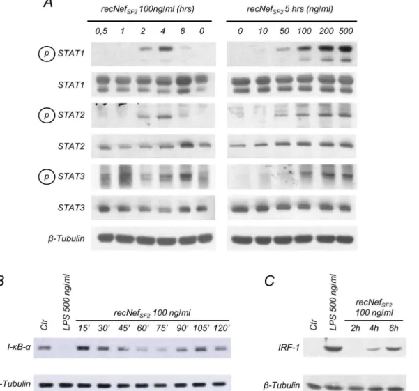
Extracellular Nef induces STATs phosphorylation, I
κ
-B degradation and
IRF-1 expression in BV-2 microglial cells
Two main transcription factors are responsible for iNOS/NOS2 induction in murine as well as human phagocytic cells,i.e. IRF-1 and NF-κB. Studies of the iNOS murine promoter revealed the presence of two NF-κB and one IRF-1 binding sites [13–15]. We previously reported that Nef activates the NF-κB signaling pathway, and synthesis of IRF-1 both in primary human MDMs and murine macrophages [28–30,32,36]. Therefore, we assessed whether this was also the case in microglial cells. First, we confirmed that myr+NefSF2treatment induced tyrosine
phosphorylation of STAT-1, -2 and -3 in BV-2 cells in a time- and dose-dependent manner. In particular, tyrosine phosphorylation signal increased starting from 2 h of cell treatment with 100 ng/ml of the viral protein and was clearly detectable after 5 h of treatment with 50 ng/ml myr+NefSF2(Fig 1A).
Subsequently, cells were challenged for different times (from 15 to 120 min) with myr+ NefSF2and total cellular lysates were analyzed for the expression of I-κB-α, the alpha isoform
of the inhibitor ofκB. As shown inFig 1B, myr+NefSF2treatment induced I-κB-αdegradation
that became clearly evident after 60 to 75 minutes. At later time points, protein levels started to recover, likely due to the neo-synthesis of I-κB which is itself encoded by a NF-κB regulated gene [42,43]. After 4/6 h of myr+NefSF2treatment the expression of IRF-1, encoded by a
STAT-1 responsive gene, was clearly increased (Fig 1C). These results indicate that in murine microglial cells Nef positively affects the two transcriptional factors involved in iNOS regulation.
Nef treatment of murine microglial cells induces iNOS expression and
NO
2-production
Based on the above results, we asked whether extracellular Nef was able to regulate iNOS ex-pression. First, BV-2 cells were exposed to myr+NefSF2and iNOS mRNA levels were evaluated
by Real Time RT-PCR from 1 to 24 h post-treatment. As shown inFig 2A, iNOS mRNA ex-pression reached a peak at 6 h post-treatment, declined thereafter and increased again at 16 and 24 h, similarly to the kinetics of iNOS mRNA induction in cytokines-stimulated DLD-1 cells [44]. Induction of iNOS mRNA expression was also confirmed in primary murine micro-glial cells (Fig 2B). To test whether iNOS expression and NO2-production were both affected,
BV-2 cells were treated for 24 h with 100 to 500 ng/ml myr+NefSF2. iNOS protein levels were
measured in total cellular extract, whereas quantification of NO2-released into the
superna-tants was performed using the Griess reagent. As depicted inFig 2C and 2D, iNOS expression and NO2-release were induced in a dose-dependent manner by myr+Nef treatment of
micro-glial cells. The effect required the integrity of the protein, as heat-denaturated Nef barely in-duced iNOS synthesis and NO2-release. As a positive control, we evaluated NO2-production
induced by LPS which is not sensitive to heat inactivation. To verify iNOS induction in a human context, we polarized monocytes into M1 inflammatory macrophages by cultivating them for 6 days in the presence of GM-CSF (Fig 2E). M1 macrophages were then treated with myr+Nef in the presence of IFNγfor 24, 48 or 72 h. We were unable to detect nitrite accumula-tion in the supernatants of Nef-treated M1 macrophages, due to the well known low NO2-/
NO3-ratio production in human cells [45], nevertheless we found that iNOS was slightly
iNOS upregulation induced by extracellular Nef requires both NF-
κ
B and
IFN
β
As previously reported, iNOS expression is regulated by both NF-κB and IRF-1. To test wheth-er the NF-κB pathway was involved in Nef-dependent iNOS induction, BV-2 cells were treated with recombinant myr+NefSF2in the presence of BMS-345541, a highly specific IKKα/IKKβ
in-hibitor. Pre-treatment with BMS-345541 at 5μM concentration greatly reduced
Nef-depen-dent induction of iNOS mRNA levels (Fig 3A) and completely inhibited iNOS expression (Fig 3B). However, due to BMS-345541 cytotoxicity upon prolonged cell treatment, NO2-
produc-tion was evaluated in supernatant of BV-2 cells treated with Nef for 24 h in the presence of lower doses of the inhibitor (i.e. 1 and 2.5μM). Nonetheless, these doses were still able to
re-duce NO2-production in a dose-dependent manner (Fig 3C).
Fig 1. Nef treatment induces STATs tyrosine phosphorylation, I-B degradation and IRF-1 expression in BV-2 microglial cells.(A) BV-2 cells were left untreated or incubated with 100 ng/ml wild type myristoylated Nef derived from HIV-1 SF2 strain (myr+Nef
SF2) for different times (left panel) or for 5 h with
different amounts of myr+Nef
SF2(right panel). Total cellular extracts were analyzed by Western Blot to evaluate STAT-1, -2 and -3 tyrosine phosphorylation or
protein expression using specific antibodies as described in materials and methods. (B,C)Cells were incubated for the indicated time with myr+NefSF2(100
ng/ml) or, as a positive control, with LPS (500 ng/ml) for 30’in (B) and 6 h in (C). Whole cell lysates were analyzed by Western Blot for I-κB-α(B) or IRF-1 (C) expression.β-tubulin expression was used as an internal loading control. Results reported in the figure are from four independent experiments.
In human MDMs we observed that Nef treatment induces both the synthesis and the release of IFNβfollowed by IRF-1 production [30,32]. We also reported that Nef treatment induced IRF-1 in RAW264.7, a murine macrophage cell line [36] and in BV-2 microglial cells (Fig 1C). Based on these findings, we hypothesized that IFNβblockade could affect iNOS upregulation. BV-2 cells were incubated with myr+NefSF2in presence of anti-IFNβneutralizing antibodies
(NAb). As shown inFig 4, anti-IFNβNAb started to inhibit the induction of iNOS mRNA at 8 h post-treatment (Fig 4A). This is in agreement with the kinetics of IRF-1 protein induction, that follows Nef-induced degradation of Iκ-B (seeFig 1B). Consequently, iNOS expression was also affected by anti-IFNβNAb starting from 6 h (Fig 4B, compare signals at 6 h in anti-IFNβ
treated and untreated cells). NO2-detected in the culture supernatants appeared also reduced
after 24 h of treatment (Fig 4C). Taken together, these results demonstrated that Nef-induced iNOS upregulation is primarily dependent on NF-κB signalling and, to a lesser extent, on IFNβ
production.
Extracellular Nef cooperates with IFN
β
to induce iNOS expression
It is well recognized that stimuli that induce IRF-1 expression do not induceper seneither iNOS expression nor NO2-production, rather, they“prime”the cells to respond to NF-κB-acti-vating stimuli, improving their effect on iNOS regulation. This is, paradigmatically, the case of LPS and IFNγcombined treatment [46–48]. Therefore, we sought to test whether IFNβhas a priming effect in promoting Nef-induced iNOS expression and function. The results shown in Fig 5demonstrate that combined treatment induced iNOS mRNA expression (Fig 5A), iNOS protein levels (Fig 5B) and NO2-production (Fig 5C) to a greater extent compared to what is
observed in cells exposed to myr+Nef alone.
Nef myristoylation and conserved acidic cluster are essential to induce
iNOS
We previously demonstrated that Nef-mediated interference with cell signalling in human and murine macrophages treated with the viral protein required the integrity of both the N-termi-nal myristoylation site and the acidic cluster (AC) which consists of four glutamates [28,32, 36]. To determine whether these motifs are also required for iNOS induction, BV-2 cells were treated with wild type myr+NefSF2, a mutant in the myristoylation site (i.e. G2A), two different
preparations of a myristoylation positive, AC-mutated viral protein (4EA’and 4EA”), and a Nef-deletion mutant lacking the N-terminal anchor domain (ΔN-term). The results shown in Fig 6indicate that all the mutants tested failed to induce iNOS mRNA expression (Fig 6A), iNOS protein (Fig 6B), and NO2-production alone or in IFNβcombined treatments (Fig 6C).
Conversely, other Nef mutants we tested retained their pro-inflammatory capabilities and Fig 2. Nef treatment of microglial cells induces iNOS expression.BV-2 cells (A) or purified primary murine microglial cells (B) were treated for the indicated time with myr+Nef
SF2(100 ng/ml inA, 200 ng/ml inB, closed circles). Total cellular RNAs were isolated and real time RT-PCR analysis was
performed as reported in the materials and methods section. Results were expressed using the 2-ΔΔCTmethod using basal mRNA level in untreated cells
(Ctr, open circles) at T = 0 as a calibrator and GAPDH level as an internal loading control. (C,D) BV-2 cells were treated for 24 h with the indicated amounts of myr+Nef
SF2, heat inactivated myr+NefSF2(Inactiv. recNef), LPS or heat treated LPS. Total cell lysates were analyzed by Western Blot for iNOS (C, upper
panel) levels and, as internal loading control,β-Tubulin expression (C, lower panel). (D) NO2-content in the supernatants was quantified using the Griess
colorimetric assay as reported in the materials and methods section. Dark gray bars: native myr+Nef or LPS. Light gray bars: heat pre-treated (Inactiv.) myr+Nef or heat pre-treated LPS. Results from one of five independent experiments are shown. (E) Cell phenotyping by flow cytometry of human monocytes and M1 macrophages obtained as described in materials and methods. According to [77], human monocytes were CD14+/CD163+/CD86+whereas M1
macrophages were CD14-/CD163-/CD86bright. (F) M1 human macrophages were left untreated or treated for 24, 48 and 72 h with IFNγ, 100 ng/ml wild type
myr+NefSF2plus IFNγ, heat pre-treated myr+NefSF2and IFNγor 100 ng/ml LPS plus IFNγ. Total cellular extracts were analyzed by Western Blot to evaluate
iNOS expression using specific antibodies as described in materials and methods.β-tubulin expression as internal loading control. Blots are representative of two independent experiments.
therefore their ability to induce iNOS expression and RNS production alone or in combined treatment with IFNβ. We tested Nef mutants affecting functional motifs involved in the inter-action and down-regulation of CD4 (CAWL), the interinter-action with SH3-containing proteins (PxxPxP) or with proteins involved in endocytic pathways such as the V1H subunit of the vac-uolar membrane ATPase or the adaptor protein complexes APs (DD and LL), as well as a loop mutant lacking the C-terminal flexible loop (data not shown).These results assign the myris-toylation site and the AC as essential structural motifs for Nef-mediated iNOS regulation. Fig 3. Nef-dependent iNOS induction requires NF-κB.(A) BV-2 cells were pretreated for 1 h with BMS-345541 (5μM) and then incubated for 2, 4 and 6 h with myr+Nef
SF2(100 ng/ml) in presence of BMS-345541. Total mRNAs were isolated and analyzed for iNOS mRNA expression as described in the materials
and methods section. Empty circles: untreated cells (Ctr); gray empty squares: BMS-345541; filled circles: recNefSF2; gray filled squares: BMS-345541 plus
recNefSF2. (B) Cells were stimulated as in (A) for 4, 6 and 8 h, then lysed and total cell lysates analyzed by Western Blot for iNOS expression.β-Tubulin was
used as an internal loading control. (C) BV-2 cells were pre-treated with BMS-345541 (1 or 2.5μM), then stimulated for 24 h with the indicated amount of myristoylated recNefSF2in presence of BMS-345541. Supernatants were collected and NO2-content evaluated. Black bars: no inhibitor; dark gray bars:
BMS-345541 at 1μM concentration; light gray bars: BMS-345541 at 2.5μM concentration. Ctr: control cells. Data shown were from one of three independent experiments.
doi:10.1371/journal.pone.0130189.g003
Extracellular Nef induced the release of iNOS-dependent neurotoxic
factors in murine microglial cells
It is thought that both viral products and cellular derived factors are mainly responsible for the neuronal loss observed in HAD [49]. To address whether Nef-dependent iNOS induction could have neurotoxic effects, the supernatant of BV-2 cells exposed to myr+NefSF2was
collect-ed and uscollect-ed to verify induction of cell death in NB41A3 murine neuroblastoma cell line. Super-natant collected from microglial cells treated for 48 h with myr+Nef, or with LPS used as positive control, induced cell death in NB41A3 cells (Fig 7C). This effect was completely abol-ished by the pre-treatment of BV-2 microglial cells with the NOS inhibitor L-NAME (Fig 7C), that prevented the Nef-dependent NO2-production without affecting iNOS protein levels (Fig
7A and 7B). Treatment of NB41A3 cells for 24 h with myr+Nef, as well as LPS or L-NAME did not induce any cytotoxic effects (Fig 7D). Again, supernatants collected from BV-2 cells treated Fig 4. Nef-induced IFNβproduction concurs to Nef-dependent iNOS induction.(A) BV-2 cells were treated for the indicated time with myr+Nef
SF2(100
ng/ml) in the presence of anti-IFNβneutralizing antibodies as described [36]. iNOS mRNA was analyzed as reported in materials and methods section. Dark gray bars: 4 h treatment; light gray bars: 8 h treatment. (B) Cells were treated as in (A) for the indicated time and iNOS expression evaluated by Western Blot. β-Tubulin expression was used as an internal loading control. (C) Cells were treated as in (A) for 24 h, supernatants were then collected and NO2-content
measured using the Griess colorimetric assay.
with G2A or 4EA mutants did not cause cell death in neuroblastoma cells (Fig 7E), demonstrat-ing that the myr+Nef neurotoxic effects also require these domains. All together, these results show that Nef-stimulated microglial cells release NOS-dependent neurotoxic factor(s), whereas Nef itself is not neurotoxic.
Fig 5. Nef synergizes with IFNβin iNOS production.(A) BV-2 cells were treated for 6 h with myr+NefSF2(100 ng/ml), IFNβ(200 IU/ml) or a combination of
both. iNOS mRNA was measured by real time RT-PCR as reported in the materials and methods section. (B) Cells were incubated for 24 h with the indicated dose of myr+Nef
SF2with or without IFNβ(50 IU/ml). Cells were also incubated with heat-inactivated recNefSF2(inactiv. recNef, 500 ng/ml) or, as control, with
LPS or pre-heated LPS (500 ng/ml each). Total cellular lysates were analyzed by Western Blot for iNOS expression.β-Tubulin expression was used as an internal loading control. (C) Cells were treated like in (B) and NO2-content in supernatants was measured using the Griess colorimetric assay. Empty
diamond: IFNβ; filled circles: myr+Nef
SF2; gray empty circles: heat-inactivated myr+NefSF2; filled squares: recNefSF2plus IFNβ; gray empty squares:
heat-inactivated myr+Nef
SF2plus IFNβ. doi:10.1371/journal.pone.0130189.g005
Discussion
Compelling evidence indicates that the neurocognitive disorders associated with the develop-ment and/or progression of AIDS are mainly due to the presence of infected macrophages/ microglial cells characterized by a pro-inflammatory M1 profile, in the central nervous system (CNS). Indeed, neuroAIDS severity is directly correlated with the levels of pro-inflammatory cytokines and chemokines, whereas no direct correlation with the viral load has been demon-strated [50,51]. Based on the theory of the“Trojan Horse”[52], HIV-1 is carried into the CNS by infected macrophages, the only immune cells able to cross the Blood Brain Barrier; the infec-tion is then spread to other susceptible CNS-resident target cells, either competent (i.e. micro-glial cells) or defective (i.e. astrocytes) for HIV-1 replication [53]. The demonstrated presence of macrophage-tropic strains into the CNS further supports this hypothesis [54,55]. Even if to the best of our knowledge, there is no evidence of Nef secretion from HIV-1 infected microglial cells and/or astrocytes, it has been reported that HIV-1 infected cells release Nef into the Fig 6. Nef myristoylation and the acidic cluster are essential to induce iNOS.BV-2 cells were left untreated or incubated with 100 ng/ml wild type (WT), G2A, two different preparations of the EEEE!AAAA mutant (4EA’and 4EA”) andΔN-terminal deleted myr+NefSF2proteins. (A) iNOS mRNA was measured
by real time RT-PCR using total RNA isolated from cells treated for 6 h. (B) Total cellular extracts obtained from cells treated for 24h were analyzed by Western Blot using anti-iNOS antibodies, whereasβ-tubulin expression levels were used as an internal loading control. (C) Supernatants collected from cells treated as in (B) were measured for NO2-content using the Griess colorimetric assay. One out of three independent experiments is shown.
extracellular microenviromment. Nef was detected in the medium from the MOLT-4 lympho-cyte cell line transfected with a Nef-producing baculovirus vector and it was quantified in 32 patient serum samples [56]. This evidence suggests that infected cells would release Nef through a non classical secretory pathway or after lysis. Then bystander cells like uninfected microglial cells in the case of CNS, might internalize Nef via endocytosis, pinocytosis or other unknow mechanisms, as we and others already reported for MDMs, dendritic and B cells [29, 57–61]. Once internalized, Nef could provoke the production of ROS and RNS as an integral part of a pro-inflammatory response [62,63].
Our previous studies demonstrated that Nef treatment of human MDMs induces the pro-inflammatory response mainly through the activation of the NF-κB and IRF-3 transcription factors that lead to synthesis and release of IL-1β, IL-6, TNFα, CCL3/macrophage inflammato-ry protein 1a (MIP-1α), CCL4/MIP-1βand IFNβ[28,29,32]. Here we describe the ability of Nef to influence also the production of nitrogen reactive species through the induction of type 2 NOS, in anin vitromurine model of microglial cells exposed to the recombinant protein. The results reported inFig 2indicate a direct correlation between RNS production and Nef-induced upregulation of iNOS, both at the mRNA and protein level. As already reported for other ca-nonical inducers, such as LPS and IFNγ, also in the case of Nef, iNOS induction is achieved through the activation of NF-κB and is reinforced by IRF-1 upregulation (Figs3and4, respec-tively). Even if we cannot formally exclude the hypothesis that a surface receptor for Nef may activate a signalling cascade leading to iNOS induction, the observation that iNOS mRNA upregulation was inhibited by both cytochalasin D and 5-(N,N-dimethyl)-amyloride, two com-pounds which inhibit membrane ruffling and macropinocytosis [64,65] argue against this hy-pothesis (data not shown).
In line with previous results, we show that iNOS induction and RNS production require the N-terminal myristoylation of Nef that influences Nef localization to the cellular membrane as well as the integrity of its conserved acidic cluster (Fig 6), which was shown to be pivotal for pro-inflammatory macrophage activation through the interaction with TNF receptor adapter factor 2 [28]. Interestingly, protein alignment studies on neurotropic strain-derived Nef from post-mortem biopsies revealed that some conserved amino acidic substitutions characterized these viruses compared to patient-matched blood-derived samples [66]. Two of them are 71R/ K/T residue, in which the brain-derived viruses had arginine and lysine, whereas peripheral blood-derived HIVs had a mixture of threonine, lysine and arginine and 83G/A residue, in which brain-derived viruses displayed a glycine whereas blood-derived ones contained an ala-nine [66]. These residues are located immediately downstream of the Nef acidic cluster and they likely play a role in the stability and/or surface accessibility of this part of the protein that might also mediate the“pro-inflammatory potential”of the acidic cluster.
Studies performed to identify the phenotype of infected macrophages in the CNS revealed that both CD14+/CD45+perivascular macrophages and multinucleated giant cells represent the major source of infected cells in macaques [67], whereas CD14-/CD45-parenchymal microglial cells do not express any viral factor. From this point of view, the capacity of Nef to be transferred from infected cells to non-infected cells by exosomes and nanotubes or possibly Fig 7. Nef-treated microglial cells release iNOS-dependent neurotoxic factors.(A) BV-2 cells were treated for 48 h with myr+Nef
SF2(200 ng/ml) in the
presence (light gray bars) or absence (dark gray bars) of L-NAME at 10μM concentration. Supernatants were collected and NO2-content was measured
using the Griess colorimetric assay. (B) iNOS expression was evaluated on total cell lysates by Western Blot using specific anti-iNOS antibodies (upper panel) and anti-β-tubulin antibodies as an internal loading control (lower panel). (C) NB41A3 cells were incubated for 24 h with supernatants from (A). (D) NB41A3 cells were treated for 24 h with myr+Nef
SF2(200 ng/ml) or with LPS (100 ng/ml). Black line: No L-Name treatment; Gray line: L-Name (10μM). (E)
NB41A3 cells were incubated for 24h with conditioned supernatants derived from untreated BV-2 cells or treated for 48 h with 200 ng/ml of wild type (WT), G2A, 4EA’, 4EA”andΔN-terminal deleted myr+NefSF2proteins. In (C)to (E), cell viability was measured by flow cytometry using Propidium Iodide (PI) dye
exclusion and data were expressed as percentage of PI positive cells. One out of three independent experiments is shown.
via endocytosis as free protein (reviewed in [26]) should be emphasized. If this route of trans-mission is active also in the CNS, Nef-dependent iNOS induction and production of RNS might be achieved in non-infected microglial cells as well. As a consequence and regardless of the nature of infected cells in the CNS, a deregulated proinflammatory response could be in-duced, thereby promoting and/or accelerating the clinical onset of neurological disease [68]. Indeed, the production of matrix metalloproteinases MMP-2, -7, and -9, as well as IL-1, IL-6, TNFα, nitric oxide (NO), L-cysteine, and Ntox further disseminate immune activation in non-infected phagocytic cells.
Our results suggest a role of Nef in this macrophage- and/or microglial-based proinflamma-tory environment, possibly contributing to the induction of neuronal death and neuronal damage observed in the brain of AIDS patients [69]. Results obtained using the NB41A3 neu-roblastoma cell line (Fig 7) demonstrated that the secreted factors induced by Nef in BV-2 cells, rather than Nef itself, are responsible for neuronal death assessed by PI uptake. These re-sults are in conflict with reports indicating a direct neurotoxic activity of Nef due to its similari-ty to scorpion peptides [70]. L-NAME-dependent inhibition of neuronal death suggests that this phenomenon is dependent on NOS activity and NO production. Interestingly, NO2-,
provided to NB41A3 cells as NaNO2, was not toxicper seand supernatants collected from
microglia treated with myr+Nef for only 24 h were unable to induce NB41A3 cell death (data not shown). The explanation of this observation is unknown at present, as the NO2-content in
supernatants collected from BV-2 cells treated for 24 and 48 h with Nef did not differ signifi-cantly (data not shown). It is conceivable that longer incubation times are needed to allow the release of other products that in concert with NO2-induce cell death in NB41A3 cells. In this
regard, it is interesting to note that both human and murine Nef-expressing astrocytes release CXCL10/IP-10, a chemokine shown to induce neuronal death, and that its neurotoxicity is more pronounced at 48 h than at 24 h after infection [71]. Further studies will be needed to evaluate iNOS induction and production of neurotoxic factor in a bona-fide human microglial cell system upon recombinant Nef treatment and/or Nef endogenous expression.
Materials and Methods
Cells cultures, recombinant Nef preparations and reagents
BV-2 cells, derived from primary murine microglial cells immortalized by transduction with v-rafand v-mycexpressing J2 retrovirus, were characterized before [72]. BV-2 cells were cultured in Dulbecco’s modified essential medium (DMEM, Lonza, Milan, Italy) supplemented with 10% heat inactivated Fetal Calf Serum (iFCS, Lonza). NB41A3 neuroblastoma cell line [73,74] was a kind gift from Ada Maria Tata, Department of Cellular and Developmental Biology, Sapienza University of Rome, Italy and was cultured in DMEM plus 10% iFCS. Primary mouse microglia were purified as previously described [75]. In brief, mixed glial cultures were estab-lished from the forebrain of 1-day old newborn CD1-Swiss mice. After 10 daysin vitro micro-glial cells were detached from the astromicro-glial monolayer by gentle manual shaking of the culture flasks; the supernatants were collected and centrifuged, and the cells were then reseeded on plastic surfaces, at the density of 105cells/cm2. After 1 h, the medium was replaced to remove non-adherent cells and microglial cells were allowed to grow for additional 24 h before starting experiments. The experimental procedures related to the use of newborn CD1 Swiss mice for establishment of primary microglial cultures have been reviewed and approved by the Italian Ministry of Health and the "Istituto Superiore di Sanitá" according to the italian law (Article 7,
Monocytes were purified as in [28]. No ethical approval from our and University“La Sapienza”ethics committees nor formal or verbal informed consent from blood donors were necessary to use buffy coats as sources of primary monocytes. Blood samples are collected for routine medical purpose and not specifically for this study. None of the authors collected the blood samples nor had any direct contact with the donors, or had access to any identifying in-formation. M1 polarization was performed as in [76] by culturing monocytes for 6 days in RPMI, 10% FCSi supplemented with GM-CSF (Peprotech, Rocky Hill, NJ) at 50 ng/ml. Ac-cording to [77], M1 phenotype was checked by flow cytometry using the following fluorch-ome-conjugated antibodies: CD14-FITC (clone UCHM1, Becton Dickinson, Research Triangle Park, NC), CD86-APC (clone IT2.2, BioLegend, San Diego, CA) and CD163-PE (clone GHI/ 61, BioLegend). At least ten thousand events were recorded using a FACs ARIA II sorter (Bec-ton Dickinson) and the obtained data were analyzed using Flowing software (v.2.5.1, Universi-ty of Turku, Finland).
Wild-type (WT) myristoylated NefSF2, the mutant in the acidic cluster (E66EEE69!AAAA),
the myristoylation deficient mutant (G2A) and the mutant lacking the first 44 amminoacids (ΔN-Term), were co-expressed with a N-myristoyl-transferase expression vector inE.coli, in-duced in presence of myristic acid and purified as C-terminal hexahistidine-tagged fusion pro-teins as previously described [78]. WT as well as mutants preparations of the viral protein were checked by SDS-PAGE and Coomassie staining after purification and titered by extinction co-efficient measurement. Nef preparations were analyzed for the presence of endotoxin using the chromogenic Limulus amebocyte lysate endpoint assay QCL-1000 and, if required, purified using the EndoTrap endotoxin removal system (both from Lonza). Escherichia coli 0111:B4 li-popolysaccharide (LPS), used as positive control, and L-NAME were purchased from Sigma-Aldrich (Milan, Italy).
The highly specific IKKα/βinhibitor BMS-345541 [79,80] was a kind gift from Dr. James R. Burke, Department of Immunology, Inflammation and Pulmonary Drug Discovery, Bristol-Myers Squibb Pharmaceutical Research Institute, Princeton, NJ.
Both murine IFNβand anti-IFNβNAb (neutralizing titer 4x10⁶UI/ml) were a kind gift from Dr. Paola Borghi, Department of Cell Biology and Neurosciences, Istituto Superiore di Sanità, Rome, Italy.
Western blot assay
Assays were performed as previously described [28,32]. In particular, cells were washed twice with phosphate-buffered saline (PBS), pH 7.4, and lysed for 20 min on ice in Lysis Buffer: 20 mM HEPES, pH 7.9, 50 mM NaCl, 10 mM EDTA, 2 mM EGTA, 0.5% nonionic detergent IGEPAL CA-630 (Sigma-Aldrich), protease and phosphatase inhibitors (COMPLETE and phosphoSTOP, Roche, Milan, Italy). Whole-cell lysates were centrifuged at 6,000 g for 10 min at 4°C, and the supernatants were frozen at 80°C. Protein concentration of cell extracts was de-termined by the Lowry’s protein assay. Aliquots of cell extracts containing 20 to 50μg of total
(Santa Cruz, CA); mouse monoclonal antiβ-tubulin from ICN Biomedicals (Costa Mesa, CA). Immune complexes were detected with horseradish peroxidase conjugated goat anti-rabbit or goat anti-mouse antiserum (Calbiochem/Merck, Milan, Italy), followed by enhanced chemilu-minescence reaction (ECL; Amersham Pharmacia Biotech, Milan, Italy). To reprobe mem-branes with antibodies having different specificities, nitrocellulose memmem-branes were stripped for 5 min at RT with Restore Western Blot Stripping Buffer (Pierce, Rockford, IL) and then ex-tensively washed with TTBS/EDTA.
RNA isolation and real-time RT-PCR assay
Real-time reverse transcriptase-PCR assays were performed on total RNA as described else-where [28]. Briefly, RNA was isolated using the RNeasy mini kit (Qiagen, Milan, Italy) accord-ing to the manufacturer’s instructions. Five hundred nanograms of total RNA was reverse transcribed using oligo(dT)12-18 (Pharmacia-Biotech) as a primer and 50 units of Moloney murine leukemia virus reverse transcriptase enzyme (Gibco/Invitrogen, Milan, Italy). Quanti-tative real-time PCR was then performed on reverse-transcribed iNOS mRNAs using syber-green PCR master mix (Applied Biosysthems, Monza, Italy) according to manufacter’s instructions and the following primers: iNOS Forward: 5’- GGCAGCCTGTGAGACCTTTG-3’; iNOS Re-verse: 5’-GCATTGGAAGTGAAGCGTTTC-3’. The expression of GAPDH (glyceraldehyde-3-phosphate dehydrogenase) was used to normalize the mRNAs levels (GAPDH Forward primer: 5’- TGAAGCAGGCATCTGAGGG-3’; GAPDH Reverse: 5’ -CGAAGGTGGAAGAGTGGGAG-3’). Basal mRNA level observed in untreated cells was chosen as a calibrator.
NO
2-production and quantification
NO2-production was measured using the Griess colorimetric assay on supernatant of BV-2
microglial cells. Cells were treated in DMEM without red phenol plus 10% iFCS. Supernatants were collected, clarified by centrifugation (3,500 g, 5’, 4°C) and stored at -80°C. Seventy microlitres of supernatants were incubated in duplicate for 5’at RT with 10μl of 10 mM
sulfa-nilamide, 10μl of 10 mM HCl and 10μl of 10 mM NEDA (N-1-napthylethylenediamine
dihy-drochloride, all from Sigma Aldrich). Samples absorbance at 550 nm was evaluated with a ELISA reader (EL800 Bio-Tek instruments, Inc., VT). Data were expressed as the percentage of maximum NO2-release obtained by treating cells with LPS 100 ng/ml.
Neuronal cell death assay
Supernatant collected from BV-2 cells treated with wt recNef (200 ng/ml) or with G2A and 4EA Nef mutants for 48 h in presence or not of L-NAME (10μM) were centrifugated at 3,500
g, 5’, RT, filtered using a 0.22μm filter and added to 50% confluent NB41A3 cells. Twenty four
hours later cells were collected and analyzed evaluating by cytofluorimetry the percentage of propidium iodide (PI) positive cells. Ten thousand events were recorded using a DAKO Galaxy flow cytometer (Dako, Glostrup, Denmark). Data analysis was performed using Flowing soft-ware (v2.5.1, Turku Centre for Biotechnology, Finland).
Supporting Information
S1 File. Western Blot densitometric analysis of MDMs treated with Nef for 48 h.Panel of
Fig 1Fcorresponding to human MDMs treated for 48 h with IFNγ, 100 ng/ml wild type myr+Nef plus IFNγ, heat pre-treated myr+Nef and IFNγor 100 ng/ml LPS plus IFNγ
as reference. (PDF)
Acknowledgments
We thank Guido Poli, Elisa Vicenzi and Francesca Capon for suggestions and critical reading of the manuscript. G.M. was a former recipient of a fellowship from the Italian Society for Vi-rology (SIV).
Author Contributions
Conceived and designed the experiments: GM GR CA TP MG EA. Performed the experiments: MF CC CV ZAP S. Leone GF S. Lülf. Analyzed the data: GM MF S. Leone CA TP EA. Contrib-uted reagents/materials/analysis tools: CV S. Lülf CA MG. Wrote the paper: GM MG EA.
References
1. Kaul M, Garden GA, Lipton SA. Pathways to neuronal injury and apoptosis in HIV-associated dementia. Nature. 2001; 410(6831):988–94. PMID:11309629
2. Dubé B, Benton T, Cruess DG, Evans DL. Neuropsychiatric manifestations of HIV infection and AIDS. J Psychiatry Neurosci. 2005; 30(4):237–46. PMID:16049567; PubMed Central PMCID:
PMCPMC1160559.
3. Joska JA, Hoare J, Stein DJ, Flisher AJ. The neurobiology of HIV dementia: implications for practice in South Africa. Afr J Psychiatry (Johannesbg). 2011; 14(1):17–22. PMID:21509406.
4. Woods SP, Moore DJ, Weber E, Grant I. Cognitive neuropsychology of HIV-associated neurocognitive disorders. Neuropsychol Rev. 2009; 19(2):152–68. doi:10.1007/s11065-009-9102-5PMID:19462243; PubMed Central PMCID: PMCPMC2690857.
5. Resnick L, Berger JR, Shapshak P, Tourtellotte WW. Early penetration of the blood-brain-barrier by HIV. Neurology. 1988; 38(1):9–14. PMID:3422110.
6. Shi B, De Girolami U, He J, Wang S, Lorenzo A, Busciglio J, et al. Apoptosis induced by HIV-1 infection of the central nervous system. J Clin Invest. 1996; 98(9):1979–90. doi:10.1172/JCI119002PMID:
8903316; PubMed Central PMCID: PMCPMC507641.
7. Pulliam L, Herndier BG, Tang NM, McGrath MS. Human immunodeficiency virus-infected macro-phages produce soluble factors that cause histological and neurochemical alterations in cultured human brains. J Clin Invest. 1991; 87(2):503–12. doi:10.1172/JCI115024PMID:1671392; PubMed Central PMCID: PMCPMC296337.
8. Burdo TH, Lackner A, Williams KC. Monocyte/macrophages and their role in HIV neuropathogenesis. Immunol Rev. 2013; 254(1):102–13. doi:10.1111/imr.12068PMID:23772617; PubMed Central PMCID: PMCPMC3704190.
9. Genis P, Jett M, Bernton EW, Boyle T, Gelbard HA, Dzenko K, et al. Cytokines and arachidonic metab-olites produced during human immunodeficiency virus (HIV)-infected macrophage-astroglia interac-tions: implications for the neuropathogenesis of HIV disease. J Exp Med. 1992; 176(6):1703–18. PMID:
1460427; PubMed Central PMCID: PMCPMC2119464.
10. Gelbard HA, Nottet HS, Swindells S, Jett M, Dzenko KA, Genis P, et al. Platelet-activating factor: a can-didate human immunodeficiency virus type 1-induced neurotoxin. J Virol. 1994; 68(7):4628–35. PMID:
8207837; PubMed Central PMCID: PMCPMC236390.
11. Wesselingh SL, Power C, Glass JD, Tyor WR, McArthur JC, Farber JM, et al. Intracerebral cytokine messenger RNA expression in acquired immunodeficiency syndrome dementia. Ann Neurol. 1993; 33 (6):576–82. doi:10.1002/ana.410330604PMID:8498837.
12. Colasanti M, Persichini T, Di Pucchio T, Gremo F, Lauro GM. Human ramified microglial cells produce nitric oxide upon Escherichia coli lipopolysaccharide and tumor necrosis factor alpha stimulation. Neu-rosci Lett. 1995; 200(2):144–6. PMID:8614565.
13. Xie QW, Kashiwabara Y, Nathan C. Role of transcription factor NF-kappa B/Rel in induction of nitric oxide synthase. J Biol Chem. 1994; 269(7):4705–8. PMID:7508926.
15. Saha RN, Pahan K. Regulation of inducible nitric oxide synthase gene in glial cells. Antioxid Redox Sig-nal. 2006; 8(5–6):929–47. doi:10.1089/ars.2006.8.929PMID:16771683; PubMed Central PMCID: PMCPMC1963415.
16. Alderton WK, Cooper CE, Knowles RG. Nitric oxide synthases: structure, function and inhibition. Bio-chem J. 2001; 357(Pt 3):593–615. PMID:11463332; PubMed Central PMCID: PMCPMC1221991. 17. Mayer B, Hemmens B. Biosynthesis and action of nitric oxide in mammalian cells. Trends Biochem Sci.
1997; 22(12):477–81. PMID:9433128.
18. Geyer M, Fackler OT, Peterlin BM. Structure—function relationships in HIV-1 Nef. EMBO Rep. 2001; 2 (7):580–5. PMID:11463741
19. Kirchhoff F, Schindler M, Specht A, Arhel N, Münch J. Role of Nef in primate lentiviral immunopatho-genesis. Cell Mol Life Sci. 2008; 65(17):2621–36. doi:10.1007/s00018-008-8094-2PMID:18438604
20. Lamers SL, Fogel GB, Singer EJ, Salemi M, Nolan DJ, Huysentruyt LC, et al. HIV-1 Nef in macro-phage-mediated disease pathogenesis. Int Rev Immunol. 2012; 31(6):432–50. doi:10.3109/08830185. 2012.737073PMID:23215766; PubMed Central PMCID: PMCPMC3544535.
21. Kestler HWr, Ringler DJ, Mori K, Panicali DL, Sehgal PK, Daniel MD, et al. Importance of the nef gene for maintenance of high virus loads and for development of AIDS. Cell. 1991; 65(4):651–62. PMID:
2032289
22. Daniel MD, Kirchhoff F, Czajak SC, Sehgal PK, Desrosiers RC. Protective effects of a live attenuated SIV vaccine with a deletion in the nef gene. Science. 1992; 258(5090):1938–41. PMID:1470917
23. Deacon NJ, Tsykin A, Solomon A, Smith K, Ludford-Menting M, Hooker DJ, et al. Genomic structure of an attenuated quasi species of HIV-1 from a blood transfusion donor and recipients. Science. 1995; 270(5238):988–91. PMID:7481804
24. Kirchhoff F, Greenough TC, Brettler DB, Sullivan JL, Desrosiers RC. Brief report: absence of intact nef sequences in a long-term survivor with nonprogressive HIV-1 infection. N Engl J Med. 1995; 332 (4):228–32. PMID:7808489
25. Gulizia RJ, Collman RG, Levy JA, Trono D, Mosier DE. Deletion of nef slows but does not prevent CD4-positive T-cell depletion in human immunodeficiency virus type 1-infected human-PBL-SCID mice. J Virol. 1997; 71(5):4161–4. PMID:9094701
26. Baur AS. HIV-Nef and AIDS pathogenesis: are we barking up the wrong tree? Trends Microbiol. 2011. doi: S0966-842X(11)00114-4 [pii] doi:10.1016/j.tim.2011.06.002PMID:21795047.
27. Swingler S, Mann A, Jacque J, Brichacek B, Sasseville VG, Williams K, et al. HIV-1 Nef mediates lym-phocyte chemotaxis and activation by infected macrophages. Nat Med. 1999; 5(9):997–1003. PMID:
10470075
28. Mangino G, Percario ZA, Fiorucci G, Vaccari G, Acconcia F, Chiarabelli C, et al. HIV-1 Nef Induces Proinflammatory State in Macrophages through Its Acidic Cluster Domain: Involvement of TNF Alpha Receptor Associated Factor 2. PLoS One. 2011; 6(8):e22982. doi: PONE-D-10-05201 [pii] doi:10. 1371/journal.pone.0022982PMID:21886773.
29. Olivetta E, Percario Z, Fiorucci G, Mattia G, Schiavoni I, Dennis C, et al. HIV-1 Nef induces the release of inflammatory factors from human monocyte/macrophages: involvement of Nef endocytotic signals and NF-kappaB activation. J Immunol. 2003; 170(4):1716–27. PMID:12574335
30. Federico M, Percario Z, Olivetta E, Fiorucci G, Muratori C, Micheli A, et al. HIV-1 Nef activates STAT1 in human monocytes/macrophages through the release of soluble factors. Blood. 2001; 98(9):2752–61. PMID:11675348
31. Percario Z, Olivetta E, Fiorucci G, Mangino G, Peretti S, Romeo G, et al. Human immunodeficiency virus type 1 (HIV-1) Nef activates STAT3 in primary human monocyte/macrophages through the re-lease of soluble factors: involvement of Nef domains interacting with the cell endocytotic machinery. J Leukoc Biol. 2003; 74(5):821–32. doi: jlb.0403161 [pii] doi:10.1189/jlb.0403161PMID:12960275. 32. Mangino G, Percario Z, Fiorucci G, Vaccari G, Manrique S, Romeo G, et al. In vitro treatment of human
monocytes/macrophages with myristoylated recombinant Nef of human immunodeficiency virus type 1 leads to the activation of mitogen-activated protein kinases, IkappaB kinases, and interferon regulatory factor 3 and to the release of beta interferon. J Virol. 2007; 81(6):2777–91. doi: JVI.01640-06 [pii] doi:
10.1128/JVI.01640-06PMID:17182689; PubMed Central PMCID: PMCPMC1865981.
33. Swingler S, Brichacek B, Jacque JM, Ulich C, Zhou J, Stevenson M. HIV-1 Nef intersects the macro-phage CD40L signalling pathway to promote resting-cell infection. Nature. 2003; 424(6945):213–9. PMID:12853962
34. Messmer D, Bromberg J, Devgan G, Jacque JM, Granelli-Piperno A, M P. Human immunodeficiency virus type 1 Nef mediates activation of STAT3 in immature dendritic cells. AIDS Res Hum Retroviruses.
35. Stansley B, Post J, Hensley K. A comparative review of cell culture systems for the study of microglial biology in Alzheimer's disease. J Neuroinflammation. 2012; 9:115. doi:10.1186/1742-2094-9-115
PMID:22651808; PubMed Central PMCID: PMCPMC3407712.
36. Mangino G, Serra V, Borghi P, Percario ZA, Horenkamp FA, Geyer M, et al. Exogenous nef induces proinflammatory signaling events in murine macrophages. Viral Immunol. 2012; 25(2):117–30. doi:10. 1089/vim.2011.0082PMID:22413916.
37. Hanna Z, Kay DG, Rebai N, Guimond A, Jothy S, Jolicoeur P. Nef harbors a major determinant of path-ogenicity for an AIDS-like disease induced by HIV-1 in transgenic mice. Cell. 1998; 95(2):163–75. PMID:9790524
38. Hanna Z, Kay DG, Cool M, Jothy S, Rebai N, Jolicoeur P. Transgenic mice expressing human immuno-deficiency virus type 1 in immune cells develop a severe AIDS-like disease. J Virol. 1998; 72(1):21–32. 39. Leonard JM, Abramczuk JW, Pezen DS, Rutledge R, Belcher JH, Hakim F, et al. Development of
dis-ease and virus recovery in transgenic mice containing HIV proviral DNA. Science. 1988; 242 (4886):1665–70. PMID:3201255
40. Radja F, Kay DG, Albrecht S, Jolicoeur P. Oligodendrocyte-specific expression of human immunodefi-ciency virus type 1 Nef in transgenic mice leads to vacuolar myelopathy and alters oligodendrocyte phenotype in vitro. J Virol. 2003; 77(21):11745–53. PubMed Central PMCID: PMCPMC229323. PMID:
14557659
41. Acharjee S, Branton WG, Vivithanaporn P, Maingat F, Paul AM, Dickie P, et al. HIV-1 Nef expression in microglia disrupts dopaminergic and immune functions with associated mania-like behaviors. Brain Behav Immun. 2014; 40:74–84. doi:10.1016/j.bbi.2014.02.016PMID:24607605
42. Sun SC, Ganchi PA, Ballard DW, Greene WC. NF-kappa B controls expression of inhibitor I kappa B alpha: evidence for an inducible autoregulatory pathway. Science. 1993; 259(5103):1912–5. PMID:
8096091
43. Brown K, Park S, Kanno T, Franzoso G, Siebenlist U. Mutual regulation of the transcriptional activator NF-kappa B and its inhibitor, I kappa B-alpha. Proc Natl Acad Sci U S A. 1993; 90(6):2532–6. PMID:
8460169; PubMed Central PMCID: PMCPMC46122.
44. Rodriguez-Pascual F, Hausding M, Ihrig-Biedert I, Furneaux H, Levy AP, Förstermann U, et al. Com-plex contribution of the 3'-untranslated region to the expressional regulation of the human inducible ni-tric-oxide synthase gene. Involvement of the RNA-binding protein HuR. J Biol Chem. 2000; 275 (34):26040–9. doi: M910460199 [pii] doi:10.1074/jbc.M910460199PMID:10859327.
45. Tsikas D. Methods of quantitative analysis of the nitric oxide metabolites nitrite and nitrate in human bio-logical fluids. Free Radic Res. 2005; 39(8):797–815. doi:10.1080/10715760500053651PMID:
16036360.
46. Colasanti M, Persichini T. Nitric oxide: an inhibitor of NF-kappaB/Rel system in glial cells. Brain Res Bull. 2000; 52(3):155–61. PMID:10822156.
47. Stuehr DJ, Marletta MA. Induction of nitrite/nitrate synthesis in murine macrophages by BCG infection, lymphokines, or interferon-gamma. J Immunol. 1987; 139(2):518–25. PMID:3110273.
48. Colasanti M, Cavalieri E, Persichini T, Mollace V, Mariotto S, Suzuki H, et al. Bacterial lipopolysaccha-ride plus interferon-gamma elicit a very fast inhibition of a Ca2+-dependent nitric-oxide synthase activity in human astrocytoma cells. J Biol Chem. 1997; 272(12):7582–5. PMID:9065411.
49. Price RW, Brew B, Sidtis J, Rosenblum M, Scheck AC, Cleary P. The brain in AIDS: central nervous system HIV-1 infection and AIDS dementia complex. Science. 1988; 239(4840):586–92. PMID:
3277272.
50. Griffin DE. Cytokines in the brain during viral infection: clues to HIV-associated dementia. J Clin Invest. 1997; 100(12):2948–51. doi:10.1172/JCI119847PMID:9399939; PubMed Central PMCID:
PMCPMC508505.
51. Yoshioka M, Bradley WG, Shapshak P, Nagano I, Stewart RV, Xin KQ, et al. Role of immune activation and cytokine expression in HIV-1-associated neurologic diseases. Adv Neuroimmunol. 1995; 5 (3):335–58. PMID:8748077.
52. Peluso R, Haase A, Stowring L, Edwards M, Ventura P. A Trojan Horse mechanism for the spread of visna virus in monocytes. Virology. 1985; 147(1):231–6. PMID:2998068.
53. Gartner S. HIV infection and dementia. Science. 2000; 287(5453):602–4. PMID:10691542. 54. Power C, McArthur JC, Johnson RT, Griffin DE, Glass JD, Perryman S, et al. Demented and
nonde-mented patients with AIDS differ in brain-derived human immunodeficiency virus type 1 envelope se-quences. J Virol. 1994; 68(7):4643–49. PMID:8207838; PubMed Central PMCID: PMCPMC236392. 55. Korber BT, Kunstman KJ, Patterson BK, Furtado M, McEvilly MM, Levy R, et al. Genetic differences
patients: evidence of conserved elements in the V3 region of the envelope protein of brain-derived se-quences. J Virol. 1994; 68(11):7467–81. PMID:7933130; PubMed Central PMCID: PMCPMC237189. 56. Fujii Y, Otake K, Tashiro M, Adachi A. Soluble Nef antigen of HIV-1 is cytotoxic for human CD4+ T
cells. FEBS Lett. 1996; 393(1):93–6. PMID:8804432
57. Percario Z, Mangino G, Gallo G, Chiantore M, Fiorucci G, Romeo G, et al. HIV-1 Nef transfer and intra-cellular signalling in uninfected cells. In: Chang TL, editor. HIV-host interactions: InTech Open Access Publisher; 2011. p. 61–78.
58. Brigino E, Haraguchi S, Koutsonikolis A, Cianciolo GJ, Owens U, Good RA, et al. Interleukin 10 is in-duced by recombinant HIV-1 Nef protein involving the calcium/calmodulin-dependent phosphodiester-ase signal transduction pathway. Proc Natl Acad Sci USA. 1997; 94(7):3178–82. PMID:9096366
59. Quaranta MG, Tritarelli E, Giordani L, Viora M. HIV-1 Nef induces dendritic cell differentiation: a possi-ble mechanism of uninfected CD4(+) T cell activation. Exp Cell Res. 2002; 275(2):243–54. PMID:
11969293
60. Quaranta MG, Mattioli B, Spadaro F, Straface E, Giordani L, Ramoni C, et al. HIV-1 Nef triggers Vav-mediated signaling pathway leading to functional and morphological differentiation of dendritic cells. FASEB J. 2003; 17(14):2025–36. PMID:14597672
61. Qiao X, He B, Chiu A, Knowles DM, Chadburn A, Cerutti A. Human immunodeficiency virus 1 Nef sup-presses CD40-dependent immunoglobulin class switching in bystander B cells. Nat Immunol. 2006; 7 (3):302–10. PMID:16429138
62. Rubartelli A, Lotze MT. Inside, outside, upside down: damage-associated molecular-pattern molecules (DAMPs) and redox. Trends Immunol. 2007; 28(10):429–36. doi: S1471-4906(07)00207-4 [pii] doi:10. 1016/j.it.2007.08.004PMID:17845865.
63. Zhang W, Wang T, Pei Z, Miller DS, Wu X, Block ML, et al. Aggregated alpha-synuclein activates micro-glia: a process leading to disease progression in Parkinson's disease. FASEB J. 2005; 19(6):533–42. doi: 19/6/533 [pii] doi:10.1096/fj.04-2751comPMID:15791003.
64. Sallusto F, Cella M, Danieli C, Lanzavecchia A. Dendritic cells use macropinocytosis and the mannose receptor to concentrate macromolecules in the major histocompatibility complex class II compartment: downregulation by cytokines and bacterial products. J Exp Med. 1995; 182(2):389–400. PMID:
7629501; PubMed Central PMCID: PMCPMC2192110.
65. Racoosin EL, Swanson JA. Macrophage colony-stimulating factor (rM-CSF) stimulates pinocytosis in bone marrow-derived macrophages. J Exp Med. 1989; 170(5):1635–48. PMID:2681516; PubMed Central PMCID: PMCPMC2189491.
66. Lamers SL, Poon AF, McGrath MS. HIV-1 Nef Protein Structures Associated with Brain Infection and Dementia Pathogenesis. PLoS One. 2011; 6(2):e16659. doi:10.1371/journal.pone.0016659PMID:
21347424; PubMed Central PMCID: PMCPMC3036659.
67. Williams KC, Corey S, Westmoreland SV, Pauley D, Knight H, deBakker C, et al. Perivascular macro-phages are the primary cell type productively infected by simian immunodeficiency virus in the brains of macaques: implications for the neuropathogenesis of AIDS. J Exp Med. 2001; 193(8):905–15. PMID:
11304551; PubMed Central PMCID: PMCPMC2193403.
68. Lassmann H, Zimprich F, Rössler K, Vass K. Inflammation in the nervous system. Basic mechanisms and immunological concepts. Rev Neurol (Paris). 1991; 147(12):763–81. PMID:1780606.
69. Percario ZA, Ali M, Mangino G, Affabris E. Nef, the shuttling molecular adaptor of HIV, influences the cytokine network. Cytokine Growth Factor Rev. 2014. doi:10.1016/j.cytogfr.2014.11.010PMID:
25529283.
70. Werner T, Ferroni S, Saermark T, Brack-Werner R, Banati RB, Mager R, et al. HIV-1 Nef protein exhib-its structural and functional similarity to scorpion peptides interacting with K+ channels. AIDS. 1991; 5 (11):1301–8. PMID:1768378.
71. van Marle G, Henry S, Todoruk T, Sullivan A, Silva C, Rourke SB, et al. Human immunodeficiency virus type 1 Nef protein mediates neural cell death: a neurotoxic role for IP-10. Virology. 2004; 329(2):302– 18. doi: S0042-6822(04)00539-2 [pii] doi:10.1016/j.virol.2004.08.024PMID:15518810.
72. Blasi E, Barluzzi R, Bocchini V, Mazzolla R, Bistoni F. Immortalization of murine microglial cells by a v-raf/v-myc carrying retrovirus. J Neuroimmunol. 1990; 27(2–3):229–37. PMID:2332483
73. Buonassisi V, Sato G, Cohen AI. Hormone-producing cultures of adrenal and pituitary tumor origin. Proc Natl Acad Sci U S A. 1962; 48:1184–90. PMID:13874682; PubMed Central PMCID: PMCPMC220930.
74. Burn JH, Rand MJ. Acetylcholine in adrenergic transmission. Annu Rev Pharmacol. 1965; 90:163–82. doi:10.1146/annurev.pa.05.040165.001115PMID:14290806.
75. Veroni C, Gabriele L, Canini I, Castiello L, Coccia E, Remoli ME, et al. Activation of TNF receptor 2 in microglia promotes induction of anti-inflammatory pathways. Mol Cell Neurosci. 2010; 45(3):234–44. doi:10.1016/j.mcn.2010.06.014PMID:20600925.
76. Beyer M, Mallmann MR, Xue J, Staratschek-Jox A, Vorholt D, Krebs W, et al. High-resolution transcrip-tome of human macrophages. PLoS One. 2012; 7(9):e45466. doi:10.1371/journal.pone0045466
PMID:23029029; PubMed Central PMCID: PMCPMC3448669.
77. Mantovani A, Sozzani S, Locati M, Allavena P, Sica A. Macrophage polarization: tumor-associated macrophages as a paradigm for polarized M2 mononuclear phagocytes. Trends Immunol. 2002; 23 (11):549–55. PMID:12401408.
78. Breuer S, Gerlach H, Kolaric B, Urbanke C, Opitz N, Geyer M. Biochemical Indication for Myristoyla-tion-Dependent Conformational Changes in HIV-1 Nef. Biochemistry. 2006; 45(7):2339–49. PMID:
16475823
79. Burke JR, Pattoli MA, Gregor KR, Brassil PJ, MacMaster JF, McIntyre KW, et al. BMS-345541 is a high-ly selective inhibitor of I kappa B kinase that binds at an allosteric site of the enzyme and blocks NF-kappa B-dependent transcription in mice. J Biol Chem. 2003; 278(3):1450–6. PMID:12403772

![Fig 4. Nef-induced IFNβ production concurs to Nef-dependent iNOS induction. (A) BV-2 cells were treated for the indicated time with myr + Nef SF2 (100 ng/ml) in the presence of anti-IFNβ neutralizing antibodies as described [36]](https://thumb-eu.123doks.com/thumbv2/123dok_br/18428751.361868/9.918.53.870.118.688/production-dependent-induction-indicated-presence-neutralizing-antibodies-described.webp)


