CLINICAL STUDY
Meningioma and breast cancer: survival of patients
with synchronous and metachronous meningioma and breast
cancer
José Pedro Lavrador1,2,3 · Marta Valente Pinto1,4 · Luís Mascarenhas Lemos1,5 ·
Catarina Ribeiro1,6,7 · André Peralta Santos1
Received: 19 November 2016 / Accepted: 22 October 2017 / Published online: 8 November 2017 © Springer Science+Business Media, LLC 2017
95% CI 1.11–1.15, p < 0.005) and grade III meningioma (HR:4.51, 95% CI 1.90–10.69, p < 0.005) were related with lower survival. Meningioma treatment had no influence on the survival (p > 0.05). The association between surgery and radiotherapy in BC treatment improved the outcome (HR 0.37, 95% CI 0.23–0.93, p < 0.05). Grade III meningioma and receptor hormonal status influenced synchronous tumors (p < 0.05) but had no influence on metachronous tumors sur-vival (p > 0.05) on stratified analysis. Synchronous tumors were associated with lower survival. Increasing age had a negative influence on patient survival. Although surgery and radiotherapy for breast cancer had a positive influence in the outcome, meningioma treatment was not related with sur-vival. Grade III meningioma and hormonal receptor status only influenced synchronous tumors patient survival.
Keywords Meningioma · Breast cancer · Treatment ·
Survival · Epidemiology
Introduction
Meningiomas (M) are arachnoidal-cell derived tumors accounting for 20–30% of primary intracranial tumours [1,
2]. According to World Health Organization (WHO) classi-fication, most Meningiomas are grade I, with grade II lesions comprising 420–30% and grade III lesions 1.0 to 2.8%. They are more frequent in elderly patient (60–70 years) with a female predominance (3.5:1 female:male ratio).
Breast cancer (BC) is the most common malignancy in women. The incidence rate of breast cancer has been decreasing in the last decades (in 2011 about 130–135 per 100,000) [1, 2] and the survival has been increasing (84 and 89–92% 5-year progression free survival (PFS) and overall survival (OS) respectively) [3, 4]. The hormonal influence
Abstract The prognosis of the association between Breast
Cancer (BC) and Meningioma (M) is unknown. To evaluate the survival impact of tumor exposure sequence in patients with both tumors. Patients were divided in groups accord-ing to the tumors sequence: BC before M (group 1), syn-chronous BC + M (group 2) and BC after M (group 3). The SEER database was used. Demographics, meningioma and breast cancer variables were analyzed. The primary out-come was oncological survival. A total of 1715 patients were included (median follow-up:84 months). Group 2 had the shortest survival (median:32 months) and group 1 the longest (median:110 months). On the unadjusted analysis, group 2 had the shortest survival (HR:3.13, 95% CI 1.62– 6.04) and adjusted analysis confirmed this finding (HR 3.11, 95% CI 1.58–6.19), with no statistical difference between the metachronous tumors groups. Increasing age (HR:1.13,
* José Pedro Lavrador
jose.pedro.lavrador@gmail.com
1 The Portugal Clinical Scholars Research Training Program, Lisbon, Portugal
2 Neurosurgical Department, Hospital Santa Maria, Centro Hospitalar Lisboa Norte, Lisbon, Portugal
3 Department of Paediatric and Adult Neurosurgery, King’s College Hospital NHS Foundation Trust, Denmark Hill, London, UK
4 Oxford Vaccine Group, Department of Paediatrics, University of Oxford, Oxford, UK
5 Pathology Department, Centro Hospitalar Lisboa Central, Lisbon, Portugal
6 Department of Medical Oncology, Centro Hospitalar Lisboa Central, Lisbon, Portugal
7 Department of Palliative Care, Policy and Rehabilitation, Cicely Saunders Institute, King’s College of London, London, UK
of a higher number of breast cancer subtypes has an impact on treatment choices.
Schoenberg et al. in 1975 [5] described, for the first time, an increased incidence of meningiomas in the group of patients diagnosed with breast cancer. This association is still not clear since epidemiological studies and other reports show contradictory findings [5–11]. In 1979 Donnell et al. described the importance of estrogen receptors (ER) in men-ingiomas. Progesterone receptors (PR) were also identified in meningioma cells, becoming the main hormone receptor recognized in those cells (70% PR versus 30% ER [12]). PR negative meningiomas tend to be larger and are normally classified as grade II or grade III. The absence of PR’s or ER’s expression is related with a more aggressive behavior, higher risk of progression and higher recurrence [13]. PR’s, grade and mitotic index are combined in a prognostic index related with patients’ outcome where absence of PR’s and higher grade and mitotic index are related with poorer prog-nosis [11, 12].
When considering cytogenetic and genetic analysis, absence of PR’s or presence of ER’s was related with karyo-type abnormalities, namely in chromosomes 14 and 22. [14,
15] Genetic analysis has also supported the M-BC associa-tion as a variaassocia-tion in DNA-helicase BRIP1 (BRCA1 interact-ing protein C-terminal helicase 1) and ATM (ataxia-telangi-ectasia mutated) gene (phosphatidylinositol-3-kinase family related with DNA breakdowns repair) has been related with both meningioma and low-risk variants of breast cancer [16,
17]. Despite the relevance in some breast cancers, [BRCA (Breast Cancer) 1 and 2] mutations are not common in men-ingiomas [14]. Therefore the clinical impact of these find-ings has not been proven and no widely used prognostic score using genetic testing has been established.
Although a clinical association between meningioma and breast cancer has been described as well—women with both lesions have smaller-sized meningiomas and more advanced BC—[18], findings considering the impact of this association in patients’ survival are scarce. Therefore, our aim was to evaluate the survival impact of tumor exposure sequence—synchronous or metachronous—in patients with both tumors.
Materials and methods
Study population
The SEER database—SEER 18 (November 2014), 1973–2012—of the National Institute of cancer in the United States of America was used to conduct a retrospective cohort study. This collects information on Cancer incidence and survival in the United States covering around 28% of the population. [4, 19].
Adult patients with meningioma and associated breast cancer were identified. All patients diagnosed with other tumors apart from the ones specified above were excluded.
No IRB/ethics committee approval or patient consent was needed to conduct this study as per SEER Dataset require-ments. [4, 19].
Exposure
Three groups were defined: group 1—patients with menin-gioma diagnosed after the breast cancer diagnosis (BC → M), group 2—patients with synchronous meningioma and breast cancer (M + BC) and group 3—patients where men-ingioma preceded the diagnosis of breast cancer (M → BC). Group 3 was considered the reference group. The definition of synchronous meningioma and breast cancer was defined as those tumours diagnosed in the same year.
Outcome
The outcome was death attributed to one of the cancers in the study (meningioma or breast cancer). The survival was measured in months after the diagnosis of the first tumor.
Covariables
Demographic and clinical characteristics such as: age at diagnosis, gender, race/ethnicity, meningioma site, grade and associated treatment, breast cancer grade and treatment and breast cancer receptors (estrogen and progesterone) were analysed [4, 19].
Statistical analysis
Chi square test was used to compare frequency distribu-tions of variables between patient subgroups and analysis of variance was used to compare the means of continuous variables.
The individuals with missing values in the survival time were excluded from the survival analysis. To explore the survival graphically we computed a Kaplan–Meier graph. In addition, we used the Cox proportional hazards mod-els to compute hazard ratios (HR) and the 95% confidence intervals (CI) of the mortality risk. The proportionality of hazards assumption was evaluated visually and graphically using the Schoenfeld residuals. P values < 0.05 were consid-ered significant. We constructed two models, one unadjusted and the final model adjusted for the co-variables above men-tioned. We tested interactions between the co-variables and the sequence of breast cancer and meningioma. The co-variables with positive interactions were stratified. Because of its importance as a possible effect modifier we tested an age interaction in the linear, quadratic, and cubic scale. The
SEER*Stat Case Listing Session was used to retrieve all the participants that fulfilled the inclusion criteria. For the statistical analysis we used STATA 13.
Results
Population characteristics
Using the SEER database, 1796 patients (99,8% women) with both meningioma and breast cancer were analyzed, 82 (4.6%) were excluded from the survival analysis due to miss-ing data regardmiss-ing the follow-up. Group 1 (BC→M) was the group with a higher number of patients (64.5%) followed by group 3 (M → BC)—20.9%—and group 2 (M + BC)— 14.6%. The mean age at diagnosis (considering the first tumour diagnosis) was higher in group 2 (65.9 (13.2) years, p < 0.001). White patients presented with a higher number of cases in all three groups. The mean time from the first tumor to the second was 7.6 years in group 1 (BC → M) and 2.9 years in group 3 [data not shown in tables].
The clinic and pathologic features analyzed for men-ingioma were the site, grade and treatment. In all groups the most common location was the cerebral meningioma (group 1(BC → M): 84.9%; group 2 (M + BC): 86.6%; group 3 (M → BC): 85.9%) and in all groups the non-treatment approach was the most common (group 1(BC→M):59%; group 2(BC + M): 74,4%; group 3(M → BC):46.8%). The majority of patients submitted to surgery were in group 3 (M → BC)—44.4% versus 33.2% in group 1 (BC → M) and 21.4% in group 2 (BC + M). Group 2 (M + BC) was the one with less patients submitted to radiotherapy [4.2% versus 7.8% in group 1 (BC→M) and 8.8% in group 3 (M → BC)] (p value < 0.001). In the majority of the cases, the grade of the meningioma was not specified (as no surgical resection or biopsy were addressed and therefore no specimen was obtained).
Regarding breast cancer clinicopathologic characteris-tics, in all groups, the most common grade of breast cancer was “moderately differentiated” (group 1(BC → M): 35.4%; group 2(BC + M): 38.2%; group 3(M → BC): 39.9%) fol-lowed by “poorly differentiated” (group 1(BC → M): 27.2%, group 2(BC + M): 34.4%; group 3(M → BC): 30.6%). Con-sidering breast cancer stage, group 2 (BC + M) had the higher prevalence of “regional” [36.3% versus 25.6% in group 1 (BC → M) and 27.1% in group 3(M→BC)] and “distant” (19.8 versus 3.1% in group 1 and 4.0% in group 3) tumors. On the other hand, group 1 (BC → M) had the higher prevalence of “in situ and localized” tumors (71.2% versus 68.9% in group 3 (M → BC) and 43.9% in group 2 (BC + M): (p value < 0.001)]. “Surgery and radiotherapy” was the main treatment option in all groups (group 1 (BC → M)—49.9%, group 2 (BC + M)—45.0% and group 3 (M →
BC)—64.5%) while “surgery” alone was more prevalent in group 1 (BC → M) [49.8 versus 38.2% in group 2 (BC + M) and 15.7% in group 3(M → BC)] and “no treatment” had a higher prevalence in group 2 (BC + M) [14.5 versus 3.0% in group 1 (BC → M) and 8.4% in group 3(M → BC)] (p value < 0.001). Concerning breast cancer hormonal receptor status, both receptors were positive in 64.4% of group 3(M → BC), 59.2% of group 2 (BC + M) and 46.7% in group 1 (BC → M) patients (p value < 0.001) (Table 1).
Survival analysis
The study population of 1715 participants was followed for 176.437 person/month, with a total of 172 deaths. Group 1 (BC→M) had the largest number of deaths—136 (death attributed to breast cancer or meningioma). Both groups 2 (BC + M) and 3 (M→BC) had 18 deaths each; 154 deaths were attributed to breast cancer and 18 to meningioma. The median overall survival in months after the diagnosis of the first tumor was lower in group 2 (BC + M), with 32 months, and higher in group 3 (M → BC), with 66 months, and group 1 (BC → M) 110 months (Logrank Test p < 0.001).
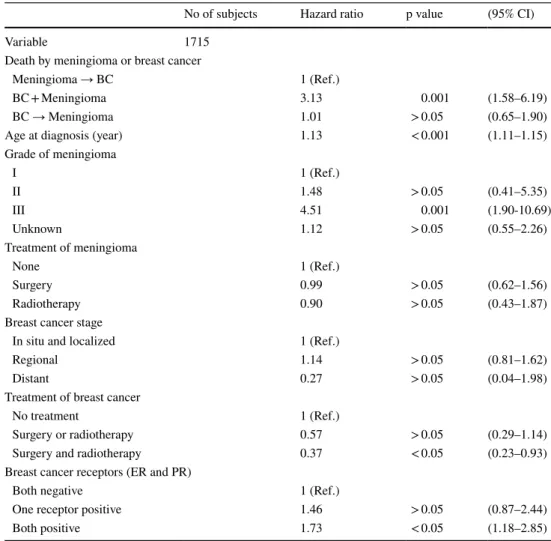
The unadjusted survival analysis showed that between group 1 (BC → M) and group 3 (M → BC), there was no statistical difference in the hazard risk of death (group 3 (M → BC) HR 0.87; 95% CI 0.52–1.46, p:0.56), opposite to what was identified in group 2 (BC + M) (HR 3.13, 95% CI 1.62–6.04, p < 0.001) (Table 2).
The characteristics described in the literature particularly associated with meningioma that showed to have influence in the increase of risk of death in the adjusted analysis were: age at diagnosis of the first tumor (HR 1.13, 95% CI 1.11–1.15, p < 0.005) and meningioma grade III (HR 4.51, 95% CI 1.90–10.69, p < 0.005).
When considering the hormonal receptor status in the breast cancer specimens, the presence of both estrogen and progesterone receptors influence survival increasing the risk of death (HR 1.73, 95% CI 1.18–2.85, p < 0.05), when compared with “both receptors negative” (reference group) and “one receptor positive” (HR 1.46, 95% CI 0.87–2.44, p > 0.05) (Table 3).
When stratifying for meningioma grade, in the first group of grade I, II and unknown (grouped together due to the insufficient number of deaths to analyze the categories sepa-rately) only the synchronous cancers posed a risk for mor-tality (HR 3.13, 95% CI 1.62–6.04, p < 0.001). In the grade III meningioma, only the synchronous cancers posed a risk for mortality but with a very high HR (HR 80.6, 95% CI 5.25–1236.9, p < 0.001) (Table 4).
When stratifying for breast cancer hormonal recep-tor status, only the synchronous cancers have a signifi-cant HR. An increased risk of death is noted in BC + M group when at least one of the tested hormonal receptor is
Table 1 Baseline characteristics of the patients with meningioma and meningioma and breast cancer at any time
NOS no other specification, ER estrogen receptor, PR progesterone receptor
BC → Meningioma—Meningioma was diagnosed after the Breast cancer diagnosis, BC + Meningioma—meningioma and breast cancer are syn-chronous cancers, Meningioma → BC—Meningioma preceeds the diagnosis of breast cancer
Meningioma and breast cancer at any time Overall
BC → Meningioma
N (%) BC + MeningiomaN (%) Meningioma → BCN (%) p value
Total n (%) 1158 (64.5) 262 (14.6) 376 (20.9) 1796
Age at the diagnosis (years) 62.1 (12.4) 65.9 (13.2) 64.2 (12.6) < 0.001 67.98
Sex Female (%) 1154 (99.7) 262 (100) 376 (100) > 0.05 1792 Race White 967 (83.5) 213 (81.3) 306 (81.4) > 0.05 1486 Black 96 (8.3) 30 (11.4) 43 (11.4) 169 Other 95 (8.2) 18 (6.9) 27 (7.2) 140 Unknown 0 1 (0.4) 0 1
Site of the meningioma
Cerebral meningioma 984 (85.0) 227 (86.6) 323 (85.9) > 0.05 1534 Spinal meningioma 49 (4.2) 11 (4.2) 20 (5.3) 80 Meningioma NOS 125 (10.8) 24 (9.2) 33 (8.8) 182 Grade meningioma I 133 (11.5) 27 (10.3) 60 (16.0) > 0.05 220 II 35 (3.0) 4 (1.5) 8 (2.1) 47 III 27 (2.3) 3 (1.2) 12 (3.2) 42 Unknown 963 (83.2) 228 (87) 296 (78.7) 1487 Meningioma treatment Surgery 385 (33.2) 56 (21.4) 167 (44.4) < 0.001 608 Radiotherapy 90 (7.8) 11 (4.2) 33 (8.8) 134 None 683 (59.0) 195 (74.4) 176 (46.8) 1054
Breast cancer grade
Well differentiated 178 (15.4) 44 (16.8) 78 (20.8) < 0.001 300
Moderately differentiated 413 (35.7) 100 (38.2) 150 (39.9) 663
Poorly differentiated 318 (27.5) 90 (34.4) 115 (30.6) 523
Undifferentiated 31 (2.7) 4 (1.5) 5 (1.3) 40
Unknown 218 (18.7) 24 (9.1) 28 (7.4) 270
Breast cancer stage
In situ and localized 825 (71.2) 115 (43.9) 259 (68.9) < 0.001 1199
Regional 297 (25.6) 95 (36.3) 102 (27.1) 494
Distant 36 (3.1) 52 (19.8) 15 (4.0) 103
Breast cancer treatment
Surgery 577 (49.8) 100 (38.2) 182 (15.7) < 0.001 859
Radiotherapy 8 (0.7) 6 (2.3) 3 (1.1) 17
Surgery and radiotherapy 538 (49.9) 118 (45.0) 169 (64.5) 825
No treatment 35 (3.0) 38 (14.5) 22 (8.4) 95
Breast cancer receptors (ER and PR)
Both negative 449 (38.8) 63 (24.0) 86 (22.8) < 0.001 598
One receptor positive 168 (14.5) 44 (16.8) 48 (12.8) 260
positive—ER+/PR- or ER-/PR + or ER+/PR+ (both recep-tors negative: HR 4.10, 95% CI 1.62–6.04, p < 0.001 and at least one receptor positive: HR 80.6, 95% CI 5.25–1236.9, p < 0.001) (Table 5).
Discussion
The previous literature has focused on epidemiological rela-tion and biological risk factors between breast cancer and
Table 2 Hazard ratios for death of breast cancer and/ or meningioma—unadjusted analysis
BC → Meningioma—Meningioma was diagnosed after the Breast cancer diagnosis, BC + Meningioma— Meningioma and breast cancer are synchronous cancers, Meningioma → BC—Meningioma preceeds the diagnosis of breast cancer
No of subjects Hazard ratio p value (95% CI)
Variable 1715
Death by meningioma and breast cancer Unadjusted
Meningioma → BC 1 (Ref.)
BC + Meningioma 3.13 0.001 (1.62–6.04)
BC → Meningioma 0.87 0.56 (0.52–1.46)
Table 3 Hazard ratios for death of breast cancer and/or meningioma—Adjusted analysis
BC → Meningioma—Meningioma was diagnosed after the Breast cancer diagnosis, BC + Meningioma— Meningioma and breast cancer are synchronous cancers, Meningioma → BC—Meningioma preceeds the diagnosis of breast cancer
No of subjects Hazard ratio p value (95% CI)
Variable 1715
Death by meningioma or breast cancer
Meningioma → BC 1 (Ref.)
BC + Meningioma 3.13 0.001 (1.58–6.19)
BC → Meningioma 1.01 > 0.05 (0.65–1.90)
Age at diagnosis (year) 1.13 < 0.001 (1.11–1.15)
Grade of meningioma I 1 (Ref.) II 1.48 > 0.05 (0.41–5.35) III 4.51 0.001 (1.90-10.69) Unknown 1.12 > 0.05 (0.55–2.26) Treatment of meningioma None 1 (Ref.) Surgery 0.99 > 0.05 (0.62–1.56) Radiotherapy 0.90 > 0.05 (0.43–1.87)
Breast cancer stage
In situ and localized 1 (Ref.)
Regional 1.14 > 0.05 (0.81–1.62)
Distant 0.27 > 0.05 (0.04–1.98)
Treatment of breast cancer
No treatment 1 (Ref.)
Surgery or radiotherapy 0.57 > 0.05 (0.29–1.14)
Surgery and radiotherapy 0.37 < 0.05 (0.23–0.93)
Breast cancer receptors (ER and PR)
Both negative 1 (Ref.)
One receptor positive 1.46 > 0.05 (0.87–2.44)
meningioma—Milano and Grossman [18] first described the clinicopathologic characteristics in the SEER cohort, but little is known about the true impact of these tumors in terms of patients’ oncological prognosis. In this context, the present study shows that synchronous breast cancer and meningioma (BC + M) is associated with a worst prognosis when compared to both breast cancer followed by meningi-oma (BC→M) and meningimeningi-oma followed by breast cancer (M → BC)—metachronous tumors.
When considering both BC + M and BC → M, the first group has a statistically significant worst unadjusted sur-vival (HR 3.13, 95% CI 1.62–6.04; Logrank Test p < 0.001) (Table 3).
So far, the literature has sustained the relation between breast cancer and meningioma is due to common risk factors
such as gender, age, hormone exposure (endogenous or exogenous), and some epidemiological variants [10]. The majority of these studies are based on national oncological databases and share the limitation of controlling for con-founding [20]. Therefore, an adjusted analysis for survival was performed considering age at diagnosis, meningioma grade, breast cancer receptors, treatment of meningioma and treatment of breast cancer.
Increased age at diagnosis (HR 1.13, 95% CI 1.11–1.15) was related with worst prognosis (Table 3). Among elderly, most breast cancers are hormone-responsive [21] and men-ingiomas are benign in this gender. However, the decreased tolerance to treatment side effects and overall physiologic reserve often result in under treatment and higher mortality in this subgroup of patients [20].
Table 4 Hazard ratios for death of breast cancer and/or meningioma, stratified by grade meningioma (unadjusted)
BC → Meningioma—Meningioma was diagnosed after the Breast cancer diagnosis, BC + Meningioma— Meningioma and breast cancer are synchronous cancers, Meningioma → BC—Meningioma preceeds the diagnosis of breast cancer
No of subjects Hazard ratio p value (95% CI)
Variable 1715
Death by breast cancer and meningioma Meningioma grade I, II and unknown
Meningioma → BC 1 (Ref.)
BC + Meningioma 3.13 0.001 (1.62–6.04)
BC → Meningioma 0.87 0.56 (0.52–1.46)
Meningioma grade III
Meningioma→ BC 1 (Ref.)
BC + Meningioma 80.6 0.001 (5.25–1236.9)
BC → Meningioma 2.61 > 0.05 (0.52–12.95)
Table 5 Hazard ratios for death of breast cancer and/or meningioma, stratified by breast cancer receptor (unadjusted)
BC → Meningioma—Meningioma was diagnosed after the Breast cancer diagnosis, BC + Meningioma— Meningioma and breast cancer are synchronous cancers, Meningioma → BC—Meningioma preceeds the diagnosis of breast cancer
No of subjects Hazard ratio p value (95% CI)
Variable 1715
Death by breast cancer and meningioma ER and PR receptors negative
Meningioma → BC 1 (Ref.)
BC + Meningioma 4.10 0.001 (1.62–6.04)
BC → Meningioma 0.55 0.56 (0.52–1.46)
ER or PR receptors positive
Meningioma→ BC 1 (Ref.)
BC + Meningioma 80.6 0.001 (5.25–1236.9)
BC → Meningioma 2.61 > 0.05 (0.52–12.95)
ER and PR receptors positive
Meningioma→ BC 1 (Ref.)
BC + Meningioma 80.6 0.001 (5.25–1236.9)
Grade III meningioma was a risk factor for lower sur-vival (HR 4.51, 95% CI [1.90–10.69) (Table 4). In the lit-erature, high recurrence and mortality rates (75 and 90% respectively in 5 years) are reported [22], which support the present results.
The biology of breast cancer cells was evaluated in terms of ER and PR receptors status. ER+/PR+ receptor status was related with a worse outcome (HR 1.73, 95%CI 1.18–2.85, p < 0.05) (Table 3). This was an unexpected result as the literature supports a better outcome in patients with ER positive breast cancer tumors and also because PR positive tumors are responsive to anti-hormonal treatment [23]. But the lack of information on the HER-2 receptors and details on the breast cancer stage does not allow any conclusions in this regard.
The treatment approach of meningiomas had no impact in overall survival (p > 0.05). On the other hand, when con-sidering breast cancer treatment (“no treatment” as refer-ence group), “surgery and radiotherapy” was related to a better survival (HR 0.37, 95% CI 0.23–0.93) but “surgery or radiotherapy” alone did not influence the outcome (HR 0.57, 95% CI 0.29–1.14) although no information concern-ing chemotherapy is available in this database. These results highlight a better outcome for a more aggressive oncological approach in breast cancer patients. These findings support the breast cancer treatment as one of the main prognostic factors in these patients, which was expected considering the overall higher aggressiveness of this tumor.
The adjusted analysis confirmed the previous results of unadjusted analysis for worst outcome of BC + M group (HR 4.10, 95% CI 1.62–6.04). (Table 3) One possible explana-tion for this finding is the expected morbility and mortality related with both tumors treatment and prognosis. This may reduce life expectancy in this group, as its effects are pre-sent for both tumors from the beginning of the exposure. On the other hand, in BC → M group the mean time between the tumors is 7.6 years where patient had been exposed to only one tumor and its effects. Nevertheless, only 18 death events where related with meningioma in the whole studied population, 2 in BC + M Group. Therefore, we can consider that the mortality due to meningioma was not responsible for this difference and hypothesize about a possible biologi-cal difference between both BC + M and BC → M groups. Indeed, BC + M had the higher prevalence of “poorly dif-ferentiated” (34.4%) breast cancer grade, “regional” (36.3%) and “distant” (19.8%) breast cancer stage and”no treatment” approach either in meningioma (74.4%) and in breast can-cer (14.5%). Unfortunately, in a significant amount of cases (1487 of meningiomas and 270 of breast cancer tumors out of 1796) the tumor grade is unknown, which does not allow further conclusions (Table 1). These results may support the fact that BC characteristics are different among these groups with a trend toward a greater aggressiveness in synchronous
tumors. In fact, a possible interaction between both tumors is supported by the fact that both meningioma and breast cancer are able to produce peptides released in circulation with known promoter oncogenic role [24].
In this study, we looked for the influence in oncological survival of the biology and histology of meningioma and breast cancer in each group. The stratification of the study sample according to meningioma grade revealed no influ-ence in BC → M group but only in BC + M group. (Table 4) On the other hand, when we considered the stratification according to hormonal receptor status, a negative influence in the outcome was observed in the BC + M group with no influence in the BC → M group (Table 5). These findings may provide an indirect support of an increased interaction and aggressiveness of synchronous tumors, as the oncologi-cal survival is dependent of meningioma grade- even though the missing data raises concerns about the power of this covariate analysis and the true impact of the meningioma grade in survival—and breast cancer biology, which does not happen in metachronous tumors. Nevertheless, hormonal receptor positivity is usually related with longer survival rates [25]. For unknown reasons, this was a paradoxal effect in this study.
Our results support for the first time the sequence of exposure as an important factor of prognosis in patients with both tumors. Even though dual-cancer patients outside of known syndromic or familial clinical situations are quite uncommon, a coherent statistical analysis was performed according to the dimension of study population.
Nevertheless, some limitations are noted. The used defini-tion of synchronous tumors as tumors diagnosed in the same year may be questioned. However, this was the possible approach according to the provided year of diagnosis in the database. It should also be noted that the higher frequency of imaging exams performed during breast cancer staging procedures, may also be responsible for a higher number of diagnoses of synchronous tumors. However, this effect exists in all the 3 subgroups, as the follow-up for both malignances implies increased number of imaging exams. The diagnostic definition of meningiomas can also be questioned as 1188 cases have no histology to document (1054 of cases has no treatment and 134 had only radiotherapy) and the similar rate of grade II and III meningiomas doesn’t find its paral-lel in the remaining literature which may highlight different diagnostic criteria along the recruitment time (1973–2012) and the need of a central review of the histological diagno-sis. Nevertheless, magnetic resonance imaging has accurate methods to provide a proper diagnosis of meningioma in the majority of the cases (sensivity 83.3%, specificity 100% and accuracy 93.3%) [25]. Considering the majority of cases were collected in the last years (despite the database started in 1973), the diagnostic assumptions are valid for this study purposes. Finally, a concern towards residual confounding
in this retrospective study exists as the adjustment for all the risk factors mentioned in the literature was not feasible. In this sense, we would like to point that no reliable infor-mation was found in database concerning chemotherapy. Indeed, this is not a major issue for meningiomas as the use of chemotherapy is residual. Nevertheless, it is one of the mainstays of therapy for disseminated breast cancer that should be considered in future studies.
Despite these limitations, this study provides an adjusted survival analysis for consensual risk factors in patients with both breast cancer and meningioma. In future studies, treat-ment-related morbidity should be considered and hormonal receptors should be evaluated not only for breast cancer but also for meningioma. New treatment approaches should be tested for different tumor exposure sequences, as this study suggests this as an important factor for oncological survival.
Conclusion
In this study, synchronous breast cancer and meningioma had shorter survival when compared with metachronous tumors, either in unadjusted or adjusted analysis. Increas-ing age and grade III menIncreas-ingioma were related with lower survival rates. Concerning the treatment approach to breast cancer, both surgery and radiotherapy were related to a bet-ter survival, whereas in meningioma treatment no particular approach proved to be superior when compared with the others. Meningioma grade and hormonal receptor status influenced only synchronous tumors (grade III and hormo-nal receptor positivity related with lower survival rates). A better understanding of this group of patients can provide valuable information for clinicians in order to provide a tai-lored clinical approach.
Acknowledgements The authors would like to thank to Clinical Scholars Research Training Harvard Medical Scholl Portugal Faculty for all the support given to the research included in this paper. They would also like to thank to Michel Milano, MD PhD, Ines Lains MD and Carla Bartosh MD for the help in sharing the methods used in their research which help the authors of this paper in designing this study. Funding The authors had no funding to do the current study. Compliance with ethical standards
Conflict of interest The authors have no conflict of interest to de-clare.
References
1. Backer-Grøndahl T, Moen BH, Torp SH (2012) The histopatho-logical spectrum of human meningiomas. Int J Clin Exp Pathol 5(3):231–242
2. Chen HL, Ding A, Wang FW (2015) Prognostic effect analysis of molecular subtype on young breast cancer patients. Chin J Cancer Res 27(4):428
3. National Cancer Institute, Surveillance, Epidemiology and end results (SEER): SEER limite-use data 1973 – 201 (9 registries databases) [Internet]. [cited 2015 Oct 10];Available from http:// seer.cancer.gov
4. Shoenberg BS, Christine BW, Whisnant JP (1975) Nervous system neoplasms and primary malignancies of other sites The unique association between meningiomas and breast cancer. Neurology 25(8):705–705
5. Smith FP, Slavik M, Macdonald JS (1978) Association of breast cancer with meningioma. Report of two cases and review of the literature. Cancer 42(4):1992–1994
6. Malmer B, Tavelin B, Henriksson R, Grönberg H (2000) Primary brain tumours as second primary: a novel associa-tion between meningioma and colorectal cancer. Int J Cancer 85(1):78–81
7. Ahsan H, Neugut AI, Bruce JN (1995) Association of malig-nant brain tumors and cancers of other sites. J Clin Oncol 13(12):2931–2935
8. Rao G, Giordano SH, Liu J, McCutcheon IE (2009) The associa-tion of breast cancer and meningioma in men and women. Neu-rosurgery 65(3):483–489
9. Custer BS, Koepsell TD, Mueller BA (2002) The association between breast carcinoma and meningioma in women. Cancer 94(6):1626–1635
10. Criscitiello C, Disalvatore D, Santangelo M, Rotmensz N, Bazolli B, Maisonneuve P, Curigliano G (2014) No Link between breast cancer and meningioma: results from a large monoinstitutional retrospective analysis. Cancer Epidemiol Biomark Prevent 23(1):215–217
11. Hsu DW, Efird JT, Hedley-Whyte ET (1997) Progesterone and estrogen receptors in meningiomas: prognostic considerations. J Neurosurg 86(1):113–120
12. Konstantinidou AE, Korkolopoulou P, Mahera H, Kotsiakis X, Hranioti S, Eftychiadis C, Patsouris E (2003) Hormone recep-tors in non-malignant meningiomas correlate with apoptosis, cell proliferation and recurrence-free survival. Histopathology 43(3):280–290
13. Kirsch M, Zhu JJ, Black PM (1997) Analysis of the BRCA1 and BRCA2 genes in sporadic meningiomas. Genes Chromosom Can-cer 20(1):53–59
14. Laugé A, Lefebvre C, Laurent-Puig P, Caux V, Gad S, Eng C, Stoppa-Lyonnet D (1999) No evidence for germline PTEN muta-tions in families with breast and brain tumours. Int J Cancer 84(3):216–219
15. Bethke L, Murray A, Webb E, Schoemaker M, Muir K, McKin-ney P, Lönn S (2008) Comprehensive analysis of DNA repair gene variants and risk of meningioma. J Natl Cancer Inst 100(4):270–276
16. Malmer BS, Feychting M, Lönn S, Lindström S, Grönberg H, Ahlbom A, Kiuru A (2007) Genetic variation in p53 and ATM haplotypes and risk of glioma and meningioma. J Neurooncol 82(3):229–237
17. Bussels B, Maes A, Flamen P, Lambin P, Erven K, Hermans R, Van den Bogaert W (2004) Dose–response relationships within the parotid gland after radiotherapy for head and neck cancer. Radiother oncol 73(3):297–306
18. Wiemels J, Wrensch M, Claus EB (2010) Epidemiology and etiol-ogy of meningioma. J Neurooncol 99(3):307–314
19. Milano MT, Grossman CE (2017) Meningioma in breast cancer patients: population-based analysis of clinicopathologic charac-teristics. Am J Clin Oncol 40(1):11–16
20. Dimitrakopoulos F. I. D., Kottorou A, Antonacopoulou AG, Makatsoris T, Kalofonos HP (2015) Early-stage breast cancer in
the elderly: confronting an old clinical problem. J Breast Cancer 18(3):207–217
21. Cain SA, Smoll NR, Van Heerden J, Tsui A, Drummond KJ (2015) Atypical and malignant meningiomas: Considerations for treatment and efficacy of radiotherapy. J Clin Neurosci 22(11):1742–1748
22. Hefti MM, Hu R, Knoblauch NW, Collins LC, Haibe-Kains B, Tamimi RM, Beck AH (2013) Estrogen receptor negative/pro-gesterone receptor positive breast cancer is not a reproducible subtype. Breast Cancer Res 15(4):1
23. Harz C, Ludwig N, Lang S, Werner TV, Galata V, Backes C, Rettig J (2014) Secretion and immunogenicity of the
meningioma-associated antigen TXNDC16. J Immunol 193(6):3146–3154
24. Grann VR, Troxel AB, Zojwalla NJ, Jacobson JS, Hershman D, Neugut AI (2005) Hormone receptor status and survival in a population-based cohort of patients with breast carcinoma. Cancer 103(11):2241–2251
25. Tantawy HI, Mohamad FF, Ismael AM (2010) The role of advanced MRI techniques in differentiating typical, from atypi-cal and malignant meningiomas. Egypt J Radiol Nucl Med 41(3):411–419