DOI: 10.1111/ajco.12722
O R I G I N A L A R T I C L E
Interleukin-18 (IL-18) is equally expressed in inflammatory
breast cancer and noninflammatory locally advanced breast
cancer: A possible association with chemotherapy response
Marco Antonio Nasser Aguiar
1Carlos Wagner S. Wanderley
2Lívia Maria Soares Nobre
2Mateus Rolim Mendes Alencar
2Maria do Perpétuo Socorro Saldanha
3Alceu Machado Souza
4Deysi Viviana Tenazoa Wong
4Paulo Goberlânio Barros
3,5Paulo Roberto Carvalho Almeida
4Roberto Cesar Pereira Lima-Júnior
2Ronaldo Albuquerque Ribeiro
2,31Department of Surgery, Faculty of Medicine,
Federal University of Ceará, Brazil
2Department of Physiology and Pharmacology,
Faculty of Medicine, Federal University of Ceará, Brazil
3Cancer Institute of Ceará, Brazil 4Department of Pathology and Forensic
Medicine, Faculty of Medicine, Federal Uni-versity of Ceará, Brazil
5Department of Dentistry, University Center
Christus
Correspondence
Professor Roberto Cesar Pereira Lima Junior, Departamento de Fisiologia e Farmacologia, Faculdade de Medicina, Universidade Federal do Ceará, Rua Cel Nunes de Melo, 1315, Rodolfo Teófilo, 60430-270, Fortaleza, Ceará, Brazil. Email: [email protected]
Marco Antonio Nasser Aguiar and Carlos Wag-ner de S. Wanderley contributed equally to the development of this work.
Funding Information
Conselho Nacional de Desenvolvimento Científico e Tecnológico, Grant Number: 307143/2014-7, 458872/2014-8; Coordenação de Aperfeiçoamento de Pessoal de Nível Supe-rior, Grant Number: CAPES-PROEX 2862/2013; Fundação Cearense de Apoio ao Desenvolvi-mento Científico, Grant Number: PRONEX PR2-0101-00054.01.00/15; de Apoio ao Desen-volvimento Científico
Abstract
Aim:Inflammatory breast cancer (IBC) is the most aggressive form of locally advanced breast can-cer. The signs of inflammation such as hyperemia and hyperthermia might suggest the possible
participation of inflammatory mediators. This study investigates stromal and tumor expression
of nuclear factor-kappa B (NF-�B) and interleukin-18 (IL-18) in samples obtained from IBC and
noninflammatory locally advanced breast cancer (LABC) and the influence of these markers on
patients’ prognosis.
Methods:Demographic data, tumor molecular characteristics and overall survival in both groups were also assessed. Furthermore, in this study, we evaluated the expression of IL-18 and p50
nuclear fraction of NF-�B by immunohistochemistry in specimens from IBC and LABC (T4b).
Results:We observed that 24.6% of women were diagnosed with IBC up to age 40. In addition, the patients with IBC showed a lower overall survival when compared to LABC. In regard to
molecu-lar markers, ER+, C-erbB2−or triple negative IBC patients showed a significantly reduced
over-all survival. In addition, a higher IL-18 immunostaining in stroma of IBC and LABC was observed
in comparison with tumor cells, but stromal immunoexpression was similar between IBC and
LABC. Besides, IL-18 positivity seemed be related with a better clinical response to neoadjuvant
chemotherapy. However, NF-�B expression was identical in both groups.
Conclusion:The IL-18 is present in tumor stroma of IBC and LABC and seems to be associated with the complete response to neoadjuvant chemotherapy.
K E Y W O R D S
inflammatory breast cancer, interleukin-18 (IL-18), non-inflammatory locally advanced breast
cancer, nuclear factor-kappa B (NF-�B)
1
I N T RO D U C T I O N
Breast cancer is the most frequent type of cancer in women
world-wide, representing 25% of all cancers diagnosed in women.1
Currently, tumor size, number of affected axillary nodes, the
type and histological grade and hormonal receptors (ER, PR and
C-erbB2) are widely used in clinical practice for determining
prognosis and treatment response.2 However, some specific
sub-types of advanced breast cancers remain associated with poor
prognosis.
The most aggressive type of locally advanced breast cancer is
clini-cally classified as inflammatory breast carcinoma (IBC).3Women
diag-nosed with IBC have an average five-year survival of 30–40%, whereas
patients with noninflammatory locally advanced breast cancer (LABC)
the average survival in five-year is 70–80%.4
The IBC has been called “inflammatory” because the breast often
shows signs of inflammation such as redness, hyperthermia and
edema.5The presence of inflammatory signs support the hypothesis
that immune cells are involved in the carcinogenesis and/or tumor
pro-gression, and seem to be relevant to aggressiveness of IBC. In this
con-text, tumor microenvironment-infiltrating cells possibly play an
impor-tant role by secreting cytokines and other cellular mediators that can
contribute to tumor progression.6
Nuclear factor-kappa B (NF-�B) is a cell-transducing transcription
factor that regulates the inflammatory response, cytokine production
and cell proliferation.7–9Dysregulated activation of NF-�B may result
in an amplified inflammatory response, altered balance of pro- and
anti-apoptotic proteins and cell survival favoring tumor progression.10
Interleukin-18 (IL-18) is one of the cytokines whose expression
par-tially involves NF-�B signaling.11 The IL-18 is a proinflammatory
cytokine that modulates the expression of adhesion molecules, nitric
oxide synthase and chemokine production.12Interestingly, serum
lev-els of IL-18 were found increased in breast cancer patients.13 In
addition, IL-18 expression was described in tumor and surrounding
tumor tissues.13However, whether IL-18 expression accounts for a
worsen prognosis in IBC and LABC breast cancer subtypes is still to be
investigated.
Then, we aimed to investigate the expression of IL-18 and NF-�B in
tumor samples of IBC and LABC and the contribution of these
inflam-matory markers as prognostic factors and the impact on neoadjuvant
chemotherapy response.
2
M AT E R I A L S A N D M E T H O D S
2.1
Study characterization
This was an retrospective study including a series of 75 patients
diag-nosed with inflammatory breast cancer (IBC) and 60 with LABC stage
T4b admitted to Instituto do Cancer do Ceará and Hospital Geral de
Fortaleza, Brazil, between 2012 and 2015. The inclusion criteria were
(a) patients diagnosed with IBC or LABC, (b) enough sample size for
immunohistochemistry studies and (c) previous
immunohistochemi-cal characterization for estrogen receptor (ER), progesterone
recep-tor (PR) and human epidermal growth facrecep-tor 2 (C-erbB2, HER2/neu).
Exclusion criteria were (a) stage IV (metastatic) IBC or LABC patients,
(b) insufficient tissue samples for immunohistochemistry studies and
(c) lack of molecular characterization of tumor samples for ER, PR and
C-erbB2 (HER2/neu).
The study was approved by the Ethics in Research Committee of
Instituto do Câncer do Ceará (Protocol Number 073/2008) Hospital
Geral de Fortaleza (Protocol Number 290201/12), and is accordance
to the Helsinki Declaration (1983).
2.2
Tumor samples
The tumor samples of patients with breast cancer were collected
by core biopsy. The collected material was fixed in 10% formalin,
embedded in paraffin, sectioned with 3-�m thick and then stained
with hematoxylin–eosin (H&E). Then, immunohistochemistry assay
was performed for 29 and 28 samples of IBC and LABC, respectively.
2.3
Tissue Microarray
Tissue samples were obtained from the donor paraffin block with a
manual puncher and arrayed in a matrix of six rows and three columns
with up to 18 samples per recipient block. Liquid paraffin was gently
added to melt the wax cylinders at 60°C. Then, typical H&E staining
and immunohistochemistry assay were performed. Two independent
observers who were unaware of clinicopathological data conducted
the analysis. The following score criteria for tumor and stromal cells,
adapted from the previous publication,14were employed: zero
expres-sion (0), mild reaction (1+), moderate reaction (2+) and strong
reac-tion (3+). Score analysis resulted in a high level of concordance (>90%).
Cases out of concordance were defined by consensus. The average
value obtained for each sample was used to statistical analyzes. Slides
were observed in an Olympus BX41 microscope.
2.4
Immunohistochemistry assay
Paraffin sections were dewaxed and rehydrated. Treatment with a 3%
H2O2solution in methanol, for 10 min, was used to block endogenous
peroxidase. An EnVision FLEX buffer target retrieval solution (Agilent
Technologies, Santa Clara, CA, USA) in 96°C warm water, for 20 min,
was applied to unmask the antigens. Envision Flex Peroxidase-Blocking
Reagent was utilized for 10 min to inhibit unspecified ground reactions.
The slides were incubated with rabbit monoclonal antibodies
anti-NF-�B (Santa Cruz Biotechnology, Dallas, TX, USA; 1:200) or anti-IL-18
(Sigma-Aldrich, St. Louis, MA, USA, 1:400); liver and pancreas tumor
samples were used as positive controls at 1:50 dillution. Negative
con-trols in which no primary antibody was applied were included.
Fol-lowing primary antibody incubation, a secondary biotinylated antibody
donkey anti-rabbit IgG (Agilent Technologies, Santa Clara, CA, USA)
1:100 was applied. Then, the streptavidin-coupled peroxidase
com-plex (TS-125-HR; LabVision, Sao Paulo, Brazil) was used for 15 min. An
automated immunostainer (Agilent Technologies, Santa Clara, CA,
USA) was utilized to process the reactions, and diaminobenzidine was
used as the chromogen. The sections were counterstained with
hema-toxylin, dehydrated, diaphanized, mounted and analyzed.
2.5
Statistical analysis
Patient and tumor characteristics were tabulated and stratified by
breast cancer type (IBCvsLABC) and compared across groups with the
chi-square test or Fisher’s exact test when appropriate. For each
cross-ing, the odds ratios with 95% of confidence interval were calculated.
Overall survival was calculated from the time of diagnosis to the date of
death or the time of last follow-up. The survival curves were estimated
test for differences between groups. The curves were confronted with
variables RE, RP, C-erbB2 and triple negative by log-rank test. In
addi-tion, we used a multivariate analysis by the Cox regression model to
identify the prognosis factors that independently affect the survival of
the patients.P<0.05 was considered significant. All data were
ana-lyzed using GraphPad Prism software version 6.01 and SPSS Statistics
22.0.
3
R E S U LT S
3.1
Clinico-pathological characterization of IBC
versus LABC
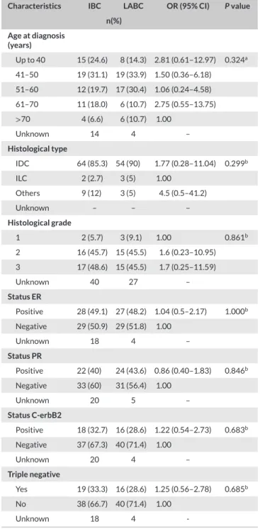
As shown in Table 1, there was no statistical difference (P=0.324)
between the IBC and LABC groups regarding the age of diagnosis.
The mean age observed for the IBC and LABC groups was 49 and 51,
respectively. The characteristics of tumor specimens are also shown in
Table 1. The histology analysis showed that most of the tumors were
invasive ductal carcinoma (IDC), which represents 85.3% of IBC cases
and 90% of LABC. A similar distribution among histological grades was
also observed, with approximately 90% of the IBC and LABC tumors
classified as grades 2 and 3. Regarding the hormone receptors
expres-sion (ER, PR, C-erbB2 and triple negative), no statistical difference
between the two groups was found.
3.2
Overall survival
The overall survival analysis showed that patients diagnosed with IBC
showed a significant (P<0.001) reduced survival when compared to
patients with LABC (Figure 1). The odds ratio of two-year survival from
diagnosis of patients with IBC was 14.4 (95% confidence interval 3.5–
58.9; Table S1) compared to patients with LABC. For five-year survival,
the odds ratio was 6.61 (95% interval confidence 1.7–25.3; Table S1).
Despite no difference concerning locoregional recurrence (data not
shown), patients with IBC clearly survived less (45.5% of patients died
in two years) than patients with LABC (20.4%,P=0.0002).
Consider-ing a five-year survival, 12.3% of patients with IBC are alive, whereas in
the group of LABC this percentage was 37.6% (Figure 1).
3.3
Overall survival according to the expression of
tumor markers (ER, PR and C-erbB2) or triple negative
patients
Figure 2A shows that LABC patients positive for estrogen receptor
(ER+) have a higher survival compared to the ER+ IBC group. We
observed that 50% of ER+LABC patients survived 4.4 years, whereas
ER+IBC individuals survived only 2.3 years (
P=0.006). Regarding PR
expression, no difference between the two populations was observed
(P>0.05, Figure 2B). The contribution of C-erbB2 on patients’
sur-vival was also described. As observed in Figure 2C, IBC patients
that were negative to C-erbB2 (C-erb2−) showed a worse prognosis
(2.1-year median survival) compared to C-erb2- LABC group (4.4-year
survival,P<0.001). Triple negative patients were also assessed. As
TA B L E 1 Clinical and pathological characterization of inflammatory
breast cancer (IBC)vsnoninflammatory locally advanced breast cancer (LABC)
Characteristics IBC LABC OR (95% CI) Pvalue
n(%)
Age at diagnosis (years)
Up to 40 15 (24.6) 8 (14.3) 2.81 (0.61–12.97) 0.324a
41–50 19 (31.1) 19 (33.9) 1.50 (0.36–6.18)
51–60 12 (19.7) 17 (30.4) 1.06 (0.24–4.58)
61–70 11 (18.0) 6 (10.7) 2.75 (0.55–13.75)
>70 4 (6.6) 6 (10.7) 1.00
Unknown 14 4 –
Histological type
IDC 64 (85.3) 54 (90) 1.77 (0.28–11.04) 0.299b
ILC 2 (2.7) 3 (5) 1.00
Others 9 (12) 3 (5) 4.5 (0.5–41.2)
Unknown – – –
Histological grade
1 2 (5.7) 3 (9.1) 1.00 0.861b
2 16 (45.7) 15 (45.5) 1.6 (0.23–10.95)
3 17 (48.6) 15 (45.5) 1.7 (0.25–11.59)
Unknown 40 27 –
Status ER
Positive 28 (49.1) 27 (48.2) 1.04 (0.5–2.17) 1.000b
Negative 29 (50.9) 29 (51.8) 1.00
Unknown 18 4 –
Status PR
Positive 22 (40) 24 (43.6) 0.86 (0.40–1.83) 0.846b
Negative 33 (60) 31 (56.4) 1.00
Unknown 20 5 –
Status C-erbB2
Positive 18 (32.7) 16 (28.6) 1.22 (0.54–2.73) 0.683b
Negative 37 (67.3) 40 (71.4) 1.00
Unknown 20 4 –
Triple negative
Yes 19 (33.3) 16 (28.6) 1.25 (0.56–2.78) 0.685b
No 38 (66.7) 40 (71.4) 1.00
Unknown 18 4
-CI, confidence interval; C-erbB2, growth factor receptor human epidermal 2; ER, estrogen receptor; IDC, invasive ductal carcinomas; ILC, invasive lob-ular carcinomas; OR, odds ratio; PR, progesterone receptor.
aFisher’s exact test. bChi-square test.
shown in Figure 2D, triple negative IBC patients also presented the
worse prognosis (2-year survival) when compared to triple negative
LABC group (3.4-year survival,P=0.049). In multivariate analysis, the
independent prognosis factors affecting the survival of patients with
breast cancer the histologic grade 3 (P=0.021) showed a survival rate
1.001 worse and the triple negative immune staining (P=0.002) with a
F I G U R E 1 Kaplan–Meier curve to overall survival of patients with inflammatory breast cancer (IBC) and noninflammatory locally advanced breast cancer (LABC). Kaplan–Meier method was applied to evaluate survival of patients the log-rank test was used to compare sur-vival curves between groups [Colour figure can be viewed at wileyon-linelibrary.com]
3.4
Immunohistochemistry to NF-
�
B and IL-18
As shown in Table S3, positive immunostaining to NF-�B (score 2–
3) was observed in 46.4% of IBC cases and 36.3% of LABC group
(P>0.05). Depicts representative photomicrographs of
immunostain-ing to NF-�B. The arrowheads indicate a strong immunoexpression of
NF-�B in the tumor tissue (Figure S1). Furthermore, no association
was found between the NF-�B expression with molecular markers (ER,
PR, C-erbB2 or triple negative tumors), neoadjuvant chemotherapy
response, or overall survival in IBC and LABC (data not shown).
IL-18 immunostaining was also investigated (Tables S4 and S5). A
stronger positivity (score 2–3) was found in the stroma (27.6% of
sam-ples) when compared to IL-18-positive tumor cells (0%,P=0.004;
Table S3) in IBC group. Similar findings were observed in LABC group
(50% in the stromavs3.5% in the tumor,P<0.001; Table 4). However,
when IL-18 immunostaining was compared in tumor (IBC: 0%vsLABC:
3.5%,P>0.05) or in stroma (IBC: 27.6%vsLABC: 50%,P=0.093),
no statistical differences were detected (Table S4). A representative
photomicrograph is depicted in Figure 3. The arrows indicate the mild
or moderated IL-18 expression in tumor cells and arrowheads shows
the marked IL-18 expression in stromal cells. In addition, no statistical
association between IL-18 expression with molecular markers (ER, PR,
C-erbB2 or triple negative,P>0.05), overall survival (data not shown)
or for neoadjuvant chemotherapy response (P=0.061; Table 2) was
detected for both breast cancer subtypes IBC and LABC.
4
D I S C U S S I O N
In this study was evaluated the expression of transcription factor
NF-�B and IL-18 in tissue samples obtained from patients with IBC and
LABC. These inflammatory markers were investigated for association
with patients’ prognosis and with the expression of breast cancer
biomarkers (ER, PR and C-erbB2).
Demographic data analysis showed that there was no difference
between the IBC and LABC regarding the prevalence of breast
can-cer subtypes according to age groups. The average age at diagnosis
for IBC and LABC groups was 49 and 51 years old, respectively.
How-ever, younger women (up to 40 years old) seemed to be more affected
by IBC than by LABC. Such finding is in accordance with the
litera-ture, because the IBC seems to be more frequent in younger women,
average age at diagnosis is 59 years old, when compared to 66 years
old for other types of breast cancer.2In a series of 74 patients with
IBC reported by SEER (Surveillance, Epidemiology, and End Results)
showed a mean age at diagnosis, which did not differ between the
groups, although being higher than that observed in the present study,
57.2 years for IBC and 57.4 years for LABC.15These differences
con-cerning the age at diagnosis might involve the population in study,
genetic variability and environmental factors.
In regard to positivity for biomarkers (ER, PR and C-erbB2), no
sig-nificant difference was observed between the two groups, which
cor-roborates previously published data.15 However, it is worth to note
that positivity to ER represented a positive prognostic factor to LABC
group compared with IBC ER+tumors, because a significantly higher
overall survival was found in that group when compared to the IBC ER+
group. Data from the SEER with a series of 2237 patients diagnosed
with IBC and 7985 cases of LABC showed that ER+-expressing LABC
patients have a significantly higher survival (80 months) compared to
patients IBC that express ER, which survive an average of 60 months.15
It is important to mention two aspects based on SEER study: (1)
posi-tivity to ER enhanced patients’ survival of both LABC and IBC groups,
which differed from the present study; (2) survival of North American
patients are significantly higher than patients form this study. Such
dif-ference seems to rely on the time of diagnosis and access to primary
health care. In Brazilian population, cancer diagnosis is commonly on
advanced stage.
Here, we also found a similar C-erbB2 positivity for IBC and LABC
(32.7% and 28.6%, respectively), which is in agreement with data
observed in the study from the Royal Marsden Hospital in London that
showed a 38% of C-erbB2 expression in IBC patients,4and the study
from the MD Anderson Cancer Center identified 44.3% of IBC
sam-ples also positive to C-erbB2.16The absence of oncoprotein C-erbB2
expression has a significant impact on patient survival in IBC group
compared to LABC. Interestingly, other studies have suggested that
lack of C-erbB2 along with ER and PR negativity also significantly
wors-ened the survival of patients in the IBC group when compared to LABC
group.17,18
We also observed a mean survival of only 25 months (2.3 years)
in IBC group, but a 51-month survival (4.3 years) in LABC group.
The worse survival of IBC patients compared to LABC individuals is
F I G U R E 2 Kaplan–Meier curves to overall survival of patients with inflammatory breast cancer (IBC) and noninflammatory locally advanced breast cancer (LABC) stratified by biological breast cancer markers (ER, PR and C-erbB-2). (A) Patients IBC or LABC positive or negative to estrogen receptor (ER). (B) Patients IBC or LABC positive or negative to progesterone receptor (PR). (C) Patients IBC or LABC positive or negative to C-erbB2 receptor (C-erbB2). (D) Patients IBC or LABC triple negative (Trip. Neg.) or not. Kaplan–Meier method was applied to evaluate survival of patients the log-rank test was used to compare survival curves between groups [Colour figure can be viewed at wileyonlinelibrary.com]
F I G U R E 3 Immunostaining for IL-18 in inflammatory breast can-cer (IBC) and noninflammatory locally advanced breast cancan-cer (LABC). The arrows indicate the mild or moderated IL-18 expression in tumor cells and arrowheads shows the marked IL-18 expression in stromal cells. Representative photomicrographs, increase of 400×. [Colour fig-ure can be viewed at wileyonlinelibrary.com]
observed a 3.3-year survival for IBC patients. The study of Low et al.19
reported similar findings (a 3.8-year survival for IBC and 5.8 years for
LABC). Despite the differences in time of survival, the outcome is quite
identical whatever the population.
Taking into account the aforementioned information, the
underly-ing pathology of IBC is likely different of LABC, which is probably
unre-lated to expression of ER, PR and C-erbB2. Therefore, the search for
new biomarkers opens the perspective for potential therapeutic
tar-gets for breast cancer subtypes. Then, we evaluated the expression of
NF-�B and IL-18 in tumors of IBC and LABC.
However, in spite of the broad description of the important role
of NF-�B in the initiation and progression of breast cancer20 and
the positive association of this transcription factor with C-erbB2,
but negative with ER expression,21 we did not identify any
associa-tion between these variables and prognostic factors (ER, PR and
C-erbB2), whatever the type of breast cancer investigated. However,
we observed a high IL-18-positivity in stromal cells in both IBC and
TA B L E 2 Association of IL-18 expression with the clinical neoadjuvant chemotherapy response
Clinical response to neoadjuvant chemotherapy IL-18 OR (95% CI) Pvalue
Negative (0–1) Positive (2–3)
Complete response 3 (11.1%) 6 (30%) 0.5 (0.05–5.51) 0.061a
Partial response 16 (59.3%) 12 (60%) 1.33 (0.16–10.87)
Stable disease 2 (7.4%) 2 (10%) 1.00
Disease progression 6 (22.2%) 0 (0%) 13 (0.4–377.8)
CI, confidence interval; OR, odds ratio.
aFisher’s exact test, invasive ductal.
tumor microenvironment.13 In our opinion, the presence of IL-18
in the stroma might have an important role in the activation of an
inflammatory antitumor response. Despite the lack of improved
sur-vival, other factors together with IL-18 seem to be necessary to
over-come tumor immunosurveillance escape. The use of
chemotherapeu-tic agents is known to activate antitumor immune response.22 As
reviewed by Zitvogel et al. chemotherapy can mediate a multipronged
immunostimulatory effect, thereby reinstating anticancer
immuno-surveillance, increasing the antigenicity of malignant cells, improving
their immunogenicity, and augmenting their susceptibility to immune
attacks.22Interestingly, we found a likely enhanced complete
neoadju-vant response (P=0.061) in association with IL-18. Then, the precise
contribution of IL-18 to improvement of chemotherapy effectiveness
merits further investigation.
Apparently, the aggressiveness of the IBC seem also involve other
immune system mechanisms, such as, IL-10, TGF-� (Transforming
Growth Factor Beta), regulatory T cells and M2 macrophages. These
cells and mediators are responsible to regulate immune response
tumor escape, leading to disease progression and metastasis.6
There-fore, understanding the role of these mediators could help to identify
specific markers for the IBC, what could explain the faster tumor
pro-gression and the shorter disease-free interval.
Immunity is now a major target for treating cancer. This study
attempted to show IL-18 and NF-�B as potential prognostic markers
of neoadjuvant chemotherapy response in patients with IBC and LABC.
The lack of positive correlation might be related to small sample size or
the population of study. Despite no statistical significance for the
mark-ers studied, the present research might be an important step for
effec-tive clinical management of breast cancer, because the role of other
inflammatory markers that regulate immune function must be
investi-gated. Additional studies are highly encouraged also with other ethnic
groups.
5
C O N C L U S I O N
In conclusion, the chemotherapy neoadjuvant complete response
seems to be partially associated with IL-18 expression in stroma of IBC
and LABC.
AC K N O W L E D G M E N T S
This study is dedicated to the loving memory of Prof. Dr. Ronaldo
Albu-querque Ribeiro (in memoriam). R.C.P. Lima-Júnior received a research
Grant from CNPq (Conselho Nacional de Desenvolvimento
Cientí-fico e Tecnológico), Grant Nos.: 307143/2014-7 and
458872/2014-8. This work was also supported by Grants from CAPES (Fundação
Coordenação de Aperfeiçoamento de Pessoal de Nível Superior, Grant
CAPES-PROEX 2862/2013) and FUNCAP (Fundação Cearense de
Apoio ao Desenvolvimento Científico, Grant PRONEX
PR2-0101-00054.01.00/15). de Apoio ao Desenvolvimento Científico.
C O N F L I C T S O F I N T E R E S T
The authors indicate that they have no potential conflicts of interest.
R E F E R E N C E S
1. Torre L, Bray F, Siegel RL, et al. Global Cancer Statistics.CA Cancer J Clin2015;65:87–108.
2. Taghian A, El-Ghamry M, Merajver SD. Overview of the
treat-ment of newly diagnosed, non-metastatic breast câncer.
Up to date [online], Available at: http://www.uptodate.com/
contents/overview-of-the-treatment-of-newly-diagnosed-non-metas tatic-breast-cancer?source=search_result&search=cancer+de+mam
a&selectedTitle=1~150. [Accessed on 16 December 2016].
3. Yamauchi H, Woodward WA, Valero V, et al. Inflammatory breast
cancer: what we know and what we need to learn. Oncologist
2012;17:891–899.
4. Sutherland S, Ashley S, Walsh G, et al. Inflammatory breast cancer
– The Royal Marsden Hospital experience.Cancer2010;116:2815–
2820.
5. Merajver S, Sabel MS. Inflammatory breast cancer. In: Harris JR,
Lipp-man ME, Morrow M, Osborne CK.Diseases of the breast. 3rd ed.
Philadelphia: Lippincott Williams and Wilkins, 2004.
6. Hanahan, Weinberg. Hallmarks of cancer: the next generation.Cell
2011;144:646–674.
7. Giuliani C, Napolitano G, Bucci I, et al. NF-�B transcription factor: role in the pathogenesis of inflammatory, autoimmune, and neoplastic dis-eases and therapy implications.Clin Ter.2001;152:249–253.
8. Biswas DK, Shi Q, Baily S, et al. NF-�B activation in human breast can-cer specimens and its role in cell proliferation and apoptosis.Proc Natl Acad Sci.2004;6:10137–10142.
9. Baker RG, Hayden MS, Ghosh S. NF-�B, inflammation, and metabolic disease.Cell Metabolism2011;13:11–22.
10. Chariot KSA. NF-�B, stem cells and breast cancer: the
links get stronger. Breast Cancer Research 2011;13:https://
doi.org/10.1186/bcr2886.
11. Liu L, Ke Y, Jiang X, et al. Lipopolysaccharide activates ERK-PARP-1-RelA pathway and promotes nuclear factor-�B transcription in murine
12. Dinarello CA. Interleukin 1 and interleukin 18 as mediators of inflam-mation and the aging process.Am J Clin Nutr.2006;83:447–455.
13. Srabović N, Mujagić Z, Mujanović-Mustedanagić J, et al. Interleukin 18 expression in the primary breast cancer tumour tissue.Off Publ Med Assoc Zenica-Doboj Canton Bosnia and Herzegovina2011;8:109–115.
14. Meteoglu I, Erdogdu IH, Meydan N, et al. NF-KappaB expression corre-lates with apoptosis and angiogenesis in clear cell renal cell carcinoma tissues.J Exp Clin Cancer Res.2008;27:53–62.
15. Anderson WF, Chu KC, Chang S. Inflammatory breast carcinoma and noninflammatory locally advanced breast carcinoma: distinct clinico-pathologic entities?J Clin Oncol.2003;21:2254–2259.
16. Robertson FM, Bondy M, Yang W, et al. Inflammatory Breast Can-cer: The Disease, the Biology, the Treatment.CA Caner J Clin.2010;60: 351–375.
17. Dawood S, Broglio K, Gong Y et al. Prognostic significance of
HER-2 status in women with inflammatory breast cancer. Cancer
2008;112:1905–1911.
18. Gianni L, Eiermann W, Semiglazov V et al. Neoadjuvant chemother-apy with trastuzumab followed by adjuvant trastuzumab versus neoadjuvant chemotherapy alone, in patients with HER2-positive locally advanced breast cancer (the NOAH trial): a randomised con-trolled superiority trial with a parallel HER2-negative cohort.Lancet
2010;375:377–384.
19. Low JA, Berman AW, Steinberg SM, et al. Long-term follow-up for
locally advanced and inflammatory breast cancer patients treated with multimodality therapy.J Clin Oncol.2004;22:4067–4074.
20. Zubair A, Frieri M. Role of nuclear factor-ĸB in breast and colorectal cancer.Curr Allergy Asthma Rep.2013;13:44–49.
21. Ahmed KM, Cao N, Li JJ, et al. HER-2 and NF-kappaB as the targets for therapy-resistant breast cancer.Anticancer Res.2006;26:4235–4243.
22. Zitvogel L, Galluzzi L, Smyth MJ, et al. Mechanism of action of conven-tional and targeted anticancer therapies: reinstating immunosurveil-lance.Immunity2013;39:74–88.
S U P P O RT I N G I N F O R M AT I O N
Additional Supporting Information may be found online in the
support-ing information tab for this article.
How to cite this article: Aguiar MAN, Wanderley CWS, Nobre LMS, et al. Interleukin-18 (IL-18) is equally expressed
in inflammatory breast cancer and noninflammatory
locally advanced breast cancer: a possible association with
chemotherapy response. Asia-Pac J Clin Oncol. 2017;0:1–7.