1
Applications of Polyoxometalates in
Medicine and their Putative Mechanisms of
Action
Licenciatura em Bioquímica
Universidade do Algarve
Faculdade de Ciências e Tecnologia
Projeto realizado por Tatiana Marine Authier Orientador: Manuel Aureliano Alves
2
Abstract
Polyoxometalates (POMs) are inorganic metal-oxide assemblies presenting a wide range of possible structure, size and metal-combination, as well as a high solubility in water and large redox chemistry. These properties are of a great interest in medicine and polyoxometalates are investigated to produce inorganic-drugs against virus, bacteria and tumour cells. Results showed that POMs, such as the PM-8 ([NH3Pr]6[Mo7O24]•3H2O) induced inhibition of tumour cell proliferation, whereas
others POMs inhibit DNA- or RNA-virus activation or bacterial growth. While the mechanisms of action of the anti-viral and –bacterial activities are quite well understood, the same does not go for the anti-tumour activity. Nevertheless, the inhibition of tumour proliferation seems to be correlated with the inhibition of certain enzymes such as histone deacetylases and ecto-nucleotidases, or with the programmed death, apoptosis. In this report, these several hypothesis were approach, in a way to explain the anti-tumour, -bacterial and -viral activities of some polyoxometales, but nothing as yet been concluded and further studies are needed for a better understanding.
3
Resumo
Os polioxometalates (POMs) são complexos inorgânicos constituídos por óxido de metais que apresentam uma grande variedade de estrutura, tamanho e combinações de metais, assim como uma grande solubilidade em meio aquoso e numerosas reações de redox. Estas propriedades são de grande interesse na medicina e os polioxometalates são estudados para desenvolver drogas eficazes contra vírus, bactérias e tumores. Os resultados mostraram que alguns POMs, tal como o PM-8 ([NH3Pr]6[Mo7O24] •3H2O) ,
podiam provocar a inibição de proliferações tumorais, enquanto que outros inibiam a activação de DNA/RNA virais ou ainda, o crescimento bacteriano. Embora os mecanismos de ação da actividade antiviral e antibacteriana serem bem conhecidos, não podemos dizer o mesmo da actividade anti-tumoral. Pórem, a inhibição da proliferação tumoral parece ser correlacionada com a inibição de enzimas, tais como as histone deacetilases e as ecto-nucleatidase, ou com a morte programada chamada apoptose. Neste trabalho, várias hipóteses foram abordadas para poder determinar as actividades anti-tumoural, -bacteriana e -viral de certos polioxometalates, mas nada ainda foi concluído e estudos adicionais são necessários para uma melhor compreensão.
4
Contents
Abstract ... 2 Resumo ... 3 I – Introduction ... 5 1 – Objective ... 8II – Applications of Polyoxometalates in Medicine ... 10
II.1 – Anti-viral Activity ... 10
II.2 – Anti-bacterial Activity ... 12
II.3 – Anti-tumour Activity ... 14
III – Putative Mechanisms of Action of Polyoxometalates ... 18
III.1 – Inhibition of enzymes ... 19
III.1.1 – Ecto-nucleotidase ... 19
III.1.2 – Histone deacetylases ... 21
III.1.3 – Sialyl- and sulfotransferase ... 23
III.2 – Formation of POM-flavinmononucleotide complex ... 24
III.3 – Apoptosis : activation of caspase and formation of DNA ladder. ... 24
III.4 – Autophagy ... 25
III.5 – Inhibition of viral binding to host cells and/or its penetration ... 26
III.6 – Inhibition of translation or transcription processes and inhibition of mRNA synthesis 27 III.7 – Oxidation of cellular components ... 27
III.8 – Bacteria Morphological Changes ... 28
III.9 – Inhibition of other proteins ... 28
IV – Methodology ... 29
V – Conclusion ... 29
5
I – Introduction
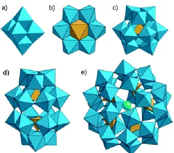
Polyoxometalates (POMs) are inorganic oligomeric assemblies of transition-metal (preferably such as vanadium, molybdenum and tungsten) and oxygen atoms (Fig. 1) having sizes in range of 1 to 3 nm. As illustrated in figure 2, POMs can be represented in various structures, each one consisting on a singular chemical architecture possessing metal oxide clusters with or without heteroatoms.
Figure 1. Two different structural representations of the same polyoxometalate [1].
POMs can be classified in two different families: the isopoly compounds (Fig. 2a), composed by only one type of d0 metal cations, forming a metal oxide cluster, and the heteropoly compounds (Fig. 2b-e) that contain a metal-oxide framework with one or more p-, d- or f-block hetero atoms [2, 3]. The heteropolyoxometalates are by far most studied and researched POMs in medicine than the isopoly compounds. Indeed, they are frequently employed in enzyme inhibition due to their structural and electronic properties, which are easier modified [4]. Nevertheless the isopolyoxometalate have interesting physical properties and are habitually used as building blocks in nanotechnology [1].
Since 1966, the interest in POMs has been growing with an important increase in the last 15 years [5, 6]. Thanks to the progress of new technologies, such as computational chemistry, a better understanding of POM complex chemistry and
6
structure has been achieved [5]. Besides their high solubility in water and wide scale of redox chemistry and photochemistry, polyoxometalates have an overwhelming diversity of different structures and size, with a versatile combination of metals as it can be observed in Fig. 2. Thanks to these properties, POMs are found in an ample range of activity, presenting many different mechanisms of action, some of which are listed in Table 1.
Figure 2. Example of POM’s structure: a) Lindqvist (isopoly compound); b) Anderson (hetero atoms on the surface); c) Keggin (hetero atom in the core); d) Wells-Dawson (2 hetero atoms); e) Preyssler (various type of hetero atoms). Each octahedron (bleu) is represented by a metal atom in its centre cornered by oxygen atoms. At the core of this metal oxide clusters resides the hetero atom (yellow and green) [7].
Even if POMs are soluble in water, the stability of these aggregates has to be assured for their biological applications. Several POMs structures are thermodynamically and kinetically unstable at physiological pH, rendering them as easy targets for reactions which will lead to the production of different structures [4]. Metal substitution in a given POM can provide an improvement in their hydrolytic stability as well as encapsulation by biopolymers or nanoparticles. Meanwhile, several studies
7
have shown a structural combination between a polyoxometalate and another molecule that can stabilize POMs and confer synergetic activities [8-11].
The Mo- and W-based POMs are far more abundant than the V- ones (Table 1). V-based POMs possess a small group of known structures due to their +5 oxidation state rendering the compound more negatively charged. Consequently, this oxidation state affects the redox and basicity properties [7]. Besides anti-bacterial and anti-tumour activities, V-based POMs, such as decavanadate, can exert other physiological functions by interacting with proteins such as myosin, actin and Ca2+-ATPase and affecting mitochondria functions, among other subcellular targets [12-13]. Mo-based POMs are the most studied and used in the anti-tumour activity, whereas the W-based ones are mostly known for their anti-viral effect (Table 1).
Table 1. Examples of POMs applications.
Examples of POMs Application Biological targets Mechanisms of action Ref. α2 -[NMe3H]7[CH3C5H4TiP2W17O61] Na16[Mn4(H2O)2(P2W15O56)2] [PriNH3]6H[Pti2W10O38(O2)2]H2 O Anti-viral HIV, herpes simplex virus, influenza virus,respiratory syncytial virus Inhibition of binding virus/host cell and/or penetration of virus
into the cell
[14-17] K6[P2W18O62]14H2O K4[SiMo12O40] 3H2O K7[PTi2W10O40]6H2O Anti-bacterial methicillin-resistant Staphyloccocus aureus, Streptococcus pneumoniae, Eschericihia coli, H. pylori inhibition of mRNA transcription or translation, inhibition of folding, morphological changes, interference or inhibition of molecular/ionic transport [8, 14, 18-20] K6[P2W18O62]14H2O [Me3NH]6[H2Mo V 12O28(OH)12( MoIVO3)4]2H2O (Na4Co(H2O)6V10O28)18H2O Anti-tumour gastric cancer, breast cancer, kidney cancer, ovaries cancer, liver cancer, pancreatic cancer Apoptosis; inhibition of enzymes [14, 18-20]
8
Furthermore, the low toxicity toward human body and the high solubility in water are main factors that contributed to the development of POMs as drugs. For a polyoxometalate-based drug, it is essentially required that its activity against a biological target must be higher than an already approved drug [14]. Besides the effectiveness and resistance effect, tumour chemotherapeutic agents, as cisplatin or gemcitabine, are also known for their high toxicity. Also, cancer incidence is growing every year all around the world. Indeed, in 2012 the global burden of cancer rose to 14,1 million of new cases and 8,2 million of death per cancer comparing with 12,7 million and 7,6 million respectively in 2008 [23]. Taking this into account, polyoxometalates have been selected by some researchers as alternative anti-tumour substances with promising results in tumour growth suppression [14, 21, 22, 24-34]. Antimicrobial resistance and viral infections also present a global health problem that urge the development of new drugs, such as POMs [8, 9, 16-19, 35, 36].
As the anti-viral, -bacterial and –tumour activities of POMs are put forward, their mechanism of actions are not yet fully understood. Nevertheless it seems that inhibition of enzymes or other proteins has been frequently observed in the anti-tumour, -viral and -bacterial activities [8-10, 14, 30-32, 35]. Inhibition of viral binding to the host cell and/or its penetration and the inhibition of translation/transcription processes have been elucidated in the anti-viral and –bacterial activities respectively. Apoptosis and oxidation of cellular components have also been reported in the tumour anti-proliferation activity.
9
In this work we report recent studies of polyoxometalate applications, such as anti-viral, -bacterial and –tumour activities and some of their putative mechanisms of actions.
10
II – Applications of Polyoxometalates in Medicine
II.1 – Anti-viral Activity
The anti-viral activity of POMs was initially studied and tested in vivo in France in 1972 by Raynaud and Jasmin group. They demonstrated that POMs, such as [NH4]17Na[Na(SbW7O24)3 (SbO7)2]•14H2O (HPA-23), exhibited inhibition of
RNA-dependent DNA polymerases of retroviruses. This discovery led to the hypothetical activity against the human immunodeficiency virus (HIV) that was later demonstrated by some polyoxotungstates [9, 16, 36].
Several years after, Witvrouw et al. have demonstrated that the anti-HIV activity of heteropolytungstates had a structure-activity relationship and could inhibit the replication of HIV-1 and HIV-2 proteases [16]. Data showed that the increase of the number of Keggin or Dawson structures per compound (single, double and triple) diminished the anti-HIV-2 activity. For instance, the triple Keggin structures had lower half inhibitory concentration (IC50)* (> 71,8 µg/mL) than the single Keggin structures (<
4 µg/mL). Nevertheless, the anti-HIV-1 activity was not affected by the structure type, as all POMs exhibited an IC50 between 0,4 to 17 µg/mL [16].
Interestingly, Flütsch et al. have also analysed the structure/activity relation of different POMs in the inhibition of HIV-1 protease [9]. The protease substrate was dissolved in an organic solvent, the dimethyl sulfoxide (DMSO), and then applied in two different buffer solutions. The buffer 1 contained a higher final concentration of DMSO than buffer 2, with 10% and 1% respectively. No significant inhibition was observed in buffer 1 for the 28 different POMs used. Nevertheless, in buffer 2, between water-soluble POMs (POM 1-5 and 18-28), the Dawson structure showed higher rates
11
of protease inhibition, with a total inhibition for POMs 21 (α/β-[P2W18O62]6-), 24 (α1
-[P2W17(NbO2)O61]7-) and 25 (α2-[P2W17(NbO2)O61]7- at 300 nM of concentration.
Regarding DMSO-soluble POMs with organic side chains, it seemed that the inhibitory effect was more correlated to the side chain of the POM than its structure type. Indeed, the Dawson type POM 9 (α2-[P2W17O62SnR]7-, R=C4H9) and the Keggin type POM 12
(α2-[PW11O39SnR]4-, R=C4H9), both with a butyl side chain, could totally inhibit the
protease activity at 300 nM and 3µM respectively. Dawson POM 10 (α2
-[P2W17O62SnR]4-, R=C18H32N3O5) and Keggin POM 13 (α2-[P2W17O62SnR]4-, R=
C18H32N3O5), presenting the same side chain could exhibit a similar protease inhibition
at 300 nM [9].
Shigeta et al. studied the anti-viral activity of POMs containing V/W atoms for several RNA viruses in human T lymphocyte cells (MT-4) [33]. One Keggin-type POM, PM-43 (K5[SiVW11O40]) and two double Keggin- type POMs, PM-1001
(K10Na[(VO)3(SbW9O33)2]26H2O) and PM-1002 (K11H[(VO)3(SbW9O33)2]27H2O)
showed toxicity, by inhibition, against dengue virus (DFV), influenza virus (FluV A), respiratory syncytial virus (RSV), parainfluenza virus (PfluV 2), distemper virus (CDV) and HIV [35]. PM-1001 and PM-1002 (Fig. 3) are the compounds that showed higher toxicity, as their median effective concentration values (EC50)* were lower than 1 µM,
comparing with PM-43 (0,3 < EC50 < 10,7 µM) [35].
* Measures the drug potency by representing the concentration of a compound where 50% of its
12 Figure 3. Example of structural representation of the double Keggin type POM-1001 and POM-1002 [7].
Another Keggin-type structure polyoxotungstate could displayed antiviral activity against an enveloped RNA viruses. In 2013, Y. Qi et al. identified a potent anti-hepatitis C virus in a dose-dependent manner. The Cs2K4Na[SiW9Nb3O40]•H2O
Keggin-type POM (POM-12) abolished completely the Hepatitis C Virus at 50 µM (with a EC50 of 0,8 µM), and around 85% of cell viability remained at such
concentration [36].
Recently, Hosseini et al. have studied the effect of a novel polyoxometalate derivate, [P2W18O62]6- (POM-4960) on the influenza virus (FluV) in MDCK cell line.
Results showed that POM-4960 had a median cytotoxic concentration (CC50)* of 100
µM, and no effective cytotoxic effect on MDCK cells was observed for concentration up to 50 µM while the viral infectivity has been reduce [17].
II.2 – Anti-bacterial Activity
Most of studies, reporting the antibacterial activity of POMs, have been conducted against resistence-adquired bacteria [8, 14, 18-20]. The antibiotic resistance represents a real threat to the world public health and demand taking measures across all sectors of government and society [37]. A high proportion of bacteria are resistant to
13
antibiotics, among which we found those responsible for infectious diseases such as urinary and blood infections, pneumonia. Staphilococcus aureus is one of the most resistant bacteria around the world as well as some Gram negative bacteria who are multi drug resistant [37].
In 2006, Yamase group have considered the antibacterial effect of β-lactam antibiotics by three different POMs against the meticillin-resistant Staphylococcus
aureus (MRSA) and the vancomycin-resistant Staphylococcus aureus (VRSA) [18].
Molybdenum- and tungsten-based POMs (Fig. 4) such as K6[P2W18O62]•14H2O
(P2W18), K4[SiMo12O40] •3H2O (SiMo12) and K7[PTi2W10O40] •6H2O (PTi2W10), were
used in combination with oxacillin antibiotic against 3 strains of MRSA (ATCC43300, SR3605, MRS394-1) and against 2 strains of VRSA (Mu3 and Mu50).
Figure 4. Structural representation of P2W18 (A), PTi2W10 (B) and SiMo12 (C) [14].
They reported that P2W18 and PTi2W10 had a growth inhibitory zone against
almost all strains except Mu3 and SR 3605 at concentration lower than their minimal inhibitory concentration (MIC)*. SiMo12 was the one who showed higher efficiency
since it inhibits growth of all strains at concentrations inferior to its respective MIC [18].
* Represents the lowest concentration of an antimicrobial that will inhibit the visible growth of a
14
Chen et al. have studied a chitosan-POM complex membrane antibacterial activity against Eschericihia coli and Staphylococcus aureus [8]. The polyoanion used in this interesting paper, V10O28(NH4)6 (Fig. 5), can assemble with the cationic chitosan
through non-covalent bonds (electrostatic forces and hydrogen-bond). This polyoxovanadate seems to agglomerate on the membrane surface of the chitosan polymer, presenting some shortcomings which are overcame by the addition of an inorganic cation such as Ca2+. The complex demonstrated a good antibacterial activity with a MIC of 12,5 µM against both bacteria and was able to perform the activity of both component.
Figure 5 - Structural representation of decavanadate [5].
In paralel, Inoue et al. have investigated the antibacterial activity of various types of POMs against Helicobacter pyroli (H. pyroli). The highly negative charged polyoxotungstates, K27[KAs4W40O140] and K18[KSb9W21O86], and the Keggin-type
polyoxotungstate were the ones who showed higher antibacterial activity with MIC values of less than 256 µg/mL, whereas most of polyoxomolybdates exhibited lower activities with MIC values higher than 256 µg/mL [19].
II.3 – Anti-tumour Activity
Many papers have been published regarding the anti-tumour activity of POMs [14, 21, 22, 24-34]. As cancer incidence is growing every year all around the world it
15
has become crucial to find drugs with low toxicity and high efficiency on normal and tumour cells respectively [23]. Japanese have been vigorously working on the anti-tumour activity of polyoxomolybdate [22, 24-26].
In 1992, Fujita et al. have studied the anti-tumour effect of various polyoxomolybdates against human cancer xenografts, on nude mice, such as human colon cancer (Co-4), human breast cancer (MX-1) and human lung cancer (OAT) [22]. Among the 50 polyoxometalates tested, four of them dispatch an anti-tumour activity. But, [NH3Pr]6 [Mo7O24]•3H2O (PM-8), an isopolyoxometalate illustrated in figure 6,
was the most effective with 54,3%, 51% and 61,9% of tumour growth inhibition for Co-4, MX-1 and OAT respectively, at 200 mg/mL of concentration. Two chemotherapeutic drugs were used as a control: cis-platin exhibited 46,6% of inhibition on Co-4 cells, whereas fluracil (5-FU) showed inhibition of 44%, 56% and 44,8% on Co-4, MX-1 and OAT cells [22].
Figure 6. Structural representation of PM-8 [14].
PM-8 was also used as a tumour growth suppressor against gastric cancer cells (MKN45) on bearing mice and on pancreatic cancer cells (AsPC-1) in vitro [24, 25]. Interestingly, after a 48h of treatment with PM-8, MKN45 and AsPC-1 showed the similar IC50 values of 500 µg/mL. The tumour growth suppression of MKN45 was
followed during 70 days of incubation and results showed that PM-8 could reduce tumour growth to almost 35%.
16
While PM-17 was described in 1992 as a toxic compound [22], Ogata et al. synthesized a new PM-17, [Me3NH]6[H2MoV12O28(OH)12(MoVIO3)4]2H2O, obtained by
photo-reduction of the former compound, and tested its anti-tumour activity on AsPC-1 transplanted in nude mice (Fig 7) [26]. This new POM has demonstrated interferences with the solid tumour growth with inhibition rates of 33,5 and 68,3% for concentration of 125 and 500 µg/body/day in a dose-dependent manner. The inhibition in vitro of PM-17 showed an IC50 of 175 and 40 µg/ml against AsPC-1 and MKN45 cells respectively.
Figure 7. Observation of tumour after 41 days of implantation in nude mice (left side represents the saline control; right side represents treated mouse with PM-17) [26].
A new cobalt-based polyoxometalate, (Himi)2[Bi2W20O66(OH)4CO2(H2O)6Na4
(H2O)14]•17H2O (BWCN) (Fig. 8) was studied by Wang et al. on human cancer cells
[28]. The inhibitory effect of BWCN, tested on three different cell lines, was higher in the gastric cancer (SGC-7901) than in colon cancer (HT-29) and HepG2, the less affected. Indeed the inhibitory rates of BWCN at 160µmol/L were approximately 85, 78, and 57% for SGC-7901, HT-29 and HepG2 respectively. Comparative results
17
between BWCN, 5-FU and oxaliplatin showed that the cobalt-based POM exhibited greater inhibitory action with rates of 78, 25 and 42% respectively on HT-29 cells.
Figure 8. Structural representation of BWCN compound (with organic himi molecule in the center) [28].
Another heteropolyoxometalate, K7Na3[Cu4(H2O)2(PW9O34)2]20H2O (PW9O34),
illustrated in figure 9, has been studied against human osteosarcoma derived cell line (MG-63) [34]. PW9O34 revealed good anti-tumour activity as its IC50 value against
MG-63 was 22 µM. Furthermore, its potency as tumour agent was tested against the reference drug cis-platin: at 25 µM, our POM induced a decrease of 60% in cell viability whereas cis-platin only exhibited 15%.
A decavanadate was studied for anti-tumour activity. Zhai et al. have synthesized a cobalt-based POM, (Na4Co(H2O)6V10O28)18H2O (CoV10) and tested its
biological activity against human liver cancer (SMMC-7721) and ovary cancer cell lines (SKOV-3) [21]. Results showed that CoV10 exhibited good anti-tumour activity as it
could inhibit 94,79 % and 90,41 % of SMMC-7721 and SKOV-3 respectively at 6,25 µg/mL, and its IC50 value for both cancer cell lines was estimated lower than 0,26
18
µg/mL. The potency of CoV10 as an anti-tumour drug was compared with the already
used drug 5-FU and proved to be more efficient. At a dose of 1,5625 µg/mL, the activity of CoV10 against SMMC-7721 was similar to 5-FU at a dose of 25 µg/mL.
Figure 9. Structural representation of PW9O34. Legend: Green spheres represent copper
atoms, black spheres are tungsten atoms, red spheres are oxygen atoms and blue spheres represent phosphorus atoms [34].
Another vanadium-based compound was synthesized with carnitine and proved to exhibit anti-tumour activity against various cancer cell lines [33]. The POM {(Me3
N-CH2-CH-(OH)-CH2-COOH)2}{Na4(H2O)16[V10O28]}MeOH (compound 1) presented
IC50 values of 0,72 and 1,8 µM against human lung adenocarcinoma cell line (A549)
and the human breast adenocarcinoma cell line (MDAMB321) respectively. This POM was compared to cis-platin and results showed that compound 1 exhibited lower cytotoxicity against A549 than the reference anti-tumour drug, while its IC50 for
MDAMB321 cells was 1,798 µM against 700 µM of cis-platin.
III – Putative Mechanisms of Action of Polyoxometalates
The mechanisms of action of POMs are not yet fully understood, nonetheless the inhibition of enzymes and proteins are frequently described in bacterial, viral and tumour activities. In this section we will summarize some of POMs’s putative mechanisms of action.
19 III.1 – Inhibition of enzymes
III.1.1 – Ecto-nucleotidase
Ecto-nucleotidases catalyse the hydrolysis of extracellular nucleotides, regulating their levels and leading to the formation of nucleosides and inorganic phophates. Such reactions are drove by four families of nucleotidases: ecto-nucleoside triphosphate diphosphohydrolases (E-NTPDase), ecto-5´-diphosphates, alkaline phosphatases (APs) and ecto-nucleotide pyrophosphatases/phosphodiesterases. The E-NTPDases are able to hydrolyze extracellular nucleotide such as ATP, ADP, UTP and UDP. APs possess a low substrate-specificity and are then able to induce dephosphorylation of several molecules such as nucleosides and proteins resulting in the liberation of inorganic phosphate [32]. This process is important for accelerating the cellular absorption of complex molecules, regulating the activity of other enzymes and providing phosphate groups for various cellular functions. Nucleotides and nucleosides regulate several tissue functions via specific receptors called P2 and P1 respectively and their activation trigger multiple processes affecting, among other things, the cellular metabolism (Fig. 11) [38].
20
Figure 11. Schematic representation of E-NTPDase (CD39) and AP (CD73) activities [39]
Abnormal levels of APs and NTPDases are detected in cancer cells since tumours are abnormal cellular growth proliferating faster than a normal cell [40]. Therefore the inhibition of NTPDases and APs will affect tumour cell metabolism and function.
III.1.1.1 – Ecto-nucleoside triphosphate diphosphohydrolase
Müller et al. have demonstrated that 6 different POMs, presenting different size, shape and charge, could inhibit, differently, three kind of NTPDase [30]. Among all these POMs, only two Keggin-type structure, compound 5 (H3[PW12O40]•H2O) and 7
(K6H2[TiW11CoO40]•13H2O), non-selective inhibitors, have proved anti-tumour activity
[30]. Moreover, compound 7 was the one who had higher inhibition against all three NTPDases with values of inhibition constant (Ki)* inferior to 1 µM. This might suggest that the anti-tumour activity of those two POMs could be correlated to the inhibition of ecto-nucleotidases which will enhanced P2 receptors activation as the levels of extracellular nucleotides increase.
III.1.1.2 – Alkaline phosphatases
Seven polyoxotungstates, illustrated in figure 12, were assessed for their inhibitory effect on APs and as putative anti-tumour agent [32]. Results showed that for a concentration of 100 µM, all compounds (A1, A2, A3, A4, A5, A6, and A7) demonstrated more than 70% inhibition on intestinal alkaline phophatase (IAP) and tissue nonspecific alkaline phosphatase (TNAP). Two POMs, A6 and A3, enhanced
* Defines the concentration of the inhibitor that is required in order to decrease the maximal rate of the
21
higher inhibition on IAP and TNAP respectively, with Ki values of 313 nM (A6) and 135 nM (A3). A3 polyoxotungstate may provide most optimum interaction with TNAP since it possesses the largest number of tungsten and phosphorus atoms [32].
Figure 32. Structural representation of polyoxotungstates studied for their ALP inhibitory activity [32].
Raza et al. have also evaluated the cytotoxicity of these seven polyoxotungstates on lung carcinoma (H157) cell lines and on human corneal epithelial cells (HCEC). Once more, all compound showed high anti-tumour activity in a dose-dependent manner on H157, with 65% of cytotoxicity at 100 µM of A4, being the most efficient. Almost no cytotoxic effect was detected against normal HCEC [32].
III.1.2 – Histone deacetylases
In eukaryotic cells, histones molecules form a complex with cellular DNA called chromatin. Chromatin has a compact organization in which most DNA sequences are structurally inaccessible and functionally inactive [41-42]. Histones are submitted to different kinds of covalent modifications (acetylation, methylation, phosphorylation) creating an epigenetic state. Indeed, the acetylation (induced by histone acetylase) of lysine in the N-terminal tails of histones is related with a looser chromatin state and a
22
gene transcription activation, whereas the deacetylation (induced by histone deacetylase) of the same lysine residues correspond to a more condensed state and a gene silencing (Fig.13).
Figura 13. Schematic representation of histone acetylation and deacetylation [43].
Tumour progression can be induced by epigenetics modifications. Studies have reported that an aberrant recruitment and altered expression of histone deacetylases (HDAC) are responsible for the repression of tumour-suppressor genes leading to malignancies [44]. HDAC inhibitors are able to inhibit tumour cell growth causing a transcriptional changing in genes that regulate biological processes in tumour proliferation [44].
Dong et al. have discovered POM-based HDAC inhibitors, illustrated in figure 14, exhibiting strong anti-tumour activity [31]. Three different germanotungstate Keggin-types (PAC-304, PAC-334 and PAC-320) and one phosphotungstate Dawson-type (PAC-128) were used against several tumour cell lines (A549, SW620, HepG2, MM-231, MGC-803). Results showed that PAC-320 exhibits higher inhibitory effect against HDAC than others POMs. Besides being the stronger HDAC inhibitors,
PAC-23
320 has demonstrated good anti-tumour activity (IC50 of 19,2 µg/ml on HepG2) and low
cytotoxicity (IC50 of 42,8 µg/ml on human hepatic cells). Tests in vivo have proved that
PAC-320 could induce hyperacetylation of histone H3 in a dose-dependent manner.
Figure 14. Structural representation of POM-based HDAC inhibitors [31]. III.1.3 – Sialyl- and sulfotransferase
The sialyltransferase is a glycosyltransferase that catalyse the transfer of a sialic acid residue to the terminal position of an oligosaccharide chains of glycoproteins and glycolipids [45], whereas the sulfotransferase catalyse the transfer reaction of a sulfate group to an acceptor sugar chains on the surface of cells [46]. These modifications of carbohydrate chains play a part in cell-cell recognition serving as a target for bacterial and viral infections. Therefore it is conceivable that the inhibition of these catalytic reactions could lead to the inhibition of biological activities of these sugar chains.
Seko et al. have pointed out the effective non-competitive inhibition of sialyl- and sulfotransferases by POMs [47] in their anti-bacterial activity. Among 20 POMs, three tungstate-based POMs, K6[H2SiNiW11O40]xH2O (POM 6),
K12[Cu3(PW9O34)2]xH2O (POM 13) and K5[SiVW11O40]xH2O (POM 14) proved to
have higher inhibition with IC50 values of 0,2 nM for the α,2,3sialyltransferase
(ST3Gal-I), whereas three vanadium-based POMs K5[H6KV13O31(MePO3)3]16H2O
24
(POM 18) proved to have higher inhibition for the galactose-3-O-sulfotransferase (Gal3ST-2) with IC50 values and 3 nM [47].
III.2 – Formation of POM-flavinmononucleotide complex
Some authors believed that when the polyoxomolybdate enters in mitochondria it can asssemble with a flavimononucleotide (FMN) and yield a 1:1 POM-FMN complex [24]. FMN, also known as riboflavin-5’-phosphate, functions as a prosthetic group serving as an electron carrier by being alternately oxidized (FMN) and reduced (FMNH2). FMN is found in mitochondria and participates in the electron transfer process by carrying electron from NADH to coenzyme Q. The electrochemical proton gradient resulting from these transfers drives ATP synthesis. Therefore by forming a complex with FMN, PM-8 (anti-tumour agent) inhibits the production of ATP in the cell, which might results in cell degeneration and apoptosis [24].
III.3 – Apoptosis : activation of caspase and formation of DNA ladder.
Caspase-3 activation is one of the most current cellular responses to apoptosis [26, 48-50].The caspase-3 zymogene (procaspase) has essentially no activity until it is cleaved by initiator caspases during apoptosis [51, 52]. Apoptosis manifest itself under the influence of different factors. These factors destroy the Bcl-2 family proteins responsible for the activation or inactivation of the mitochondrial permeability transition pore. Once activated, the cytochrome C is released into the cytoplasm and will complex with other molecules forming the apoptosome. The latest will then contribute to the activation of caspase cascade.
25
Zhao et al. have studied the expression of Bax, Bim, Bcl-2 and β-actin (as a control) in HepG2 cells treated with tungsten-based POM K6[P2W18O62]14H2O (POM
Wo) [27]. Results showed that Bax and Bim (two apoptotic promoting family members of Bcl-2) expression were affected by Wo. Meanwhile Bcl-2 expression was not as much affected. In the other hand, Ogata et al. have demonstrated that PM-17 induced caspase-3 activation on both MKN45 and AsPC-1 cells [26]. Wang et al. also measured the cleaved-caspase-3 expression on HT-29 cells treated with BWCN. Results showed an increased expression of procaspase-3 and cleaved-caspase-3 in a dose-dependent manner [28].
Moreover morphological analyses have been conducted on AsPC-1 and MKN45 treated PM-17 and PM-8 and enhanced the detection of apoptotic bodies due to the formation of DNA ladder [25, 26]. Besides, the translocation of phosphatydilserine and the disruption of cytoplasmic membrane have been determined showing results of 77,8% after 48h for MKN45 cells [26].
III.4 – Autophagy
The autophagy is a degradation process were obsoletes parts of the cell are digested inside the lysosomes. This process seems to begin with the confinement of an organelle through unknown membranes, originating an autophagosome which then merges with a lysosome (Fig 15) [53].
26 Figure 15. Autophagy mechanism
Ogata et al. have observed the presence of vesicles containing cytosolic components and organelles, described as figurative autophagosomes, in MKN45 and AsPC-1 cells treated with PM-8 and PM-17 [26]. In order to confirm the hypothesis of an autophagic pathway, they try to localise LC3, a protein conjugation system required for the autophagosome formation as well as its transport and maturation. Results showed that in AsPC-1 cells the LC3-II (truncated form of LC3) expression increased proportionally with time [26].
III.5 – Inhibition of viral binding to host cells and/or its penetration
It is known that HIV specially targets CD4 molecules present in T lymphocytes, monocytes and macrophage lineage. A glycoprotein, denominated gp120 which is located on the surface of the virus, is the principal weapon of HIV because it allows its binding on CD4 cells and the injection of the viral material into the cell [54]. The single Dawson structure compound 13 (α2-[NMe3H]7[CH3C5H4TiP2W17O61]) and the double
Dawson structure compound 24 (Na16[Mn4(H2O)2(P2W15O56)2]) both inhibited the
27
cells, in a concentration dependent manner [16]. It was also demonstrated that PM-1001 strongly inhibits the binding viral gp-120 antibodies [14].
Hosseini et al. confirmed that POM-4960 is likely to have a dual mechanism of action in the inhibition of FluV replication: it inhibits the virus hemaglutination (HA), responsible for the first stage of viral attachment and inhibits fusion of viral particles into the cell [17].
III.6 – Inhibition of translation or transcription processes and inhibition of mRNA synthesis
It is known that the β-lactam resistance of bacteria is acquired by the presence of a peptidoglycan-synthetic enzyme that possesses a low affinity with β-lactam, called PBP2’. This protein is encoded by the mecA gene. Yamase group has investigated the PBP2’expression and data showed that all polyoxometalates (except for SiMo12)
diminished the mec-A-induced mRNA band intensity in the electrophoresis, thus suggesting that POMs were affecting the transcription process [18]. As for the polyoxomolybdate compound, it seemed that the decrease of PBP2’expression was related to another mechanism such as the inhibition of the translation from mecA-induced mRNA to PBP2’ or of the folding of the polypeptide chain [14].
III.7 – Oxidation of cellular components
The redox balance is very important in living organism since most of cellular functions are dependent of these processes, and is well regulated. During mitochondrial oxidative phosphorylation, a leak of electron by the respiratory chain produces ROS. Although they are normal processes occurring in all living cells [55], increased levels of OH•, O2•and H2O2 are commonly found in neoplastic tissues because cancer cells are
28
characterized by an altered pH and an imbalance in the cellular redox homeostasis causing disturbance in mitochondrial function. An overproduction of ROS and a depletion of the antioxidant defence system (resulting in an increase of ROS level in the cell) were observed by León et al. in their study of anti-proliferative tumour effect of POM PW9Cu [34].
Also, the reduction of some POMs allows their penetration and uptake into the bacterial cell, going right after the electron transfer system. Due to the redox potential of POMs SiMo12 and P2W18, the oxidation of some compounds involved in the respiratory
mechanism, leading to reduction of others, could direct to a possible inhibition of ATP synthesis, thus inducing a cell death [18].
III.8 – Bacteria Morphological Changes
POMs are highly negative charged compound that can stimulate bacterial cells in changing their morphological structure leading them to death. Inoue et al. have reported that POMs As4W40 and Sb9W21 could enable morphological changes of H. pyroli from
bacillary to a U-shaped or coccoid form [19]. Fiorani et al. have also revealed extensive degradation of an Escherichia coli strain in the disruption of their rod-shaped morphology by a complex formed of a chitosan compound and POM Mo10V2 [10].
III.9 – Inhibition of other proteins
POM-12, from Qi et al. study, can act directly on Hepatitis C Virus (HCV) virion particles and destabilize the integrity of its structure [36]. Data suggested that it could specifically inhibit HCV infection at an early stage of life cycle. The inhibition of HCVcc and HCVpp, surface proteins of the HCV envelope, with an EC50 of 0,8 µM and
29
proteins. Furthermore, it seemed that POM-12 could endorse the degradation of HCV genomic RNA [36].
Other studies reported that POMs could inhibit proteases in a non-competitive manner at low micro molar concentrations [4, 9, 16]. The HIV-1 protease is important for the maturation of protein components of an infectious HIV virion, thus its inhibition could be responsible for the anti-HIV effect of POMs.
In the other hand, inhibition of proteins has been reported in anti-bacterial activity of POMs [8, 47]. Disturbance in the molecular transport across the membrane, thus devastating the bacteria metabolism, was reported by Chen et al. [8]. Indeed V10O286- is known for its inhibition of Na+/K+-ATPase [56], causing a disturbance in the
molecular transport across the membrane and consequently devastating the bacteria metabolism.
IV – Methodology
Among all the studies reported, several methodologies and techniques were used in a view to evaluate and observe the anti-viral, -bacterial and –tumour activity of polyoxometalates. Some of these are indicated in this section such as: MTT assay, MTS assay and Western blot. Cell apoptosis was evaluated by propidium iodide staining and flow cytometry. Dilatation of nuclear membrane, DNA fragmentation and presence of vacuoles in the cytoplasm were observed by TEM.
30
Polyoxometalates are interesting compounds possessing a rich diversity of structure and size with large redox chemistry and a high solubility in aqueous solutions. Thanks to these properties and others, POMs are studied and used in a large scale of fields, and have become of a great interest in medicine. Indeed many polyoxometalates have proved to be efficient against virus, bacteria and tumour cells.
Anti-viral activity is one of the most known and studied effect of POMs. HPA-23 was the first and only polyoxometalate to have been tested in clinical trials in 1972, but was also one of the most toxic. Despite its noxious effect, HPA-23 gave hope for further studies regarding POMs and their anti-viral activity against HIV and several others virus. It seems that polyoxometalates are able to inhibit the DNA- or RNA-virus activities by preventing the virus-cell host binding and penatration.
On the other hand, several polyoxometalates have demonstrated good activity against antibiotic resistant-bacteria. Some molybdenum- and tungsten-based POMs can disable the β-lactam resistance of MRSA and VRSA by inhibiting the transcription or translation of the mecA-gene, thus preventing the production PBP2’. In other cases, polyoxometalates seem to dispatch their anti-bacterial activity by inhibiting some enzyme, such as sialyl- and sulfotransferase, but also by inducing morphological changes or interfering with the ionic transport systems, leading bacteria to their death.
Besides the high anti-tumour activity of polyoxometalates against a large number of tumour cells, it looks like their mechanisms of action are still difficult to understand. In general, polyoxometalates are able to inhibit tumour growth by inducing apoptosis. Several studies have investigated the possible pathways leading to this cellular death. Some thinks that POMs, once entered into the mitochondrion, can form a complex with the flavin mononucleotide leading to the inhibition of ATP-synthesis.
31
Generation of oxidative stress, activation of caspase-3 and presence of DNA ladder were also factors that contribute to apoptosis. In the other hand, others studies demonstrate that the anti-tumour activity of polyoxometalates is correlated with the inhibition of certain enzymes such as the ecto-nucleotidases (E-NTPD and AP) and histone deacetylases.
Based on all these sutdies, the anti-viral, -bacterial and -tumour activities seems to depend on the polyoxometalate structure and nature, as well as its target. For example, in the study of polyoxometalates against HIV, the single and double Dawson structures were the ones who enhanced higher inhibition among 28 others compounds. In contrast, PM-8 and decavanadate, two isopolycompounds, enhanced different anti-tumour activity. Plus, PM-8 and PM-17 were both tested against AsPC-1 cell lines and both of them exhibit different rate of inhibition. Furthermore, some structures might present appropriate size and conformation for inhibition.
Despite promising results against virus, bacteria and tumour cells, polyoxometalates are not yet tested in clinical trials. This may be due to the lack of understanding of these compounds. To be approved as a drug, the polyoxometalate must show higher activity against its biological target and very low toxicity toward normal cells compared to an already approved drug, if it exists. Additionally, its mechanism of action has to be quite well understood. For instance, as the stability of POMs varies from structures to another it is difficult to say if the activity of a POM is not due or contributed by their fragmentation. Also, the mechanism of inhibition are not always studied, and the binding site on the substrate by the POM are not known.
In the end, we know that certain POMs acts on certain cells and produce a certain activity, and for that we conclude that polyoxometalates are promising inorganic
32 drugs. Nevertheless, further studies must be conducted to better understand the interaction of POMs with and/or within the cell as well as the conjugation of POMs with organic/inorganic compounds that can be used to enhance biological targeting and activity.
Bibliography
[1] D. L. Long and L. Cronin, Chem. Eur. J., 2006, 12, 36.
[2] D-L. Long, E. Burkholder and L. Cronin, Chem. Soc. Rev., 2007, 36, 105-121. [3] J. T. Rhule, C. L. Hill, D. A. Judd, Chem. Rev., 1998, 98, 327-357.
[4] H. Stephan, M. Kubeil, F. Emmerling, C. E. Müller, Eur, J. Inorg. Chem., 2013, 1585-1594.
[5] J. M. Poblet, X. López and C. Bo, Chem. Soc. Rev., 2003, 32, 297-308. [6] Dimitris E. Katsoulis, Chem. Rev., 1998, 98, 359-387.
[7] X. López, J. J. Carbó, C. Bo and J. Poblet, Chem. Soc. Rev., 2012, 41, 7537-7571. [8] S. Chen, G. Wu, D. Long, Y. Liu, Carboh. Polym., 2006, 64, 92-97.
[9] A. Flütsch, T. Schroeder, M. G. Grütter, G. R Patzke, Bioorg. Med. Chem. Lett., 2011, 21, 1162-1166.
[10] G. Fiorani, O. Saoncella, P. Kanner, S. A. Altinkaya, A. Figoli, M. Bonchio, M. Carraro, J. Clust. Sci., 2014, 25, 839-854.
[11] E. Kioseoglou, C. Gabriel, S. Petanidis, V. Psycharis, C. P. Raptopoulou, A. Terzis, A. Salifoglou, Z. Anorg. Allg Chem., 2013, 639, (8-9), 1407-1416.
[12] M. Aureliano, D.C. Crans, J. Inorg. Biochem., 2009, 103, 536-546. [13] M. Aureliano, C.A. Ohlin, J.Inorg. Biochem., 2014, 137, 123-130.
33
[14] Toshihiro Yamase, J. Mater. Chem., 2005, 15, 4773-4782.
[15] D. K. Haapala, C. Jasmin, F. Sinoussi, J. C. Chermann and M. Raynaud, Biomedicine, 1973, 19, 7.
[16] M. Witvrouw, H. Weigold, C. Pannecouque, D. Schols, E. De Clercq and G. Holans, J. Med. Chem., 2000, 43, 778-783.
[17] S.M. Hosseini, E. Amini, M. Tavassoti Kheiri, P. Mehrbod, M. Shahidi, E. Zabihi, IJMCM, 2012, vol.1, No 1.
[18] M. Inoue, T. Suzuki, Y. Fujita, M. Oda, N. Matsumoto, T. Yamase, J. Inorg. Biochem, 2006, 100, 1225-1233
[19] M. Inoue, K. Segawa, S. Matsunaga, N. Matsumoto, M. Oda, T. Yamase, J. Inorg. Biochem., 2005, 99, 1023-1031.
[20] T.Yamase, N. Fukuda, Y. Tajima, Biol. Pharm. Bull., 1996, 19, 459-465.
[21] F. Zhai, X. Wang, D. Li, H. Zhang, R. Li, L. Song, Biomed. & Pharmacoth., 2009, 63, 51-55.
[22] H. Fujita, T. Fujita, T. Sakurai, T. Yamase and Y. Seto, Tohoku J. Exp. Med., 1992, 168, 421-426.
[23] IARC, 2013, Press release nº223.
[24] A. Ogata, S. Mitsui, H. Yanagie, H. Kasano, T. Hisa, T. Yamase, M. Eriguchi, Biomed. Pharmacother., 2005, 59, 240-244.
[25] S. Mitsui, A. Ogata, H. Yanagie, H. Kasano, T. Hisa, T. Yamase, M. Eriguchi, Biomed. Pharmacother., 2006, 60, 353-358.
[26] A. Ogata, H. Yanagie, E. Ishikawa, Y. Morishita, S. Mitsui, A. Yamashita, K. Hasumi, S. Takamoto, T. Yamase and M. Eriguchi, British J. Can, 2008, 98, 399-409. [27] W. Zhao, C. Wang, S. Dong, Y. Li, D. Zhang and L. Han, Int. Confer. Hum. Health and Biomed. Eng., 2011, China.
[28] L. Wang, K. Yu, B-B. Zhou, Z-H. Su, S. Gao, L-L. Chu and J-R. Liu, Dalton Trans., 2014, 43, 6070-6078.
34
[29] Y-T. Li, C-Y. Zhu, Z-Y. Wu, M. Jiang and C-W Yan, Transition Met. Chem., 2010, 35, 597-603.
[30] C. E. Müller, J. Iqbal, Y. Baqi, H. Zimmermann, A. Röllich and H. Stephan, Bioorg. & Med. Chem Let, 2006, 16, 5943-5947.
[31] Z. Dong, R. Tan, J. Cao, Y. Yang, C. Kong, J. Du, S. Zhu, Y. Zhang, J. Lu, B. Huang and S. Liu, Eur. J. Med. Chem., 2011, 2477-2484.
[32] R. Raza, A. Matin, S. Sarwar, M. Barsukova-Stuckart, M. Ibrahim, U. Kortz and J. Iqbal, Dalton Trans., 2012, 41, 14329.
[33] A. Galani,V. Tsitsias, D. Stellas, V. Psycharis, C. P. Raptopoulou, A. Karaliota, J. Inorg., Biochem., 2015, 142, 109-117.
[34] I. E. León, V. Porro, S. Astrada, M. G. Egusquiza, C. I. Cabello, M. Bollati-Fogolin, S. B. Etchevery, Chemico-Biol. Interac., 2014, 142, 222, 87-96.
[35] S. Shigeta, S. Mori, T. Yamase, N. Yamamoto, N. Yamamoto, Biomed. Pharmacoth., 2006, 60, 211-219.
[36] Y. Qi, Y. Xiang, J. Wang, Y. Qi, J. Li, J. Niu, J. Zhong, Antiviral Res., 2013, 100, 392-398.
[37] World Health Organization, 2014, fact sheet n°194.
[38] S. C. Robson, J. Sévigny, H. Zimmermann, Purinerg. Sign., 2006, 2, 409-430. [39] S. Velasquez and E. A. Eugenin, Front. Physiol., 2014, Vol. 5, Art. 96.
[40] B. Katrine, D. Whitaker, D. Eckland, H. J. F. Hodgson, S. Saverymuttu, G. Williams, and D. W. Moss, Clin. Chem., 1982, 28, 374-377.
[41] B. Alberts, A. Johnson, J. Lewis, M. Raff, K. Roberts, P. Walter, Biologie moléculaire de la cellule, 4ème édition, Médecine-Sciences Flammarion, 207-216.
[42]B. Lewin, Genes IX, Jones & Bartlett Publishers, 758-813.
[43] A. Ramanathan, The Newest Cupid on the Block : Epigenetics, Knowing neurons website, 2013.
35
[45] A. Harduin-Lepers, R. Mollicone, P. Delannoy, R. Oriol, Glycobiol., 2005, 15, 805-817.
[46] M: Negishi, L. G. Pedersen, E. Petrotchenko, S. Stevtsov, A. Gorokhov, Y. Kabuta, L. C. Pedersen, Arch. Bioch. Biophysio., 2001, vol. 390, 149-157.
[47] A. Seko, T. Yamase, K. Yamashita, J. Inorg. Chem, 2009, 103, 1061-1066.
[48] I.E. Leon, A.L. Di Virgilio, V. Porro, CI. Muglia, LG. Naso, PA Williams, M. Bollati-Fogolin, SB Etcheverry, Dalton Trans., 2013, 43, 11868-11880.
[49] M. Stianese, A. Basile, A. Mazzone, S. Morello, MC. Turco, C. Pellecchia, J. Cell. Physiol., 2013, 228.
[50] C. Zhang, C. Liu, D. Li, N. Yao, X. Yuan, A. Yu, C. Lu and X. Ma, J. Cell. Physiol., 2010, 222, 444-455.
[51] J. Walters, C. Pop, F. L. Scott, M. Drag, P. Swartz, C. Mattos, G. S. Salvesen and A. C. Clark, Biochem. J., 2009, 424, 335-345.
[52] N. Katunuma, A. Matsui, Q.T. Le, K. Utsumi, G. Salvesen and A. Ohashi, Advan. Enzyme Regul., 2001, vol. 41, 237-250.
[53] B. Alberts, A. Johnson, J. Lewis, M. Raff, K. Roberts, P. Walter, Biologie moléculaire de la cellule, 4ème édition, Médecine-Sciences Flammarion, p745.
[54] C. Tran, C. Hanh, G. R. Dressman and R. C. Kennedy, Proc. Natl. Acad. Sci. USA, 1987, vol.84, 3891-3895.
[55] D. E hamdy, and J. Loscalzo, Antioxidants and Redox signalling, 2012, vol.16, 1323-1367.
[56] D. Krstić, M. Čolović, N. Bošnjaković-Pavlović, A. Spasojević-De Bire and V. Vasić, Gen. Physiol. Biophys., 2009, 28, 302–308.
![Figure 1. Two different structural representations of the same polyoxometalate [1].](https://thumb-eu.123doks.com/thumbv2/123dok_br/18052601.863115/5.892.264.640.395.574/figure-different-structural-representations-polyoxometalate.webp)

![Figure 4. Structural representation of P 2 W 18 (A), PTi 2 W 10 (B) and SiMo 12 (C) [14]](https://thumb-eu.123doks.com/thumbv2/123dok_br/18052601.863115/13.892.226.687.605.779/figure-structural-representation-p-w-pti-w-simo.webp)
![Figure 5 - Structural representation of decavanadate [5].](https://thumb-eu.123doks.com/thumbv2/123dok_br/18052601.863115/14.892.380.558.501.653/figure-structural-representation-of-decavanadate.webp)
![Figure 6. Structural representation of PM-8 [14].](https://thumb-eu.123doks.com/thumbv2/123dok_br/18052601.863115/15.892.354.533.666.822/figure-structural-representation-of-pm.webp)
![Figure 7. Observation of tumour after 41 days of implantation in nude mice (left side represents the saline control; right side represents treated mouse with PM-17) [26]](https://thumb-eu.123doks.com/thumbv2/123dok_br/18052601.863115/16.892.356.536.411.802/figure-observation-tumour-implantation-represents-control-represents-treated.webp)
![Figure 8. Structural representation of BWCN compound (with organic himi molecule in the center) [28]](https://thumb-eu.123doks.com/thumbv2/123dok_br/18052601.863115/17.892.221.681.211.513/figure-structural-representation-bwcn-compound-organic-molecule-center.webp)
![Figure 32. Structural representation of polyoxotungstates studied for their ALP inhibitory activity [32]](https://thumb-eu.123doks.com/thumbv2/123dok_br/18052601.863115/21.892.286.664.295.521/figure-structural-representation-polyoxotungstates-studied-alp-inhibitory-activity.webp)
![Figura 13. Schematic representation of histone acetylation and deacetylation [43].](https://thumb-eu.123doks.com/thumbv2/123dok_br/18052601.863115/22.892.310.636.291.555/figura-schematic-representation-histone-acetylation-deacetylation.webp)
