ADENOSINE RECEPTORS IN VASCULAR SYMPATHETIC
NEUROTRANSMISSION: THE ROLE OF ENDOTHELIUM AND OF
NITRIC OXIDE-MEDIATED EFFECTS AND THEIR RELATION
WITH HYPERTENSION
JOANA BEATRIZ ALVES DA SILVA PINHEIRO DE SOUSA
TESE DO 3º CICLO DE ESTUDOS CONDUCENTE AO GRAU DE DOUTORAMENTO EM CIÊNCIAS FARMACÊUTICAS NA ESPECIALIDADE DE FARMACOLOGIA E FARMACOTERAPIA
Trabalho realizado sob a orientação da Professora Doutora Carmen Diniz Pereira, Professora Auxiliar da Faculdade de Farmácia da Universidade do Porto e co-orientação da Professora Doutora Paula Maria Façanha da Cruz Fresco, Professora Associada da Faculdade de Farmácia da Universidade do Porto.
DE ACORDO COM A LEGISLAÇÃO EM VIGOR, NÃO É PERMITIDA A REPRODUÇÃO DE QUALQUER PARTE DESTA TESE
DECLARAÇÃO
Ao abrigo do nº2 do artigo 8º do Decreto-Lei nº388/70, declara-se que fazem parte integrante desta tese os seguintes trabalhos publicados ou submetidos. Para esses trabalhos, o autor da tese contribuiu maioritariamente na execução das experiências laboratoriais, interpretação de resultados e preparação dos manuscritos.
PUBLICAÇÕES ACEITES OU SUBMETIDAS DURANTE O DOUTORAMENTO Capítulos de livros
Leitão-Rocha A, Sousa JB and Diniz C. Adenosinergic System in the Mesenteric Vessels. In book: The Cardiovascular System - Physiology, Diagnostics and Clinical Implications, 2012. David Gaze (Ed.). Source: InTech
Sousa JB, Fresco P and Diniz C. Imaging receptors with Laser Scanning Confocal Microscopy: qualitative and quantitative analysis. Microscopy: advances in scientific research and education, 2014. A. Méndez-Vilas (Ed.)
Artigos Científicos
Rocha-Pereira C, Sousa JB, Vieira-Rocha MS, Fresco P, Gonçalves J, Diniz C. “Differential inhibition of noradrenaline release mediated by inhibitory A₁-adenosine receptors in the mesenteric vein and artery from normotensive and hypertensive rats.” Neurochemistry International. 62, 399-405 (2013).
Sousa JB, Fresco P, Diniz C. “Endothelial nitric oxide modulatory role on vascular sympathetic reactivity.” Published in Medimond International Proceedings, 43-47 (2013).
Sousa JB, Vieira-Rocha MS, Sá C, Ferreirinha F, Correia-de-Sá P, Fresco P, Diniz C. Lack of endogenous adenosine tonus on sympathetic neurotransmission in spontaneously hypertensive rat mesenteric artery. Accepted for publication on PLoS One, 2014.
Sousa JB, Fresco P, Diniz C. Endothelial dysfunction impairs vascular neurotransmission in tail arteries. Submitted to Neurochemistry International.
Sousa JB, Vieira-Rocha MS, Arribas SM, González MC, Fresco P, Diniz C. Nitric oxide production in mesenteric and tail arteries: differential role of eNOS and nNOS in sympathetic neurotransmission. Submitted to Nitric Oxide: Biology and Chemistry.
AGRADECIMENTOS
“A coisa mais bela que o homem pode experimentar é o mistério. É essa emoção fundamental que está na raiz de toda ciência e toda arte.”
Albert Einstein
Uma Tese de Doutoramento é uma longa viagem, com muitas tribulações pelo caminho. Ao concluir esta tese de doutoramento, quero deixar meu o reconhecimento a pessoas e instituições pelo auxílio que me prestaram e que, de forma decisiva, contribuíram para a sua realização.
Começo por agradecer à Professora Doutora Carmen Diniz pelo seu entusiasmo, apoio, tolerância, disponibilidade, motivação e espirito crítico na orientação desta tese, mas sobretudo pelos alicerces de tudo isto…a amizade que foi crescendo ao longo destes 8 anos em que já trabalhamos juntas.
À Professora Doutora Paula Fresco pelo incentivo e apreciação científica dos resultados experimentais e pelo seu apoio sempre que precisei mas sobretudo pela sua amizade e carinho.
Ao Professor Paulo Correia de Sá por disponibilizar o microscópio confocal para a visualização e aquisição de imagens das artérias em estudo e pelo apoio científico na elaboração do manuscrito resultante da colaboração que estabelecemos.
À Doutora Fátima Ferreirinha pela imensa paciência e disponibilidade na “aventura de explorar” uma nova área para ambas, no “universo da microscopia confocal”. A dificuldade inicial foi ultrapassada graças às suas sugestões, boa vontade e amizade que se criou nas “intermináveis sessões” que jamais esquecerei.
À Professora Silvia Arribas e Maria Carmen González do Departamento de Fisiologia, da Faculdade de Medicina, da Universidade Autónoma de Madrid, pela possibilidade de realizar parte do trabalho desta tese no seu laboratório, pela simpatia e boa vontade com que me receberam e acompanharam o trabalho lá realizado.
Ao Professor Carlos Sá do Centro de Materiais da Universidade do Porto pela elaboração das macros desenvolvidas especificamente para o trabalho de quantificação das imagens de confocal desta tese.
À Ana, à Diana e à Sofia pela amizade, entreajuda e partilha dos bons e maus momentos do nosso dia-a-dia, e pelos momentos de descontração, tão importantes nesta vida de investigação.
A todos os membros do Serviço de Farmacologia da Faculdade de Farmácia da Universidade do Porto pelo bom ambiente, simpatia e sentido de entreajuda que juntos criamos.
Passo agora a agradecer às pessoas mais importantes da minha vida, e que me ajudam a crescer como ser humano.
À minha tão amada família, de modo especial à minha mãe, ao meu pai e aos meus irmãos, por todo o carinho, apoio e ensinamentos em todos os momentos da minha vida, com eles ao meu lado, tudo é mais fácil!
E, por fim, todas as palavras são poucas para agradecer ao Pedro pelo seu amor, incentivo diário e apoio incondicional, por tornar os meus dias mais leves e pela capacidade de transformar uma lágrima num sorriso….
A candidata desenvolveu o seu trabalho experimental com o apoio de uma bolsa de Doutoramento (SFRH/BD/64911/2009) financiada pela Fundação para a Ciência e Tecnologia (FCT) que também apoiou os estudos desenvolvidos em Madrid.
Este trabalho foi, também, financiado pelo Fundo Europeu de Desenvolvimento Regional (FEDER) através do Programa Operacional de Factores de Competitividade (COMPETE) sem os quais a realização desta tese não teria sido possível.
O Laboratório de Farmacologia do Departamento de Ciências do Medicamento da Faculdade de Farmácia da Universidade do Porto e o laboratório associado REQUIMTE providenciaram as instalações e apoio logístico necessário ao trabalho desenvolvido.
ABSTRACT
Increased sympathetic activity has been implicated in hypertension. Adenosine provides a link between local mechanisms of blood flow autoregulation and systemic mechanisms of autonomic cardiovascular regulation. Endothelial cells interact with adenosine mechanisms in many different ways. Endothelium is widely recognized as an important regulator of blood vessel tone via release of various endothelium-derived endogenous substances, such as nitric oxide. Endothelial cells are known to have a very active adenosine metabolism, characterized by a large capacity for uptake and release of this nucleoside. Adenosine may modulate endothelial function via activation of cell membrane receptors. The precise nature of the interaction between adenosine receptor subtypes and endothelial cells and their role in the regulation of endothelial function is not completely understood. However, adenosine-induced production of nitric oxide by endothelial cells is recognized.
The present study intends to clarify the neuromodulatory role exerted by endogenous adenosine and nitric oxide (generated by endothelial and neuronal nitric oxide synthases) in sympathetic neurotransmission, to evaluate if endothelium dysfunction impairs vascular sympathetic neurotransmission and to explore the distribution profiles of adenosine A1 and A2A receptors and of neuronal nitric oxide synthase isoform in the
adventitia layer of mesenteric and tail arteries of both normotensive and hypertensive rats. Electrically-evoked tritium overflow (100 pulses/5 Hz) was evaluated on Wistar Kyoto and spontaneously hypertensive rats mesenteric and tail arteries, pre-incubated with [3H]-noradrenaline. The role of endogenous adenosine and nitric oxide in sympathetic
neurotransmission was studied in the presence of adenosine receptor agonists and antagonists; enzymes substrates and inhibitors; nitric oxide donors and a nucleoside transporter inhibitor. Purine content was determined by HPLC with fluorescence detection. The distribution profiles of adenosine A1 and A2A receptors and of neuronal nitric oxide
synthase isoform in the adventitia layer of mesenteric and tail arteries of Wistar Kyoto and Spontaneously Hypertensive rats was investigated by Laser Scanning Confocal Microscopy. Results indicate a higher electrically-evoked noradrenaline release from hypertensive mesenteric and tail arteries comparatively to those from normotensive vessels. In intact Wistar Kyoto arteries, a facilitation of tritium overflow was observed in the presence of an selective adenosine A1 receptor antagonist. This effect can be ascribed to a
tonic inhibitory effect mediated by A1 receptors. In denuded and hypertensive arteries, in
the presence of an adenosine A2A receptor antagonist a reduction of tritium overflow was
observed. Moreover, when endogenous adenosine bioavailability was increased by the nucleoside transporter inhibitor, tritium overflow increased in intact, denuded and
higher comparatively to normotensive ones.
Nitric oxide donors reduced noradrenaline release in mesenteric arteries while, in tail arteries, caused the opposite effect. In mesenteric arteries, both a neuronal nitric oxide synthase inhibitor and an endothelial nitric oxide synthase inhibitor, increased noradrenaline release. However, in intact tail arteries, inhibition of endothelial nitric oxide synthase lead to a reduction of noradrenaline release whereas inhibition of neuronal nitric oxide synthase was devoid of effect showing that only endothelial nitric oxide synthase is involved in vascular sympathetic neuromodulation. Nitric oxide modulatory role on sympathetic neurotransmission differs in the mesenteric and tail arteries depending on the nitric oxide synthase isoform, endothelial nitric oxide synthase and/or neuronal nitric oxide synthase, involved in its production.
Immunohistochemistry revealed an adenosine A1 receptors redistribution from
sympathetic fibers to Schwann cells, in hypertensive mesenteric arteries adventitia which can explain, at least in part, the absence of effect of these receptors in hypertensive arteries. Moreover, immunohistochemical studies also revealed neuronal nitric oxide synthase locatization presence in Schwann cells which seem to be the main source of nitric oxide to perivascular sympathetic nerves. Moreover, lower amounts of neuronal nitric oxide synthase were found in tail arteries comparatively to those observed in mesenteric arteries.
In the experimental conditions of this work, the inhibition of enzymes involved in reactive oxygen species or prostaglandins production (with apocynin and allopurinol or indomethacin, respectively) failed to alter tritium overflow.
Data highlight the role of purines in hypertension by revealing that an increase in sympathetic activity in hypertensive arteries is occurring due to a higher noradrenaline and ATP release from sympathetic nerves and the loss of endogenous adenosine inhibitory tonus. The observed nerve-to-glial redistribution of inhibitory A1 adenosine receptors in
hypertensive arteries may explain the latter effect. Furthermore, among the endothelium-derived factors studied that could alter vascular sympathetic transmission, adenosine and adenosine receptor mediated mechanisms were clearly impaired by endothelium injury/dysfunction.
Keywords: mesenteric artery, tail artery, adenosine, nitric oxide, noradrenaline, hypertension.
RESUMO
O aumento da atividade simpática tem sido implicado na hipertensão. A adenosina fornece uma ligação entre os mecanismos locais de autoregulação do fluxo sanguíneo e mecanismos sistémicos de regulação cardiovascular do sistema nervoso autónomo. O endotélio é um componente vascular amplamente reconhecido como sendo um importante regulador do tónus vascular através da libertação de diversas substâncias endógenas, como por exemplo o monóxido de azoto. As células endoteliais são reconhecidas como tendo um metabolismo de adenosina muito ativo, caracterizado por uma grande capacidade de captação e libertação do nucleosídeo. A adenosina pode, ainda, modular a função endotelial por meio da ativação de recetores de membrana. O tipo de interação entre os subtipos de recetores de adenosina e as células endoteliais bem como o seu papel na regulação da função endotelial está longe de estar esclarecido. No entanto, a produção de monóxido de azoto pelas células endoteliais induzida pela adenosina é amplamente reconhecida.
Com o presente estudo pretendeu-se esclarecer o papel neuromodulador exercido pela adenosina endógena e pelo monóxido de azoto na neurotransmissão simpática, avaliar se a disfunção endotelial afeta a neurotransmissão simpática vascular e explorar o perfil de distribuição dos recetores da adenosina A1 e A2A e da sintase neuronal do monóxido azoto na camada adventícia de artérias mesentéricas e artérias da cauda de ratos normo e hipertensos.
No trabalho experimental deste trabalho, realizaram-se ensaios de libertação de trítio por estimulação elétrica (100 pulses/5 Hz) com a artéria mesentérica e artéria da cauda de ratos Wistar Kyoto e de ratos espontaneamente hipertensos, pré-incubadas com [3H]-noradrenalina. O papel da adenosina endógena e do monóxido de azoto na
neurotransmissão simpática foi estudada na presença de agonistas e antagonistas de recetores; substratos e inibidores de enzimas; dadores de monóxido de azoto e um inibidor dos transportadores de adenosina. O teor de purinas libertado foi, ainda, determinado por HPLC com deteção por fluorescência.
O perfil de distribuição dos recetores de adenosina A1 e A2A e da sintase neuronal do
monóxido de azoto, na camada adventícia da artéria mesentérica e da artéria da cauda de ratos Wistar Kyoto e espontaneamente hipertensos foi investigado por microscopia confocal.
Os resultados revelaram ocorrer uma maior libertação de noradrenalina por estimulação elétrica na artéria mesentérica e na artéria da cauda de ratos hipertensos comparativamente com os normotensos. Em artérias intactas de ratos normotensos, foi observado um aumento da libertação de trítio na presença do antagonista dos recetores de adenosina A1. Este efeito pode ser atribuído a um efeito tónico inibitório mediado pelos
foi aumentada pelo inibidor dos transportadores de adenosina, verificou-se um aumento da libertação de trítio em artérias intactas, em artérias sem endotélio e em artérias de ratos hipertensos, sugerindo a ocorrência de uma ativação tónica dos recetores de adenosina A2A.
A modulação inibitória tónica de libertação de noradrenalina é mediado pelos recetores de adenosina A1 que parece estar ausente em artérias de animais hipertensos, apesar do seu teor em purinas ser maior comparativamente ao dos normotensos.
Por outro lado, os dadores de monóxido de azoto promoveram uma redução na libertação de noradrenalina na artéria mesentérica, enquanto na artéria da cauda, causaram o efeito oposto. Na artéria mesentérica, tanto o inibidor específico da enzima sintase neuronal do monóxido de azoto como o inibidor específico da sintase endotelial do monóxido de azoto, promoveram um aumento da libertação de noradrenalina. No entanto, na artéria da cauda intacta, a inibição da sintase endotelial do monóxido de azoto reduziu a libertação de noradrenalina enquanto a inibição da sintase neuronal do monóxido de azoto não teve qualquer efeito, mostrando que apenas a sintase endotelial do monóxido de azoto está envolvida na neuromodulação simpática da artéria da cauda. Assim sendo, o papel modulador do monóxido de azoto na neurotransmissão simpática difere nas duas artérias em estudo e depende da isoforma da sintase do monóxido de azoto, endotelial ou neuronal, envolvida na sua produção.
Estudos imunohistoquímicos revelaram uma redistribuição dos recetores A1 de
adenosina das fibras simpáticas para as células de Schwann, na camada adventícia de artérias mesentéricas de ratos hipertensos, o que pode explicar, pelo menos em parte, a falta de efeito destes recetores nestas artérias. Para além disso, os estudos imunohistoquímicos revelaram a presença da sintase neuronal do monóxido de azoto em células de Schwann parecendo ser esta enzima a fonte principal de monóxido de azoto em nervos simpáticos perivasculares. Além disso, verificou-se uma menor quantidade de sintase neuronal do monóxido de azoto na artéria da cauda, comparativamente com a observada na artéria mesentérica.
Nas condições experimentais deste estudo, a inibição das enzimas envolvidas na produção de espécies reativas de oxigénio ou de prostaglandinas (com apocinina e alopurinol ou indometacina, respetivamente) não conseguiu alterar a libertação de noradrenalina.
Os resultados deste trabalho destacam o papel das purinas na hipertensão, revelando que um aumento na atividade simpática em artérias de ratos hipertensos ocorre devido a uma maior libertação de noradrenalina e ATP de nervos simpáticos e a perda do
tónus inibitório da adenosina endógena. A redistribuição nervo-glial observada dos recetores de adenosina A1 inibitórios em artérias de ratos hipertensos pode explicar este
aumento da libertação de noradrenalina e ATP observado. Para além disso, entre as substâncias/fatores derivados do endotélio estudados, que poderiam alterar a transmissão simpática vascular, a adenosina e os mecanismos mediados por este nucleósido estão claramente comprometidos pela disfunção ou lesão endotelial.
Palavras-chave: artéria mesentérica, artéria da cauda, adenosina, monóxido de azoto, noradrenalina, hipertensão.
TABELA DE CONTEÚDOS
PUBLICAÇÕES ACEITES OU SUBMETIDAS DURANTE O DOUTORAMENTO………...iii
AGRADECIMENTOS ... v
ABSTRACT ... ix
RESUMO ... xi
LIST OF FIGURES ... xviii
LIST OF TABLES ... xix
ABBREVIATIONS AND SYMBOLS ... xx
CHAPTER I - General Introduction ... 1
INTRODUCTION Vessels ... 3
Autonomic Nervous System ...6
Sympathetic Neurotransmission ... 8
Sympathetic Neurotransmission: NA/ATP co-transmission ...10
Adenosine ...10
Nitric Oxide ... 13
Hypertension ... 18
Introduction References ... 20
AIMS ... 25
CHAPTER II - Experimental section ... 27
BOOK CHAPTER “Imaging receptors with laser scanning confocal microscopy: qualitative and quantitative analysis” ... 29
MANUSCRIPT I “Lack of endogenous adenosine tonus on sympathetic neurotransmission in spontaneously hypertensive rat mesenteric artery” ... 47
MANUSCRIPT III
“Endothelial nitric oxide modulatory role on vascular sympathetic
reactivity”……….………..95
MANUSCRIPT IV “Nitric oxide production in mesenteric and tail arteries: differential role of eNOS and nNOS in sympathetic neurotransmission” ... 107
CHAPTER III - General Discussion and Conclusions ... 129
Conclusions ... 135
LISTA DE FIGURAS CHAPTER I
GENERAL INTRODUCTION
Figure 1 – Schematic representation of vessels wall ...……….3 Figure 2 – Schematic representation of autonomic nervous system regulation of cardiac and vascular function ………..……….………..7 Figure 3 – Noradrenaline synthesis, storage, release and action………..………..………..9 Figure 4 – Schematic representation of adenosine formation, metabolism and transport, and G-protein coupled adenosine receptors………..………11 Figure 5 – Synthesis of nitric oxide by Nitric Oxide Synthases isoforms………..……14 Figure 6 – Neuronal isoform of nitric oxide synthase……….……15 Figure 7 – Potential mechanisms by which cardiovascular risk factors lead to endothelial dysfunction……….….17
CHAPTER II BOOK CHAPTER
Figure 1 – Laser scanning confocal immunofluorescence images of rat mesenteric artery adventitia immunostained for adenosine A1 receptors, tyrosine hydroxylase and DAPI…...35
Figure 2 – Laser scanning confocal immunofluorescence images of rat mesenteric artery adventitia: a) immunostained for adenosine A1 receptors, DAF-FM DA and DAPI; b)
immunostained for adenosine A1, DHE and DAPI……….37
MANUSCRIPT I
Figure 1 – Influence of endogenous adenosine on tritium overflow modulation from WKY and SHR MA………..…………57 Figure 2 – Effects of NBTI or ITU in the absence or presence of SCH 58261 on the electrically-evoked tritium overflow from WKY and SHR MA……….58 Figure 3 – Basal and electrically-evoked ATP (A) and adenosine (B) release in MA of WKY and SHR……….……..59 Figure 4 – LSCM representative images of WKY and SHR MA exhibiting (A) A1ARs, TH and overlay of A1-TH immunoreactivities, nuclei; (B) Relative means of TH, A1 and TH-A1 overlay expressed as percentage of WKY values. (C) Mean percentage of overlay rate with TH (D) and mean percentage of overlay rate with A1 are depicted……….……..………….61
with TH (D) and mean percentage of overlay rate with A2A are depicted………..…..63 Figure 6 – LSCM images of A1ARs (panel A) and A2AARs (panel B) immunoreactivities in
GFAP-immunoreactive (Schwann) cells located in adventitia of WKY and SHR MA………..65
MANUSCRIPT II
Figure 1 – Time course of tritium outflow from: (A) intact and denuded; (B) SHR and WKY tail artery segments from typical experiments……….82 Figure 2 – Influence of NOS inhibitors on vascular sympathetic transmission in: (A) WKY intact and denuded and (B) SHR tail arteries. ………..…….….84 Figure 3– Prostaglandins and ROS involvement on vascular sympathetic transmission in: (A) WKY intact and denuded and (B) SHR tail arteries……….….……85 Figure 4 – Influence of endogenous adenosine on vascular sympathetic transmission in: (A) WKY intact and denuded and (B) SHR tail arteries……….……..87 Figure 5 – Basal and electrically-evoked adenosine release from tail arteries of WKY and SHR……….……….….…88
MANUSCRIPT III
Figure 1 – Effects of NO synthase inhibitor and NO on noradrenaline release from rat mesenteric and tail arteries……….…..…..101 Figure 2 – Effects of NO synthase substrate and NO synthase inhibitor and NO donors on noradrenaline release from rat intact and denuded tail arteries………..……102
MANUSCRIPT IV
Figure 1 – Time course of tritium outflow from intact and denuded tail artery segments, and from intact and denuded mesenteric artery segments, taken from a typical experiment………..……….116 Figure 2 – Influence of nitric oxide donors on the modulation of electrically-evoked tritium overflow in intact and denuded tail and mesenteric arteries: interaction with L-Arginine and nitric oxide donors, SNP and DEA-NONOate………..………..117 Figure 3 – Influence of nitric oxide inhibitors on the modulation of electrically-evoked tritium overflow in tail and mesenteric arteries: interaction with nitric oxide inhibitors L-NAME, Nω-Propyl-L-arginine hydrochloride and L-NIO dihydrochloride………….……….118 Figure 4 – nNOS in Schwann cells in the adventitia of tail and mesenteric arteries……...120
LISTA DE TABELAS CHAPTER II
BOOK CHAPTER
Table 1 – Description of the algorithm steps for the quantification of receptors, nerves and of the interception of receptors and nerves……….…………..………40
MANUSCRIPT II
Table 1 – Basal tritium outflow (b1), electrically-evoked tritium overflow (S1) and
S2/S1ratios from WKY and SHR segments of tail artery………..………83
MANUSCRIPT IV
Table 1 – Basal tritium outflow (b1), electrically-evoked tritium overflow (S1) and
5′-N- 5′-nucleotidase AC- Adenylyl cyclase ADA- Adenosine deaminase ADP- Adenosine diphosphate AK- Adenosine kinase
AMP- Adenosine monophosphate AR- Adenosine receptors
ATP- Adenosine triphosphate BH4 - Tetrahydrobiopterin Ca2+- calcium
cAMP- cyclic AMP
CGS 21680- 2-p–(2-carboxyethyl)phenethylamino-5´-N-ethylcarboxamidoadenosine hydrochloride CNT- Concentrative transporters COX- Ciclooxigenase CPA- N6-cyclopentyladenosine DAF-2- 4,5-Diaminofluorescein DAF-FM- 4-Amino-5-Methylamino-2',7'-Difluorofluorescein DHE- dihydroetidium DMSO- Dimethylsulphoxide DPCPX- 8-cyclopentyl-1,3-dipropylxanthine eNOS- Endothelial nitric oxide synthase ENT- Equilibrative nucleoside transporters GFP- green fluorescent protein
HPLC- High-performance liquid chromatography ITU- 5-iodotubericidin
K+- potassium
L-NAME- Nω-Nitro-L-arginine methyl ester hydrochloride LSCM- Laser scanning confocal microscopy
NA- Noradrenaline Na+- sodium
NADPH oxidase- Nicotinamide adenine dinucleotide phosphate oxidase NBTI- S-(4-Nitrobenzyl)-6-thioinosine
NO- Nitric oxide
NOS- Nitric oxide synthase ONOO−- Peroxynitrite
PGI2- Prostaglandin 12 PKA- Protein kinase A PLA2- Phospholipase A2
RCFP- reef coral fluorescent protein RNS- Nitrogen oxygen species ROS- Reactive oxygen species SAH- S-adenosyl-homocysteine
SAHH- S-adenosyl-homocysteine hydrolase
SCH 58261- 7-(2-phenylethyl)-5-amino-2-(2-furyl)-pyrazolo-[4,3-e]-1,2,4-triazolo[1,5-c] pyrimidine
SHR- Spontaneously hypertensive rats TH- tyrosine hydroxilase
CHAPTER I
1. INTRODUCTION
1.1 Vessels
Blood vessels are an intricate part of the circulatory system, and are the structures through which blood flows throughout the body. Blood vessels are involved in every pathological condition, including cancer, inflammation and cardiovascular disease. There are several types of vessels, each designed for a special purpose in various parts of the body. Arteries and veins vary largely in the thickness of their walls, the diameters of their lumen or their elasticity.
Arteries and veins have a similar structure (Figure 1). Surrounding the lumen is the innermost layer, the tunica intima, which is the thinnest layer, made up of simple endothelial cells and lines the entire circulatory system. Also included in this thin layer are circular, elastic bands, the internal elastic lamina. The second layer, the tunica media, is made up of more circular elastic bands, the external elastic lamina. The tunica media contains vascular smooth muscle which helps regulate the size of the lumen and those of arteries contains more smooth muscle than the tunica media of their counterpart, the veins, and this allows arteries to constrict and dilate to adjust the volume of blood needed by the tissues that they support.
Figure 1 – Schematic representation of vessels wall. (adapted from Copyright McGraw-Hill Companies, Inc)
When the smooth muscle found within the tunica media is stimulated, it contracts, squeezing the walls of the artery and narrowing the lumen vessel. The term vasoconstriction is used to describe the narrowing of the blood vessel due to contraction of the muscular wall. When an artery constricts, the blood flow decreases and the pressure within the vessel rises. Vasoconstriction is a mechanism that body uses to regulate important functions, such as body temperature and blood pressure. Vasodilation is the opposite of vasoconstriction and consists in the widening of the blood vessel due to relaxation of the muscular wall. When a blood vessel dilates, blood is able to flow through the vessel with less resistance. Therefore, vasodilation has the opposite effect of vasoconstriction on such functions as body temperature and blood pressure. The tunica adventitia is the outermost layer of the arteries and veins. This outer layer is composed of connective tissue that allows the blood vessel to withstand forces acting on the vessel wall. It also contains strong collagen fibers that help anchor the blood vessel to surrounding tissues, and this gives the vessel some stability.
Over the last years, endothelium has emerged as a key regulator of vascular homeostasis, and is no longer looked as having only a barrier function but also as an active signal transducer for circulating influences that modify the vessel wall phenotype. Alteration in endothelial function precedes the development of morphological atherosclerotic changes and can also contribute to lesion development and later clinical complications.[1]
Endothelium is able to respond to physical and chemical signals by production of a wide range of factors that regulate vascular tone, cellular adhesion, thromboresistance, smooth muscle cell proliferation, and vessel wall inflammation. Endothelium effect on vascular tone is achieved by production and release of several vasoactive molecules that relax or constrict the vessel, as well as by response to and modification of circulating vasoactive mediators. This vasomotion plays a direct role in the balance of tissue oxygen supply and metabolic demand by regulation of vessel tone and diameter, and is also involved in the remodeling of vascular structure and long-term organ perfusion.[2]
Veins and arteries both have a similar three-layer structure which contain smooth muscle that is regulated by the sympathetic nervous system. In veins, nerves are distributed throughout the medial smooth muscle coat, often in close apposition (50 nm) to smooth muscle cells. In arteries, nerves are located at the adventitial-medial border, few closer than 2 nm to smooth muscle cells, often with interposing connective tissue and Schwann cell processes.[3] When necessary, the sympathetic nervous system signals arteries to relax or contract, which changes the lumen size of the lumen and thereby regulates blood flow and also affects blood pressure.
The autonomic nervous system is often defined as a motor neuronal system, generally concerned with involuntary body functions, in contrast to the somatic nervous
system, which has both motor and sensory neurons responsible for voluntary muscle function and general sensation. The autonomic nervous system innervates smooth muscle, cardiac muscle, and glands. Physiologically, smooth and cardiac muscles are not completely dependent on autonomic motor neurons for contraction. The first-order neuronal cell body is located in a central nervous system nucleus, and its fiber (axon) travels peripherally to synapse with a second-order neuron, which is located in a peripheral nervous system ganglion. In the peripheral nervous system, the larger diameter axons are surrounded by a lipid-rich myelin sheath formed by the Schwann cells. Many nerve fibers in the central and peripheral nervous system are unmyelinated. In the peripheral nervous system, however, even the unmyelinated fibers are enveloped in Schwann cells. The Schwann cell’s plasma membrane does not spiral repeatedly around the fiber as it does in a myelin sheath, but folds once around each fiber and somewhat overlaps itself along the edges. Most nerve fibers travel through individual channels in the Schwann cell, but small fibers are sometimes bundled together within a single channel.[4] The Schwann cells (in the peripheral nerves) and the satellite cells (in the ganglia) are glial cells (supporting cells) of the peripheral nervous system. Glial cells provide support and protection for neurons. They are thus known as the "supporting cells" of the nervous system. The four main functions of glial cells are: to surround neurons and hold them in place, to supply nutrients and oxygen to neurons, to insulate one neuron from another, and to destroy and remove dead neurons.[5, 6]
The small arteries and arterioles are responsible for the major part of the total resistance to blood flow in the circulatory system. Normally these vessels are maintained in a state of functional constriction (vascular tone) determined by the interaction between intrinsic (myogenic tone) and extrinsic (neurotransmitters, hormones, autacoids, and products of metabolism) factors. Myogenic tone is a property of arteriolar smooth muscle that is independent of any other influence and is the reference tone for the control of vascular resistance by extrinsic factors. Neurogenic mechanisms occupy a central position in the normal regulation of vascular tone and coordinating the degree of transient changes in vascular tone in many regions and organs, enable the rapid redistribution of blood flow to areas functionally important to specific activities.
The renin-angiotensin-aldosterone system plays an important role in regulating blood volume and systemic vascular resistance, which together influence cardiac output and arterial pressure.[7] Renin, which is primarily released by the kidneys and also by other cell types,[8] stimulates the formation of angiotensin in blood and tissues, which in turn stimulates the release of aldosterone from the adrenal cortex.[9, 10] Angiotensin II facilitates noradrenaline release from sympathetic nerve endings and inhibits noradrenaline re-uptake by nerve endings, thereby enhancing sympathetic adrenergic function.[11]
1.2 Autonomic Nervous System
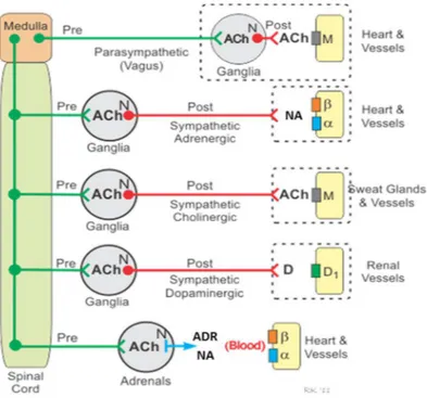
The autonomic nervous system plays an important role in the regulation of cardiac and vascular function. The sympathetic division is the most important nervous division of the autonomic nervous system influencing both the whole-body hemodynamics and the local vascular tone in many areas. The activity of sympathetic neurons usually elicits vasoconstriction roughly proportional to the level of neural activity. This vasoconstriction is determined by various combinations of noradrenaline, neuropeptides, and purines.[12] The sympathetic nerves that originate in the spinal cord run through the spinal cord, where they synapse with preganglionic cell bodies. From these cell bodies run preganglionic fibers that synapse with the cell bodies of postganglionic sympathetic fibers (Figure 2).
Acetylcholine is the neurotransmitter within these ganglia, and the acetylcholine binds to nicotinic receptors on postganglionic neurons. From these neurons arise relatively long postganglionic fibers that travel to their target organ and release noradrenaline as the primary neurotransmitter. Some of the preganglionic sympathetic fibers, instead of synapsing within paravertebral ganglia, synapse in prevertebral ganglia located within the abdomen (celiac, superior mesenteric, and inferior mesenteric ganglia). Postganglionic sympathetic fibers then travel from the prevertebral ganglia to innervate tissues, such as blood vessels, where they release noradrenaline as the primary neurotransmitter. Therefore, sympathetic postganglionic fibers can originate in either paravertebral or prevertebral ganglia (Figure 2).
Figure 2 – Schematic representation of autonomic nervous system regulation of cardiac and vascular function. (adapted from Cardiovascular Physiology Concepts, Richard E. Klabunde,
Lippincott Williams & Wilkins)
Activation of sympathetic adrenergic nerves in the heart releases noradrenaline that binds to adrenergic receptors (primarily beta-adrenoceptors), increasing heart rate, contractility and velocity of electrical impulse conduction. Together, these changes increase cardiac output and arterial blood pressure. Sympathetic adrenergic activation also constricts blood vessels, through the actions of noradrenaline binding to alpha-adrenoceptors leading to increase of arterial blood pressure. Therefore, the cardiovascular effects of vagal activation are primarily mediated through the heart, whereas sympathetic activation affects both the heart and vasculature. Preganglionic sympathetic nerves that synapse in the adrenal glands stimulate catecholamine release that circulates in the blood and affect heart, blood vessels and other organs by activating adrenergic receptors.[13, 14] Moreover, under basal resting conditions, vagal and sympathetic adrenergic nerves are tonically active. In the heart, because vagal influences override sympathetic effects, there is a resting vagal tone that is responsible for maintaining a low heart rate at rest. In contrast, most blood vessels, which have little or no vagal innervation, are dominated by sympathetic adrenergic influences the sympathetic vascular tone, at rest.
1.3 Sympathetic Neurotransmission
Noradrenaline is the classical neurotransmitter, released from sympathetic nerve terminals and is responsible for tonic and reflexive changes in cardiovascular tones. Noradrenaline is one of the catecholamine neurotransmitters that are synthesized from tyrosine by three consecutive enzymatic steps. While tyrosine hydroxylase is the enzyme responsible for the first step of catecholamine biosynthesis, converting tyrosine to l-dopa (the enzymatic rate-limiting step in catecholamine synthesis), and is expressed in all catecholamine neurons, dopamine-β-hydroxylase is the one responsible for conversion of dopamine to noradrenaline and is specifically expressed in noradrenergic neurons (Figure 3).
A presynaptically localized noradrenaline transporter retrieves released noradrenaline to limit the spread and duration of synaptic excitability and allows repackaging of noradrenaline into synaptic vesicles. During sympathetic discharge, noradrenaline produces vasoconstriction by activating α-adrenoceptors located on vascular smooth muscle.
Most of the noradrenaline contained in adrenergic nerves is stored in granular vesicles. Two types of granular vesicles, small and large dense-cored vesicles, both storing noradrenaline, have been identified. Vesicles generated near the Golgi apparatus, in cell bodies, travel by axonal transport to the nerve terminals. Noradrenergic vesicles may also be formed, by endocytosis, within the axons. Noradrenaline is believed to be stored in the vesicles in a complex with adenosine triphosphate (ATP), but the vesicles also contain enzymes involved in noradrenaline synthesis, and other proteins. It is currently accepted that the ATP released together with noradrenaline from sympathetic nerves may act as a sympathetic cotransmitter.[15, 16] Noradrenaline taken up into the axoplasm is subject to two fates, translocation into storage vesicles and deamination by monoamine oxidase. The combination of enzymatic breakdown and vesicular uptake constitute an intraneuronal way to keep axoplasmic noradrenaline concentrations very low. Sympathetic stimulation releases noradrenaline, and binding of noradrenaline to adrenoceptors on cardiovascular smooth muscle cells causes the cells to contract.
Figure 3 – Noradrenaline synthesis, storage, release and action. (adapted from Cardiovascular Pharmacology Concepts. Richard E. Klabunde. Lippincott Williams & Wilkins, Second Edition)
Sympathetic vascular innervation varies widely among vascular beds, with dense innervation present on resistance vessels. The density of sympathetic nerves increases as the arterial caliber decreases, so that small arteries and arterioles, the smallest nutrient vessels possessing smooth muscle cells, have the densest innervation. Sympathetic stimulation in these beds produces profound vasoconstriction. Different stressors can elicit different patterns of sympathoneural outflows and therefore differential noradrenaline release in the various vascular beds. Local sympathoneural release of noradrenaline also markedly affects cardiac function and glandular activity.
A search for new transmitters, other than acetylcholine and noradrenaline, was prompted by the discovery that nerves that relax some smooth muscles did not involve the release of noradrenaline or acetylcholine. A variety of nonadrenergic, noncholinergic transmitters were identified, including ATP, vasoactive intestinal polypeptide, nitric oxide (NO), and neuropeptide Y. The notion of the presence of multiple transmitters is now firmly established becoming clear that within a single neuron multiple transmitter systems may exist, and that within a given ganglion the variety and pattern of neurotransmitters may be quite extensive.
1.3.1 Sympathetic Neurotransmission: NA/ATP co-transmission
Purinergic neurotransmission expands the cotransmission subject. ATP is a primitive extracellular signaling molecule and is now established as a cotransmitter in most nerve types both in the peripheral and central nervous systems. There is purinergic cotransmission together with noradrenaline and neuropeptide Y, from sympathetic nerves; with acetylcholine, from parasympathetic nerves; with calcitonin gene–related peptide and substance P, from sensory-motor nerves, with nitric oxide and vasoactive intestinal polypeptide, from enteric nerves and with glutamate, dopamine, noradrenaline, g-aminobutyric acid, and 5-hydroxytryptamine, from subpopulations of central neurons.[17] Sympathetic purinergic cotransmission has been demonstrated in a variety of blood vessels and the proportion of noradrenaline to ATP is extremely variable in the sympathetic nerves supplying different blood vessels.[18-20] ATP contributes to vasoconstriction produced after activation of the perivascular nerves in SHR. This involves a synergistic interaction with noradrenaline to causes enhanced arterial vasoconstriction, which may contribute to the hypertension.[21]
Distinct presynaptic and postsynaptic mechanisms are observed: ATP is in high concentration in sympathetic synaptic vesicles and, after release, is rapidly catabolized to other adenosine derivatives.[22]
1.4 Adenosine
Adenosine is a naturally endogenous occurring purine nucleoside formed by the degradation of ATP (Figure 4) during energy-consuming processes. ATP is the primary energy source in cells for transport systems and many enzymes. Most ATP is hydrolyzed to ADP, which can be further dephosphorylated to AMP. Most ADP and AMP formed in the cell are rephosphorylated in the mitochondria by enzymatic reactions requiring oxygen. If there are large amounts of ATP hydrolyzed, and especially if there is insufficient oxygen available (i.e., hypoxia), then some of the AMP can be further dephosphorylated to adenosine by the cell membrane associated enzyme, 5'-ectonucleotidase.
Adenosine is rapidly transported into red blood cells (and other cell types) where it is rapidly deaminated, by adenosine deaminase, to inosine, which is further broken down to hypoxanthine, xanthine and uric acid, which is then excreted by the kidneys. Adenosine deamination also occurs in the plasma, but at a lower rate than that occurring within cells. Adenosine can be acted on by adenosine kinase and rephosphorylated to AMP. This salvage pathway helps maintain adenine nucleotide pools in the cell. Extracellular concentrations
of adenosine are increased when energy demands exceed oxygen supply that is, during ischemia.[23]
Figure 4 - Schematic representation of adenosine formation, metabolism and transport, and G-protein coupled adenosine receptors (rectangles representing A1, A2A, A2B, and A3 receptors).
Neurons can release adenosine and adenosine-tri-phosphate (ATP). All cell types express adenosine receptors, adenosine transporters (cylinder), and ecto-nucleotidases that convert ATP into adenosine. ADP, adenosine-di-phosphate; AMP, adenosine-mono-phosphate; SAH, adenosyl-homocysteine; 5′-N, 5′-nucleotidase; AK, adenosine kinase; ADA, adenosine deaminase; SAHH, S-adenosyl-homocysteine hydrolase. Activation of adenosine receptors either inhibits (A1/3 receptors)
or stimulates (A2A/2B receptors) adenylate cyclase and the cyclic AMP (cAMP) pathway. (adapted from
Landolt HP et al, 2012)
Adenosine modulates many physiological processes through activation of four subtypes of G protein–coupled membrane receptors: A1, A2A, A2B, and A3 (Figure 4).[24] It
is well established that adenosine effects occur via activation of A1, A2A, A2B, and A3
adenosine receptors classically considered to be coupled to Gi/o, Gs, Gs/Gq and Gi/o/Gq, respectively.[25] Adenosine receptors are broadly grouped into two main categories: A1 and
A3 receptors, which couple to inhibitory G proteins, and A2A and A2B receptors, which couple
to stimulatory G proteins. However, adenosine receptors are pleiotropic; they can couple with various G proteins and transduction systems according to their degree of activation and their particular cellular or subcellular location.[26] Its physiologic importance depends on the affinity of these receptors and the extracellular concentrations reached. Adenosine
may have tonic actions even during physiologic conditions, mostly through activation of high-affinity A1 and A2A receptors.[27, 28]
Membrane transporters are responsible for the uptake of essential nutrients, modulation of concentrations of physiologically relevant chemicals, and active release of substances such as signaling molecules.[29] Transmembrane transport is a critically important physiological process in all cells and is likely to have evolved early to allow for controlled uptake and release of nonlipophilic compounds. Nucleoside transporters constitute a family of membrane proteins with different pharmacological and kinetic properties.[30] To date it is accepted that there are two types of transporters: Equilibrative Nucleoside Transporters (ENT) and Concentrative Transporters (CNT). The ENT promote equilibrative bidirectional transport processes driven by chemical gradients by facilitated diffusion. ENT are present in most, possibly all, cell types. They might mediate adenosine transporter in both directions, depending on the concentration gradient of adenosine across the plasma membrane. Until the present day, there are four subtypes described: ENT1, ENT2, ENT3 and ENT4. The CNT carry out active inwardly directed concentrative processes, driven by the Na+ electrochemical gradient: Na+-dependent. CNT are expressed
in a tissue-specific fashion. Three subtypes were described: CNT1, CNT2 and CNT3.[31] After stimulation, released adenosine is quickly transported back into cells by an energy-dependent uptake mechanism, which is part of a purine salvage pathway designed to maintain intracellular levels of ATP. The intra and extracellular concentration of adenosine is determined, nearby their receptors, by the existence and function of the transporters. Adenosine transporters contribute to the intra and extracellular concentration of adenosine, modulating its concentration in the vicinity of its receptors.[32-34] The effectiveness of this adenosine transport system is species dependent. It is particularly active in humans, and it is mainly responsible for the extremely short half-life of adenosine in human blood. Adenosine mechanisms are the target of commonly used drugs acting by blockade of adenosine reuptake, thus potentiating its actions or antagonizing adenosine receptors. Adenosine receptors are ubiquitous and, depending on their localization, may mediate opposite effects. This phenomenon is particularly evident in the interaction of adenosine and the autonomic nervous system; adenosine can produce either inhibition or excitation of autonomic neurons.[35] Adenosine inhibits the release of neurotransmitters putatively through presynaptic A1 receptors both in the brain and periphery. This is true for
practically all neurotransmitters studied, including noradrenaline and acetylcholine. Interstitial levels of adenosine are elevated under conditions of increased metabolic demand (exercise) and decreased energy supply (ischemia), reaching physiologically relevant concentrations. Sympathetic activation leads to systemic vasoconstriction, increase in blood pressure, and improved perfusion pressure. This systemic vasoconstriction would
be deleterious to the ischemic organ if not for the simultaneous local inhibitory actions of adenosine, which produce vasodilation and inhibit noradrenaline release. These actions are, for the most part, circumscribed to the local ischemic tissue so that it is protected from sympathetically mediated vasoconstriction while it benefits from the improved perfusion pressure. Adenosine provides a link between local mechanisms of blood flow autoregulation and systemic mechanisms of autonomic cardiovascular regulation.[36]
Adenosine is released into the extracellular space and signals to restore the balance between local energy requirements and energy supply. Endothelial cells interact with adenosine mechanisms in many different ways. Endothelial cells are known to have a very active adenosine metabolism, characterized by a large capacity for uptake and release of the nucleoside, [37, 38] and can be an important source of adenosine released during ischemia.[39] Conversely, adenosine may modulate endothelial function via activation of cell membrane receptors. The precise nature of the interaction between adenosine receptor subtypes and endothelial cells and their role in the regulation of endothelial function is not completely understood. However, adenosine-induced endothelial cells production of nitric oxide is recognized.
1.5 Nitric Oxide
The greatest surprise in the study of autonomic neuroeffector transmission, after the discovery that transmitters other than noradrenaline and acetylcholine exist, was the role of endothelial cells in the relaxation of arterial smooth muscle. This pointed to the existence of a substance that, when released from endothelial cells, acts on vascular smooth muscle cells to relax them. The subsequent identification of this substance as nitric oxide led to the discovery that nitric oxide is an important transmitter at some autonomic neuromuscular junctions.[40, 41]
Nitric oxide is one substance proposed to act as a neurotransmitter in the autonomic nervous system and is very different from the classical neurotransmitters, noradrenaline and acetylcholine. NO is a free radical and potentially very toxic. It passes freely through membranes and, thus, cannot be stored in vesicles for release during an action potential. In addition, it cannot act in a stereospecific way on postjunctional receptors on the target membrane to produce a response. It is now known that NO does act as a neurotransmitter within the autonomic nervous system, particularly within the parasympathetic and enteric nervous systems. NO relaxes smooth muscle by activating soluble guanylate cyclase, which increases cGMP levels. Moreover, the complexity of autonomic control and the range of mechanisms available to peripheral sympathetic and parasympathetic neurons and their
targets are expanded by gaseous molecules (NO) and endothelium released peptides. Also, central nervous system centers involved in autonomic control contain NO synthase, and evidence suggests that NO can mediate either sympathoinhibition or sympathoexcitation. Peripherally, NO exerts tonic vasodilation and mediates acetilcholine-induced vasodilation, as well as catecholamine release and action. Nitrergic neurotransmission has been demonstrated in the gastrointestinal, urogenital, and cardiovascular systems and its loss has also been implicated in a variety of pathological conditions.
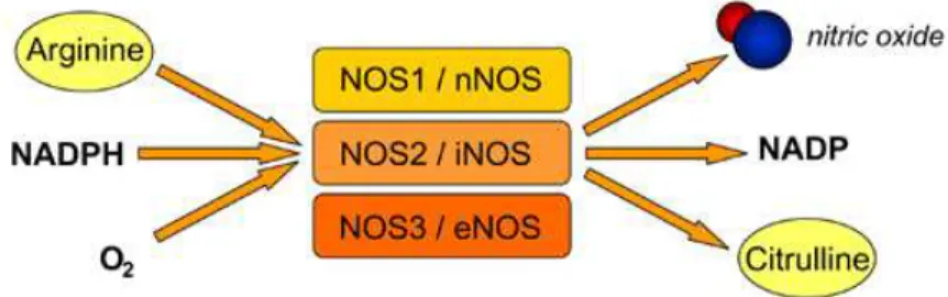
Figure 5 - Synthesis of nitric oxide by Nitric Oxide Synthases (NOS) isoforms.
NO is synthesized by the enzyme nitric oxide synthase (NOS), which can exist in three isoforms (Figure 5). The isoform involved in nitrergic neurotransmission in the autonomic nervous system is nNOS. The other sources of NO are endothelial NOS (eNOS) in the endothelium of blood vessels and inducible NOS (iNOS), which is expressed as part of an immune response.
The neuronal isoform of nitric oxide synthase (nNOS) is a highly complex enzyme (Figure 6) that exists as a dimer, in which each monomer consists of two enzymes in one. One end, the reductase domain, produces electrons during the conversion of reduced NADPH to NADP. Electrons are passed along the enzyme by flavin cofactors until they reach the oxygenase domain. An important region, the calmodulin binding region, links the two domains. In the oxygenase domain there is a heme-binding site. In the presence of electrons, heme and O2, L-arginine is converted to citrulline, resulting in NO synthesis in a
highly regulated, Ca2+-dependent, manner. At basal Ca2+ concentrations within the nerve,
nNOS is inactive.[42] During an action potential, intracellular concentrations of Ca2+
increase resulting in the binding of calmodulin to nNOS and activation of the enzyme. Once activated, nNOS converts L-arginine to citrulline and NO.
Figure 6 - Neuronal isoform of nitric oxide synthase. nNOS contains a reductase domain and a oxygenase domain which are separated by a calmodulin (CaM) binding motif. nNOS is active in dimeric form. (from Nitric Oxide Contribution in the CNS: a NO brainer, May O.)
The most important step in demonstrating nitrergic neurotransmission has been the development of L-arginine analogs that can inhibit NO synthesis such as the non-selective NG-nitro-l-arginine methyl ester (L-NAME) and the selective Nω-propyl-L-arginine
hydrochloride (nNOS inhibitor) and L-NIO dihydrochloride (eNOS inhibitor). NO cannot be stored but reaches the postjunctional target by diffusion. Drugs have been developed that act as NO donors (e.g. sodium nitroprusside and diethylamine NONOate), and these have demonstrated that exogenous NO can mimic the effects of nitrergic neurotransmission. NO reacts with hemoglobin or other reactive species such as superoxide anions, both of which inhibit neurotransmission or the response to NO donors. Once NO enters the postjunctional smooth muscle cell, it produces a response by interacting with the soluble guanylate cyclase. NO binds to its heme group, resulting in the activation of the enzyme and production of increased levels of cyclic guanosine monophosphate, which causes relaxation of smooth muscle. Once the physiologic stimulus of the action potential has ended, Ca2+ levels in the
nerve return to low resting levels and NO synthesis stops. Therefore, the amount of NO synthesized in autonomic nerves does not reach toxic levels.[42, 43] NO, itself, is difficult to detect or measure however, the capacity to synthesize NO can be readily demonstrated
in the autonomic nervous system by immunohistochemical localization of nNOS. nNOS has been localized in both sympathetic and parasympathetic preganglionic neurons and is particularly prominent in the enteric nervous system, where it is localized in the myenteric and submucosal plexuses. nNOS has also been demonstrated in perivascular nerves supplying many blood vessels. Functional studies have demonstrated nitrergic neurotransmission throughout the cardiovascular, urogenital, respiratory, and gastrointestinal systems.[44-47] NO can function either to cause direct inhibition of smooth muscle or as a neuromodulator by inhibiting excitatory transmission.
An endothelium-derived relaxing factor has been shown to be NO. The vascular endothelium synthesizes NO from L-arginine and this action is catalyzed by the action of NO synthases, of which two forms are present in the endothelium. Endothelial eNOS is highly regulated, constitutively active and generates NO in response to shear stress and other physiological stimuli and is responsible for most of the vascular NO· produced.[48] Inducible iNOS is expressed in response to immunological stimuli, is transcriptionally regulated and, once activated, generates large amounts of NO that contribute to pathological conditions. The physiological actions of NO include the regulation of vascular tone and blood pressure, prevention of platelet aggregation and inhibition of vascular smooth muscle proliferation. Many of these actions are a result of the activation by NO of the soluble guanylate cyclase and consequent generation of cyclic guanosine monophosphate.[49] An additional target of NO is cytochrome c oxidase, the terminal enzyme in the electron transport chain, which is inhibited by NO in a manner that is reversible and competitive with oxygen. The consequent reduction of cytochrome c oxidase leads to the release of superoxide anion. This may be an NO-regulated cell signalling system which, under certain circumstances, may lead to the formation of the powerful oxidant species, such as peroxynitrite, that is associated with a variety of vascular diseases.
A functional eNOS oxidizes its substrate L-arginine to L-citrulline and NO·. This normal function of eNOS requires dimerization of the enzyme, the presence of the substrate L-arginine and the essential cofactor tetrahydrobiopterin (BH4), one of the most potent naturally occurring reducing agents. Cardiovascular risk factors such as hypertension, hypercholesterolemia, diabetes mellitus, or chronic smoking stimulate the production of reactive oxygen species in the vascular wall. NADPH oxidases represent major sources of this reactive oxygen species and have been found upregulated and activated in animal models of hypertension, diabetes, and sedentary lifestyle and in patients with cardiovascular risk factors. Superoxide (O2·−) reacts avidly with vascular NO· to form
peroxynitrite (ONOO−).[50, 51] The cofactor BH4 is highly sensitive to oxidation by
ONOO−. Diminished levels of BH4 promote O2·− production by eNOS (referred to as eNOS
oxidative stress has been observed in several in vitro models, in animal models of cardiovascular diseases, and in patients with cardiovascular risk factors. In many cases, supplementation with BH4 has been shown to correct eNOS dysfunction in animal models and patients. In addition, folic acid and infusions of vitamin C are able to restore eNOS functionality, most probably by enhancing BH4 levels as well.
Figure 7 - Potential mechanisms by which cardiovascular risk factors lead to endothelial dysfunction. In many types of vascular disease, NADPH oxidases and eNOS are upregulated in parallel. Their respective products rapidly recombine to form ONOO−. This oxidizes BH4, the essential cofactor of
eNOS, and/or produces oxidative damage to the zinc-thiolate cluster of eNOS. Thus, O2 reduction by
eNOS is uncoupled from NO· formation, and a functional NOS is converted into a dysfunctional O2·−
-generating enzyme that contributes to vascular oxidative stress. (from Förstermann U , and Münzel T Circulation. 2006;113:1708-1714, Copyright © American Heart Association, Inc.)
Most cardiovascular risk factors activate molecular machinery in the endothelium that results in expression of chemokines, cytokines, and adhesion molecules designed to interact with leukocytes and platelets and target inflammation to specific tissues to clear microorganisms (Figure 7). The fundamental change involved in this process is a switch in
signaling from an NO-mediated silencing of cellular processes toward activation by redox signaling. Reactive oxygen species (ROS), in the presence of superoxide dismutase, lead to generation of hydrogen peroxide, which, like NO, can diffuse rapidly throughout the cell and react with cysteine groups in proteins to alter their function. It is intriguing that eNOS, which normally helps maintain the quiescent state of the endothelium, can switch to generate ROS in appropriate circumstances as part of endothelial activation. This is termed eNOS uncoupling, and results in superoxide formation if the key cofactor tetrahydrobiopterin is not present, or generation of hydrogen peroxide if the substrate L-arginine is deficient. Thus, the ability of eNOS to regulate both the quiescent and activated endothelial phenotype puts this enzyme at the center of endothelial homeostasis.
1.6 Hypertension
Globally cardiovascular disease accounts for approximately 17 million deaths a year, nearly one third of the total. Of these, complications of hypertension account for 9.4 million deaths worldwide every year. Hypertension is a global public health issue. Hypertension, also known as high or raised blood pressure, is a condition in which the blood vessels have persistently raised pressure. The higher the pressure in blood vessels the harder the heart has to work in order to pump blood. If left uncontrolled, hypertension can lead to a heart attack, an enlargement of the heart and eventually heart failure. Blood vessels may develop aneurysms and weak spots due to high pressure, making them more likely to clog and burst. The pressure in the blood vessels can also cause blood to leak out into the brain. This can cause a stroke. Hypertension can also lead to kidney failure, blindness, rupture of blood vessels and cognitive impairment. It contributes to the burden of heart disease, stroke and kidney failure and premature mortality and disability.
Hypertension is responsible for at least 45% of deaths due to heart disease, and 51% of deaths due to stroke. The increasing prevalence of hypertension is attributed to population growth, ageing and behavioral risk factors, such as unhealthy diet, harmful use of alcohol, lack of physical activity, excess weight and exposure to persistent stress.[52] Enhanced activity of the sympathetic nervous system contributes to the pathogenesis and maintenance of hypertension. Sympathetic nervous system activity is increased in both the developmental and chronic stages of primary hypertension.[53, 54] Animal and human studies demonstrate that sustained sympathetic stimulation of the vasculature induces smooth muscle cell hypertrophy and hyperplasia, resulting in sustained increases in peripheral resistance. These observations indicate that sympathetic activation plays a key role in both initiating and maintaining chronic increase of blood pressure.
Vascular smooth muscle cell proliferation/hypertrophy is a consequence of the complex interaction of intravascular pressure, systemic and local hormones, genetic predispositions, and environmental factors. Animal and human studies demonstrate that sustained increases in sympathetic activation directly induce vascular remodeling. Many studies report greater circulating levels of noradrenaline in patients with hypertension than in normotensive control subjects. In normotensive subjects, increased levels of circulating noradrenaline generally induce down-regulation of noradrenergic receptors. However, in subjects with hypertension, such down-regulation appears not to occur, resulting in enhanced sensitivity to noradrenaline. The combination of enhanced sensitivity to and increased circulating levels of noradrenaline likely contributes significantly to sympathetic nervous system activity-related hypertension.
Specific animal models for studying cardiovascular diseases conditions can help support research efforts towards finding the necessary cures. The Spontaneously Hypertensive Rat (SHR) model is the most commonly used genetically hypertensive rat model and also recognized a model of endothelium dysfunction.[55-57]
INTRODUCTION REFERENCES
1. Vita, J.A. and J.F. Keaney, Jr., Endothelial function: a barometer for cardiovascular risk? Circulation, 2002. 106(6): p. 640-2.
2. Deanfield, J.E., J.P. Halcox, and T.J. Rabelink, Endothelial function and dysfunction: testing and clinical relevance. Circulation, 2007. 115(10): p. 1285-95. 3. Birch, D.J., et al., Sympathetic innervation of human mesenteric artery and vein. J
Vasc Res, 2008. 45(4): p. 323-32.
4. Saladin, K.S., ed. Anatomy and Physiology. 6th edition ed. 2011, McGraw-Hill. 5. Jessen, K.R., Glial cells. Int J Biochem Cell Biol, 2004. 36(10): p. 1861-7.
6. Jessen, K.R., A brief look at glial cells. Novartis Found Symp, 2006. 276: p. 5-14; discussion 54-7, 275-81.
7. Danyel, L.A., et al., Impact of AT2-receptor stimulation on vascular biology, kidney function, and blood pressure. Integr Blood Press Control, 2013. 6: p. 153-61. 8. Persson, P.B., Renin: origin, secretion and synthesis. J Physiol, 2003. 552(Pt 3): p.
667-71.
9. Ma, X., F.M. Abboud, and M.W. Chapleau, A novel effect of angiotensin on renal sympathetic nerve activity in mice. J Hypertens, 2001. 19(3 Pt 2): p. 609-18. 10. McKinley, M.J., et al., The brain renin-angiotensin system: location and
physiological roles. Int J Biochem Cell Biol, 2003. 35(6): p. 901-18.
11. Westfall, T.C., et al., Interactions of neuropeptide y, catecholamines, and angiotensin at the vascular neuroeffector junction. Adv Pharmacol, 2013. 68: p. 115-39.
12. Macarthur, H., et al., Neuronal and non-neuronal modulation of sympathetic neurovascular transmission. Acta Physiol (Oxf), 2011. 203(1): p. 37-45.
13. Eisenhofer, G., I.J. Kopin, and D.S. Goldstein, Catecholamine metabolism: a contemporary view with implications for physiology and medicine. Pharmacol Rev, 2004. 56(3): p. 331-49.
14. Schulz, C., G. Eisenhofer, and H. Lehnert, Principles of catecholamine biosynthesis, metabolism and release. Front Horm Res, 2004. 31: p. 1-25.
15. Burnstock, G., Purinergic nerves. Pharmacol Rev, 1972. 24(3): p. 509-81.
16. Burnstock, G., Noradrenaline and ATP: cotransmitters and neuromodulators. J Physiol Pharmacol, 1995. 46(4): p. 365-84.
17. Burnstock, G., Physiology and pathophysiology of purinergic neurotransmission. Physiol Rev, 2007. 87(2): p. 659-797.
18. Pablo Huidobro-Toro, J. and M. Veronica Donoso, Sympathetic co-transmission: the coordinated action of ATP and noradrenaline and their modulation by neuropeptide Y in human vascular neuroeffector junctions. Eur J Pharmacol, 2004.
500(1-3): p. 27-35.
19. Demel, S.L., et al., Antioxidant treatment restores prejunctional regulation of purinergic transmission in mesenteric arteries of deoxycorticosterone acetate-salt hypertensive rats. Neuroscience, 2010. 168(2): p. 335-45.
20. Burnstock, G., Purinergic cotransmission. Exp Physiol, 2009. 94(1): p. 20-4. 21. Goonetilleke, L., V. Ralevic, and W.R. Dunn, Influence of pressure on adenosine
triphosphate function as a sympathetic neurotransmitter in small mesenteric arteries from the spontaneously hypertensive rat. J Hypertens, 2013. 31(2): p. 312-20.
22. Mutafova-Yambolieva, V.N. and L. Durnin, The purinergic neurotransmitter revisited: A single substance or multiple players? Pharmacol Ther, 2014.
23. Shryock, J.C. and L. Belardinelli, Adenosine and adenosine receptors in the cardiovascular system: biochemistry, physiology, and pharmacology. Am J Cardiol, 1997. 79(12A): p. 2-10.
24. Klinger, M., M. Freissmuth, and C. Nanoff, Adenosine receptors: G protein-mediated signalling and the role of accessory proteins. Cell Signal, 2002. 14(2): p. 99-108.
25. Fredholm, B.B., et al., International Union of Pharmacology. XXV. Nomenclature and classification of adenosine receptors. Pharmacol Rev, 2001. 53(4): p. 527-52. 26. Cunha, R.A., Neuroprotection by adenosine in the brain: From A(1) receptor
activation to A (2A) receptor blockade. Purinergic Signal, 2005. 1(2): p. 111-34. 27. Tawfik, H.E., et al., Role of A1 adenosine receptors in regulation of vascular tone.
Am J Physiol Heart Circ Physiol, 2005. 288(3): p. H1411-6.
28. Thomas, T., et al., Localization and action of adenosine A2a receptors in regions of the brainstem important in cardiovascular control. Neuroscience, 2000. 95(2): p. 513-8.
29. Hyde, R.J., et al., The ENT family of eukaryote nucleoside and nucleobase transporters: recent advances in the investigation of structure/function relationships and the identification of novel isoforms. Mol Membr Biol, 2001. 18(1): p. 53-63.
30. Fredholm, B.B., Adenosine receptors as targets for drug development. Drug News Perspect, 2003. 16(5): p. 283-9.