2013/2014
José Mário Martins Costa
Analysis of the Effects of
Aflibercept in Age-Related
Macular Degeneration Previously
Treated with Bevacizumab and/or
Ranibizumab
Mestrado Integrado em Medicina
Área: Oftalmologia/Departamento de Órgãos dos Sentidos
Trabalho efetuado sob a Orientação de: Doutor Manuel Alberto de Almeida e Sousa Falcão
Trabalho organizado de acordo com as normas da revista: Acta Ophthalmologica
José Mário Martins Costa
Analysis of the Effects of Aflibercept
in Age-Related Macular
Degeneration Previously Treated with
Bevacizumab and/or Ranibizumab
Dedicatória
Dedico este trabalho ao meu pai José Ferreira da Costa e à minha mãe Gracinda da Costa Martins Costa.
1
Analysis of the Effects of Aflibercept in Age-Related Macular
Degeneration Previously Treated with Bevacizumab and/or
Ranibizumab
José Mário Costa,1 João Pinheiro-Costa2,3, MD João Beato, MD,2 Paulo Freitas-da-Costa, MD,2,3 Manuel Sousa Falcão, MD,2,4 Fernando Falcão-Reis, MD, PhD,2,4 Ângela Maria Carneiro, MD, PhD2,4
1Faculty of Medicine of University of Porto
2Department of Ophthalmology, Hospital de São João
3Department of Anatomy, Faculty of Medicine of University of Porto 4Department of Sense Organs, Faculty of Medicine of University of Porto
Correspondence:
José Mário Martins Costa
Faculty of Medicine of University of Porto
Al Prof Hernâni Monteiro, 4200-319, Porto, Portugal Tel +351 225 51 2168, Fax +351 225 51 3669
2
Abstract:
Purpose: To report the clinical outcomes of intravitreal aflibercept therapy in eyes with neovascular AMD switched from intravitreal bevacizumab or ranibizumab. Methods: A retrospective review of 85 eyes treated in an as needed regimen in a clinical setting with bevacizumab or ranibizumab that were switched to aflibercept. Aflibercept was used in patients considered refractory to bevacizumab (persistent exudation despite consecutive injections) – group 1, and in patients on therapy with ranibizumab due to an institutional policy decision (controlled but requiring frequent injections) – group 2. Changes in best-corrected visual acuity (BCVA), anatomic response with the switch, central retinal thickness (CRT) and frequency of injections were compared.
Results: Eighty five eyes of 69 patients were analyzed; 39 eyes in group 1 and 46 in group 2. Mean follow-up time was 18 months prior to the switch and 8.3 months with aflibercept. Visual acuity showed stability with therapeutic switch in both groups (group1: 58.2 and 56.5, p=0.282; group2: 56.4 and 55.5, p=0.382) and the mean number of injections per month was significantly lower (0.76 vs 0.63, p<0.001). With the switch to aflibercept, 90.6% of patients showed anatomic improvement with reduction of intra and/or subretinal fluid and both groups presented significant improvement in CRT (Group 1, 65.3 µm (p=0.051); Group 2, 73.0 µm (p < 0.001)).
Conclusion: Switching patients with neovascular AMD from bevacizumab or ranibizumab to aflibercept results in anatomical improvement and stabilized vision, while allowing injection intervals to be extended.
Key Words: age-related macular degeneration, aflibercept, ranibizumab, bevacizumab, switch
3
Introduction:
Age-related macular degeneration (AMD), specially the neovascular (wet or exudative) form, is one of the major causes of visual impairment in developed countries (Bressler, Bressler et al. 2003, Klein, Peto et al. 2004).
Neovascular AMD is characterized by the development of new choroidal vessels that leak an exudative and hemorrhagic fluid, that eventually give rise to fibrotic scar tissue(Parmeggiani, Romano et al. 2012, Stewart 2012), and this pathological findings seem to be the basis of the clinical manifestations.
The demonstration of vascular endothelial growth factor (VEGF)-A as the most prominent growth factor responsible for this pathophysiology(Adamis and Shima 2005, Carneiro, Costa et al. 2012), led to a shift in the treatment options of AMD, specifically with intravitreal Anti-VEGF that significantly improved the outcome of these patients(Rosenfeld, Brown et al. 2006, Andreoli and Miller 2007, Brown, Michels et al. 2009, Carneiro, Falcao et al. 2010, Meyer and Holz 2011).
Ranibizumab (Lucentis, Genentech/Roche) is a humanized monoclonal antigen-binding fragment VEGF antibody that possesses the ability to bind all VEGF-A isoforms as well as their biologically active degradation products (Carneiro, Costa et al. 2012). Monthly injections of ranibizumab were proven efficient in the treatment of the neovascular form of AMD in the ANCOR and MARINA studies (Rosenfeld, Brown et al. 2006, Brown, Michels et al. 2009).
Bevacizumab (Avastin, Genentech/Roche) is a full-length monoclonal antibody against all VEGF-A isoforms, known for its efficacy in many malignancies, such as breast, lung and colorectal cancers (Carneiro, Costa et al. 2012). Since 2005 it has been used off-label to treat neovascular AMD (Rich, Rosenfeld et al. 2006, Bashshur, Haddad et al. 2008, Carneiro, Falcao et al. 2010, Carneiro, Mendonca et al. 2012).
Despite the same mechanisms of action, they have differences regarding molecular weight, affinity to VEGF and pharmacokinetic properties that seem to favor the use ranibizumab (Meyer and Holz 2011, Carneiro, Costa et al. 2012).
Although this theoretical advantage, multiple studies, from which the CATT and IVAN trials stand out, have shown similar effects on visual acuity of patients with neovascular AMD treated with bevacizumab or with ranibizumab(Martin, Maguire et al. 2011, Chakravarthy, Harding et al. 2012, Martin, Maguire et al. 2012).
Regardless of the therapeutic options available many patients with neovascular AMD still need continuous treatment to maintain the disease stable, others maintain some degree exudation regardless of the treatment and some develop a
4
need of higher doses to control the disease (tachyphylaxis)(Keane, Liakopoulos et al. 2008, Gasperini, Fawzi et al. 2012).
Aflibercept (Eylea, Regeneron Pharmaceuticals) is a chimeric protein that results from the fusion of the Fc segment of a human Immunoglobulin G (IgG) to the second binding domain of the VEGFR-1 receptor and the third binding domain of the VEGFR-2 receptor(Holash, Davis et al. 2002, Stewart 2012). As a soluble receptor it possesses the ability to bind all isoforms of VEGF-A, VEGF-B and placental growth factor (PIGF) (Papadopoulos, Martin et al. 2012, Stewart 2012). Aflibercept has shown a higher affinity to VEGF-A (Stewart and Rosenfeld 2008, Papadopoulos, Martin et al. 2012) and a longer half-life(MW 2011).
The randomized trials VIEW 1 and VIEW 2 proved that aflibercept was noninferior to ranibizumab, with the advantage of a more spaced need of treatment(Heier, Brown et al. 2012) which reduces the risk associated with intravitreal injections. As so, aflibercept appears to have both pharmacological and clinical advantage in the treatment of neovascular AMD, when compared with bevacizumab and ranibizumab. A drug with such higher VEGF-binding affinity may be useful in patients with persistent fluid despite treatment with ranibizumab and bevacizumab.
In Hospital de São João intravitreal bevacizumab is the first line therapy for the treatment of neovascular AMD, essentially for economic reasons. The patients considered refractory to bevacizumab were switched to intravitreal ranibizumab, but since May 2013, when aflibercept became available, the salvage molecule used in our department became aflibercept. Those patients that were already on therapy with ranibizumab were switched to aflibercept, due to this non-medical board decision.
Our purpose in this retrospective analysis was to compare the clinical outcomes after switching from intravitreal ranibizumab or bevacizumab to intravitreal aflibercept in the treatment of patients with chronic neovascular AMD.
5
Patients and methods:
This retrospective, interventional, noncomparative review was performed at Hospital de São João, Porto, Portugal, a tertiary health care center.
The study protocol was approved by the Ethics Committee of Health of Hospital de São João and followed the tents of the Declaration of Helsinki.
Data were collected from patient’s charts and analyzed from November 2013 to March of 2014. We reviewed all patients with neovascular AMD on treatment with intravitreal bevacizumab or ranibizumab that were switched to intravitreal aflibercept. All patients were followed continuously at Hospital de São João and were treated as needed. All patients enrolled in the study had a minimum of three injections of bevacizumab or ranibizumab before the switch, and at least three aflibercept injections with subsequent follow-up.
The patients analyzed had chronic neovascular AMD, with a long time of follow-up and a large number of injections before the switch to Aflibercept. We did not exclude patients that had done other therapies like photodynamic therapy with vertporfin (Visudyne, Novartis Ophtalmics) and pegaptanib (Macugen, Eyetech)) prior to the drug on the moment of the switch. Most patients that were switched from ranibizumab to aflibercept had done bevacizumab in the past, and had been unresponsive to bevacizumab. The patients who were submitted to cataract surgery during the follow-up period and patients with concomitant diseases in the study eye were excluded. Eighty five eyes of 69 patients were included and analyzed.
During the follow-up the best corrected visual acuity (BCVA) was determined using Early Treatment Diabetic Retinopathy (ETDRS) charts. On first visit all patients were submitted to slit-lamp biomicroscopy, stereoscopic fundus examination, fluorescein angiography (FA) and macular optical coherence tomography (OCT) scanning. Only two experienced ophthalmologists (AMC and MSF) performed all FA and OCT evaluations at the first visit.
The CNV lesions were classified angiographically into predominantly classic, minimally classic, occult with no classic and polypoidal choroidal vasculopathy (PCV). All angiographic lesions, even those with large areas of fibrosis, geographic atrophy, hemorrhage, or retinal pigment epithelium detachments more than 50% of the lesion’s size, were included in the study.
The BCVA was not a criterion for treatment. If the patient was judged as having some potential visual recovery, intravitreal aflibercept therapy was proposed.
6
All patients were followed with a Spectralis HRA-OCT platform (Heidelberg Engineering, Germany). Macular thickness was measured manually on a scan that the observer interpreted as being on the fovea.
At each follow-up visit all patients were reevaluated with ETDRS score, fundoscopic examination and OCT. Fluorescein angiography was repeated periodically, whenever an unexplained visual loss or an adverse event occurred. Retreatment was applied if intraretinal or subretinal fluid was present on OCT scans, if a new macular hemorrhage was present or if a new neovascular component of the lesion was identified on FA.
Depending on the time period the patients were either treated with intravitreal via pars plana injection of 1.25mg of bevacizumab, 0.5mg of ranibizumab or 2mg of aflibercept. All treatments were performed in an operating room, under aseptic conditions.
Patients were divided into two groups: patients considered refractory to bevacizumab (group 1) and patients switched from ranibizumab to aflibercept due to an institutional policy decision (group 2).
Patients refractory to bevacizumab were defined as patients with persistent exudation after 3 or more consecutive monthly bevacizumab injections, regardless of best-corrected visual acuity. Signs of persistent exudation included subretinal fluid and/or intraretinal fluid on OCT.
Patients on therapy with ranibizumab were recurrent (controlled with exudation suppressed, but requiring frequent injections).
The main clinical outcome analyzed was the variation of the BCVA with the switch to aflibercept. Other outcomes analyzed were the anatomic response with the switch to aflibercept, the change in fluid and foveal thickness on OCT, the frequency of injections while receiving both drugs and the proportion of patients with visual acuity stability after switch to aflibercept.
Statistical analysis was performed using the SPSS statistical software (Version 20.0 for Windows, SPSS Inc, Chicago, IL). Intervals, rates and visual acuities were statistically compared between ranibizumab or bevacizumab and aflibercept treatments with paired student t test. Comparisons between groups were done with two-sample t test. Values in the text will be represented by mean (± standard deviation). A p value of ˂0.05 was considered statistically significant.
7
Results:
Patient and Treatment Characteristics:
Eighty five eyes of 69 patients with neovascular AMD who were switched from bevacizumab or ranibizumab to aflibercept met the inclusion criteria and were analyzed. Patient characteristics at the moment of aflibercept conversion are summarized in table 1.
The mean age of patients was 76.6 years (range, 61-92 years). Thirty eight patients (55.1%) were female and 31 (44.9%) were male. Regarding the angiographic classification, there were 66 (77.6%) occult lesions, 7 (8.2%) predominantly classic lesions, 6 (7.1%) minimally classic lesions and 2 (2.4%) polypoidal choroidal vasculopathy (PCV) lesions. Four patients (4.7%) had large lesions with fibrosis and retinal pigment epithelial tears at the beginning of follow-up.
Thirty nine eyes (45.9%) were refractory to bevacizumab (group 1) and 46 eyes (54.1%) were switched from treatment with ranibizumab due to a non-medical board decision (group 2).
The mean time of treatment before the switch was 17.8 (±11.5) months overall, 22.5 (±4.7) months for patients of group 1 and 13.8 (±13.8) months for patients of group 2.
The mean number of bevacizumab injections in group 1 was 16.5 (±4.5) (range, 6-24) and the mean number of ranibizumab injections in group 2 was 8.9 (±8.2) (range, 3-39). The mean injection rate per month was 0.74 in group 1 and 0.77 in group 2.
Forty eyes of group 2 had done bevacizumab injections prior to the period of treatment with ranibizumab described in this report. In addition, 7 eyes also had history of previous treatment with photodynamic therapy with vertporfin (Visudyne, Novartis Ophtalmics) (3 in group 1 and 4 in group 2) and 1 eye with pegaptanib (Macugen, Eyetech) (group 2).
At the moment of the therapeutic switch all patients of group 1 had signs of persistent exudation despite of consecutive monthly bevacizumab injections, 25 eyes (64.1%) had persistent SRF, 14 eyes (35.9%) had persistent IRF and 13 eyes (33.3%) had persistent serous pigment epithelial detachment (PED). In group 2 all eyes had recurrent exudation requiring frequent injections. Serous PED were present in 6 eyes in group 2.
The mean follow-up after the switch to aflibercept was 8.3 (±2.2) months overall, 7.6 (±2.5) months for group 1 and 8.8 (±1.7) months for group 2. The mean
8
number of aflibercept injections in both groups was 5.1 (±1.7) (range, 3-9), 4.5 (±1.6) in group 1 and 5.6 (±1.6) in group 2.
Visual Outcomes:
Visual outcomes are summarized in table 2. The mean BCVA before the switch was 57.2 (±15.3) letters overall. Visual acuity showed stability with therapeutic switch in both groups (group 1: 58.2 vs 56.5, p = 0.282; group 2: 56.4 vs 55.5, p = 0.382). In group 1, 28 eyes (71.8%) maintained a stable visual acuity (within a variation of 5 letters), 5 eyes (12.8%) lost more than 5 letters and 6 eyes (15.4%) had a gain superior than 5 letters. In group 2, 24 eyes (52.2%) maintained a stable visual acuity, 13 eyes (28.3%) lost and 9 eyes (19.6%) gained more than 5 letters.
Anatomic Outcomes:
With the switch to aflibercept central retinal thickness (CRT) was significantly reduced after 1 injection and at the end of follow-up in all groups (table 2). A mean decrease in CRT of 73.3 µm was noted after 1 injection of aflibercept (375.0 µm vs 301.7 µm, p < 0.001). This improvement was maintained until the end of follow-up (375.0 µm vs 295.8 µm, p < 0.001). Group 1 and group 2 presented similar significant improvements (table 2).
Qualitatively, after evaluation of all OCT studies available, there was an anatomic improvement in 77 eyes (90.6%) with reduction of exudation on OCT, 6 eyes (7.1%) were stable and only 2 eyes (2.4%) worsened. A dry OCT, without signs of subretinal or intraretinal fluid, was present in the last visit in 25 eyes (64.1%) of group 1 and in 33 eyes (71.7%) of group 2. A tear of the retinal pigment epithelium (RPE) occurred in one patient after 1 injection of aflibercept, but visual acuity remained stable and exudation improved. An extensive subretinal hemorrhage developed in another patient, with a significant vision loss and worsening of OCT. There were no cases of endophthalmitis or systemic complications registered during the follow-up period.
Injections Outcomes:
After the switch to aflibercept the injections intervals were significantly extended in all groups (table 2). The mean number of injections per month significantly diminished from 0.76 on prior therapy to 0.63 with aflibercept (p < 0.001) overall, from 0.74 to 0.60 (p < 0.001) in group 1 and from 0.77 to 0.65 (p = 0.016) in group 2.
9
Discussion
Since the discovery of the importance of VEGF-A in the pathophysiology of neovascular AMD, the standard treatment suffered a shift towards anti-VEGF drugs.
Bevacizumab was regularly used as first treatment option, although AMD was not one of its approved indications. Ranibizumab demonstrated in the ANCHOR and MARINA trials effectiveness for treating the disease. The molecular weight, affinity to VEGF and pharmacokinetic properties gave ranibizumab a theoretical advantage. However, after the results of the CATT trial, the use of bevacizumab has become more consensual.
Our purpose was to evaluate the response to aflibercept in patients resistant to bevacizumab and in patients that needed continuous injections of ranibizumab. Therefore we evaluated the variation of the BCVA, the change in fluid and foveal thickness on OCT after the switch to aflibercept and the frequency of injections prior and after the switch.
The anatomic response was the outcome that showed better improvements, which seems to confirm the theoretical advantage of aflibercept in terms of pharmacokinetic and its ability to bind not only to VEGF-A and VEGF-B, but also placental growth factor (PIGF)(Papadopoulos, Martin et al. 2012, Stewart 2012). A statistically significant decrease in CRT occurred after the switch to aflibercept. This decrease was noted after the first injection and maintained until the end of the follow-up. Exudation on OCT was largely decreased, resulting in absence of subretinal or intraretinal fluid in majority of patients. It should be noted that these anatomical gains were present in both the group refractory to bevacizumab and the group previously treated with ranibizumab.
It is possible that the better anatomic response that we documented may be attributable to a more potent effect of aflibercept, especially in the cases that did not respond at all to bevacizumab. A controversial aspect of the treatment of neovascular AMD is the possibility of tachyphylaxis (Keane, Liakopoulos et al. 2008, Gasperini, Fawzi et al. 2012) with the long term usage of ranibizumab in patients that were previously responsive to this drug. This may be the case of some of our patients in group 2. Due to its pharmacological properties, aflibercept can bypass this problem and this may be the basis of its superior results in our second group. As ranibizumab was proven effective in the short-term treatment we can speculate that there may not be a difference in effectiveness between this two drugs, but rather that there was still not enough time to develop tachyphylaxis with aflibercept. If this was to occur, we could possibly find similar results with a switch from aflibercept to ranibizumab. A longer follow-up with our patients may
10
prove or not the development of tachyphylaxis and the worsening of anatomic parameters with long-term aflibercept.
The BCVA showed a very small variation after the switch, with both groups keeping a stable visual acuity.
Various clinical trials showed an improvement in visual acuity after treatment with both bevacizumab and ranibizumab in treatment naïve patients (Martin, Maguire et al. 2011, Martin, Maguire et al. 2012) or in a clinical setting(Carneiro, Mendonca et al. 2012). Our patients were not treatment naïve. A visual improvement was not demonstrated in our cohorts of patients with aflibercept regardless of the superior anatomic outcomes in both groups. This may be due to the fact that this is a late stage of the disease, with profound structural changes and a long history of treatment with other agents and not the inability of aflibercept. Therefore it is legitimate to think that anti-VEGF drugs may be unable to obtain any visual gain in neovascular AMD with such a long natural history. The clinical trials have shown that patients with wet AMD improve visual acuity with the first injections, but after the initial period, a plateau stage is reached with monthly injections without any visual improvements.
On the other hand we must question if on the long-term, persistence of exudation can have deleterious effects on retinal function. All the patients included in the study were either poor responders to bevacizumab or patients that needed frequent injections with ranibizumab. Even though the switch did not lead to an increase in visual acuity despite the anatomic improvement, it is plausible that if the retina is kept dry, on the long-term, visual acuity will remain stable whilst retinas that always maintain a certain level of exudation may eventually lose vision with time. On the other hand, CATT has described a higher evolution to geographic atrophic AMD in patients whose neovascular AMD was controlled with monthly ranibizumab injections (Martin, Maguire et al. 2012).
It is possible that the more pronounced effect on the neovascular activity that we describe for aflibercept, could lead to progression of geographic atrophy. However, the follow-up period of our study is too short to determine these changes.
With the switch the injections intervals were extended in all groups and the overall mean number of injections per month diminished.
The results we obtained follow the same contour as the papers published regarding this subject.
Yonekawa et al reported on 102 eyes of 94 patients with either refractory or recurrent neovascular AMD switched from bevacizumab or ranibizumab to aflibercept. A mean follow-up of 18 weeks showed that vision was stable while
11
anatomic outcomes (central macular thickness) improved. After the switch injection intervals were extended (Yonekawa, Andreoli et al. 2013).
Bakall et al analyzed 36 eyes from 31 patients that were resistant to treatment with ranibizumab or resistant to treatment with bevacizumab. Once again an anatomic improvement was established by reduction of either subretinal or intraretinal fluid and a decrease of the central macular thickness. No significant change in visual acuity was described.(Bakall, Folk et al. 2013) Cho et al and Ho et al obtained similar outcomes in their research. (Cho, Shah et al. 2013, Ho, Yeh et al. 2013)
Kumar et al analyzed 34 eyes of 33 patients with persistent subretinal and/or intraretinal fluid despite previous treatments with ranibizumab. An anatomic improvement was described with the switch to aflibercept. Unlike any other study described in this paper, there was a significant improvement in visual acuity. Visual gain was described only after 6 months of follow-up. No visual gain was obtained after the third consecutive injection.(Kumar, Marsiglia et al. 2013)
The major strength of our study is the possibility to define two different groups that represent the common problems of anti-VEGF’s treatment in neovascular AMD. With this we can evaluate the expected response to aflibercept in the normal clinical practice and the possibility of establishing it as a good alternative to the commonly used bevacizumab and ranibizumab.
In group 2, due to the institutional decision to switch from ranibizumab to aflibercept, we can assess the effect of aflibercept in the patients that were still responding to ranibizumab (although needing recurrent injections) and thus showing that patients with poor responses to ranibizumab may respond to aflibercept.
Additionally, in our study, patients were used as their own control which allowed a direct comparison between drugs within the same parameters.
The limitations of our study go beyond its retrospective noncomparative nature, and many of them are consequence of its realization in a clinical setting. One limitation is that there was a lack of standardized protocols regarding the decision to treat or not the patient as such some bias of this parameter depends on the practice of the treating physician.
Another limitation of the study is the inability to evaluate patients that had poor responses to aflibercept and could be switched back to either ranibizumab or bevacizumab, as the mean follow-up time was too short to evaluate its long term effects.
12
Most patients had a chronic neovascular AMD with a long time of follow-up and a large number of injections before the switch to aflibercept. Moreover, a lot of patients were not treatment naïve, having received other treatments prior to the drug at the moment of switch.
These biases, which arise from the chronicity of the disease and the recent availability of aflibercept, may influence the results and conceal the true effects of aflibercept.
As such, a long term follow-up of a cohort of naive patients would be the best option to obtain a more valid conclusion regarding the visual effects of aflibercept, and to elucidate us about the possible evolution of a continuously dry neovascular AMD to a condition baring a worse prognosis. Once again, it is very hard to obtain a cohort in the clinical setting that meets these criteria.
In conclusion, our results show that switching patients with neovascular AMD from bevacizumab or ranibizumab to aflibercept results in anatomical improvement and stabilized vision, while allowing injection intervals to be extended, and therefore this drug appears to be a valuable tool in the treatment of neovascular AMD. Switching between different classes of anti-VEGF drugs should be equated in non-responsive or patients with frequent recurrences.
Conflicts of Interest:
João Pinheiro-Costa and José Mário Costa had full access to all the data in the study and take responsibility for the integrity of the data and the accuracy of the data analysis.
Ângela M. Carneiro has participated in advisory boards for Alimera, Alcon, Bayer and Novartis Pharma. The other authors have no conflicts of interest to disclose.
13
References
Adamis, A. P. and D. T. Shima (2005). "The role of vascular endothelial growth factor in ocular health and disease." Retina 25(2): 111-118.
Andreoli, C. M. and J. W. Miller (2007). "Anti-vascular endothelial growth factor therapy for ocular neovascular disease." Curr Opin Ophthalmol 18(6): 502-508.
Bakall, B., J. C. Folk, H. C. Boldt, E. H. Sohn, E. M. Stone, S. R. Russell and V. B. Mahajan (2013). "Aflibercept therapy for exudative age-related macular degeneration resistant to bevacizumab and ranibizumab." Am J Ophthalmol 156(1): 15-22.e11.
Bashshur, Z. F., Z. A. Haddad, A. Schakal, R. F. Jaafar, M. Saab and B. N. Noureddin (2008). "Intravitreal bevacizumab for treatment of neovascular age-related macular degeneration: a one-year prospective study." Am J Ophthalmol 145(2): 249-256.
Bressler, N. M., S. B. Bressler, N. G. Congdon, F. L. Ferris, 3rd, D. S. Friedman, R. Klein, A. S. Lindblad, R. C. Milton, J. M. Seddon and G. Age-Related Eye Disease Study Research (2003). "Potential public health impact of Age-Related Eye Disease Study results: AREDS report no. 11." Arch Ophthalmol 121(11): 1621-1624.
Brown, D. M., M. Michels, P. K. Kaiser, J. S. Heier, J. P. Sy and T. Ianchulev (2009).
"Ranibizumab versus verteporfin photodynamic therapy for neovascular age-related macular degeneration: Two-year results of the ANCHOR study." Ophthalmology 116(1): 57-65.e55. Carneiro, A. M., R. Costa, M. S. Falcao, D. Barthelmes, L. S. Mendonca, S. L. Fonseca, R. Goncalves, C. Goncalves, F. M. Falcao-Reis and R. Soares (2012). "Vascular endothelial growth factor plasma levels before and after treatment of neovascular age-related macular
degeneration with bevacizumab or ranibizumab." Acta Ophthalmol 90(1): e25-30. Carneiro, A. M., M. S. Falcao, E. M. Brandao and F. M. Falcao-Reis (2010). "Intravitreal bevacizumab for neovascular age-related macular degeneration with or without prior treatment with photodynamic therapy: one-year results." Retina 30(1): 85-92.
Carneiro, A. M., L. S. Mendonca, M. S. Falcao, S. L. Fonseca, E. M. Brandao and F. M. Falcao-Reis (2012). "Comparative study of 1+PRN ranibizumab versus bevacizumab in the clinical setting." Clin Ophthalmol 6: 1149-1157.
Chakravarthy, U., S. P. Harding, C. A. Rogers, S. M. Downes, A. J. Lotery, S. Wordsworth and B. C. Reeves (2012). "Ranibizumab versus bevacizumab to treat neovascular age-related macular degeneration: one-year findings from the IVAN randomized trial." Ophthalmology 119(7): 1399-1411.
Cho, H., C. P. Shah, M. Weber and J. S. Heier (2013). "Aflibercept for exudative AMD with persistent fluid on ranibizumab and/or bevacizumab." Br J Ophthalmol 97(8): 1032-1035. Gasperini, J. L., A. A. Fawzi, A. Khondkaryan, L. Lam, L. P. Chong, D. Eliott, A. C. Walsh, J. Hwang and S. R. Sadda (2012). "Bevacizumab and ranibizumab tachyphylaxis in the treatment of choroidal neovascularisation." Br J Ophthalmol 96(1): 14-20.
Heier, J. S., D. M. Brown, V. Chong, J. F. Korobelnik, P. K. Kaiser, Q. D. Nguyen, B. Kirchhof, A. Ho, Y. Ogura, G. D. Yancopoulos, N. Stahl, R. Vitti, A. J. Berliner, Y. Soo, M. Anderesi, G. Groetzbach, B. Sommerauer, R. Sandbrink, C. Simader and U. Schmidt-Erfurth (2012). "Intravitreal aflibercept (VEGF trap-eye) in wet age-related macular degeneration." Ophthalmology 119(12): 2537-2548.
Ho, V. Y., S. Yeh, T. W. Olsen, C. S. Bergstrom, J. Yan, B. E. Cribbs and G. B. Hubbard, 3rd (2013). "Short-term outcomes of aflibercept for neovascular age-related macular degeneration in eyes previously treated with other vascular endothelial growth factor inhibitors." Am J Ophthalmol
156(1): 23-28.e22.
Holash, J., S. Davis, N. Papadopoulos, S. D. Croll, L. Ho, M. Russell, P. Boland, R. Leidich, D. Hylton, E. Burova, E. Ioffe, T. Huang, C. Radziejewski, K. Bailey, J. P. Fandl, T. Daly, S. J.
14 Wiegand, G. D. Yancopoulos and J. S. Rudge (2002). "VEGF-Trap: a VEGF blocker with potent antitumor effects." Proc Natl Acad Sci U S A 99(17): 11393-11398.
Keane, P. A., S. Liakopoulos, S. C. Ongchin, F. M. Heussen, S. Msutta, K. T. Chang, A. C. Walsh and S. R. Sadda (2008). "Quantitative subanalysis of optical coherence tomography after treatment with ranibizumab for neovascular age-related macular degeneration." Invest Ophthalmol Vis Sci 49(7): 3115-3120.
Klein, R., T. Peto, A. Bird and M. R. Vannewkirk (2004). "The epidemiology of age-related macular degeneration." Am J Ophthalmol 137(3): 486-495.
Kumar, N., M. Marsiglia, S. Mrejen, A. T. Fung, J. Slakter, J. Sorenson and K. B. Freund (2013). "Visual and anatomical outcomes of intravitreal aflibercept in eyes with persistent subfoveal fluid despite previous treatments with ranibizumab in patients with neovascular age-related macular degeneration." Retina 33(8): 1605-1612.
Martin, D. F., M. G. Maguire, S. L. Fine, G. S. Ying, G. J. Jaffe, J. E. Grunwald, C. Toth, M. Redford and F. L. Ferris, 3rd (2012). "Ranibizumab and bevacizumab for treatment of neovascular age-related macular degeneration: two-year results." Ophthalmology 119(7): 1388-1398.
Martin, D. F., M. G. Maguire, G. S. Ying, J. E. Grunwald, S. L. Fine and G. J. Jaffe (2011). "Ranibizumab and bevacizumab for neovascular age-related macular degeneration." N Engl J Med 364(20): 1897-1908.
Meyer, C. H. and F. G. Holz (2011). "Preclinical aspects of anti-VEGF agents for the treatment of wet AMD: ranibizumab and bevacizumab." Eye (Lond) 25(6): 661-672.
MW, S. (2011). "What are the half-lives of ranibizumab and aflibercept (VEGF Trap-eye) in human eyes? Calculations with a mathematical model." Eye Reports. 1:e5.
Papadopoulos, N., J. Martin, Q. Ruan, A. Rafique, M. P. Rosconi, E. Shi, E. A. Pyles, G. D. Yancopoulos, N. Stahl and S. J. Wiegand (2012). "Binding and neutralization of vascular endothelial growth factor (VEGF) and related ligands by VEGF Trap, ranibizumab and bevacizumab." Angiogenesis 15(2): 171-185.
Parmeggiani, F., M. R. Romano, C. Costagliola, F. Semeraro, C. Incorvaia, S. D'Angelo, P. Perri, P. De Palma, K. De Nadai and A. Sebastiani (2012). "Mechanism of inflammation in age-related macular degeneration." Mediators Inflamm 2012: 546786.
Rich, R. M., P. J. Rosenfeld, C. A. Puliafito, S. R. Dubovy, J. L. Davis, H. W. Flynn, Jr., S. Gonzalez, W. J. Feuer, R. C. Lin, G. A. Lalwani, J. K. Nguyen and G. Kumar (2006). "Short-term safety and efficacy of intravitreal bevacizumab (Avastin) for neovascular age-related macular
degeneration." Retina 26(5): 495-511.
Rosenfeld, P. J., D. M. Brown, J. S. Heier, D. S. Boyer, P. K. Kaiser, C. Y. Chung and R. Y. Kim (2006). "Ranibizumab for neovascular age-related macular degeneration." N Engl J Med
355(14): 1419-1431.
Stewart, M. W. (2012). "Aflibercept (VEGF Trap-eye): the newest anti-VEGF drug." Br J Ophthalmol 96(9): 1157-1158.
Stewart, M. W. (2012). "Clinical and differential utility of VEGF inhibitors in wet age-related macular degeneration: focus on aflibercept." Clin Ophthalmol 6: 1175-1186.
Stewart, M. W. and P. J. Rosenfeld (2008). "Predicted biological activity of intravitreal VEGF Trap." Br J Ophthalmol 92(5): 667-668.
Yonekawa, Y., C. Andreoli, J. B. Miller, J. I. Loewenstein, L. Sobrin, D. Eliott, D. G. Vavvas, J. W. Miller and I. K. Kim (2013). "Conversion to aflibercept for chronic refractory or recurrent neovascular age-related macular degeneration." Am J Ophthalmol 156(1): 29-35.e22.
15
Figures and figures legends
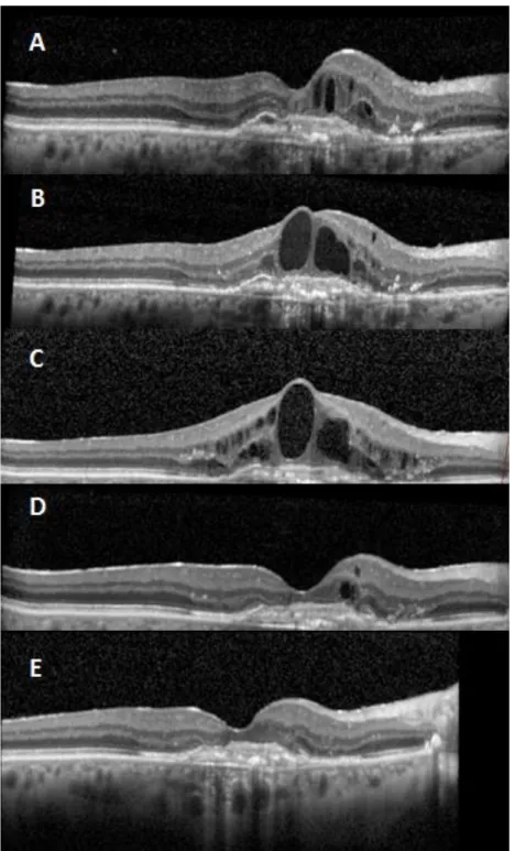
Fig. 1. Patient initially treated with bevacizumab (group 1) that ended developing resistance to the drug (A and B).
C represents the moment that it was decided to switch to aflibercept.
D is after one injection of aflibercept. E is after the three injections of aflibercept.
16
Fig. 2. Patients previously treated with ranibizumab (group 2) with a good response (A and B).
Later on had recurrence with this drug (C) and it was proposed to treatment with aflibercept.
D is after one injection of aflibercept. E is after three injections of aflibercept.
17
Tables
Table 1. Switching to Aflibercept in Patients with Chronic Neovascular AMD - Patient and Treatment Characteristics
Eyes (Patients) 85 (69)
Age, mean (range) 76.6 (61-92)
Women, n (%)
Angiographic Classification, n (%) Occult with no Classic Predominantly Classic Minimally Classic
Polipoidal Choroidal Vasculopathy
38 (55.1%) 66 (77.6%) 7 (8.2%) 6 (7.1%) 2 (2.4%)
Eyes Refractory Bevacizumab, n (%) 39 (45.9%)
Eyes on Treatment with Ranibizumab, n (%) 46 (54.1%) Months on Therapy, mean (±SD)
Prior to Switch Aflibercept
Number Injections, mean (±SD) Prior to Switch Aflibercept 17.8 (±11.5) 8.3 (±2.2) 12.4 (±7.7) 5.1 (±1.7) SD, Standard Deviation.
18
Table 2. Outcomes After Switching to Aflibercept in Patients with Chronic Neovascular AMD All (N = 86) P Group 1 Bevacizumab (N = 39) P Group 2 Ranibizumab (N = 47) P
Mean BCVA (ETDRS score) Before Switch (±SD) Final (±SD) 57.2 (±15.3) 56.0 (±17.4) 0.162 58.2 (±16.8) 56.5 (±18.4) 0.282 56.4 (±14.0) 55.5 (±16.7) 0.382 Mean CRT (μm) Before Switch (±SD) After 1 Injection (±SD) Final (±SD) 375.0 (±178.0) 301.7 (±122.8) 295.8 (±128.7) < 0.001 *< 0.001 374.1 (±188.1) 298.1 (±96.6) 308.8 (±132.4) 0.009 *0.051 375.7 (±171.1) 304.7 (±142.3) 284.7 (±125.9) < 0.001 *< 0.001 Injections per Month
Previous (±SD) Aflibercept (±SD) 0.76 (±0.26) 0.63 (±0.17) <0.001 0.74 (±0.17) 0.60 (±0.18) <0.001 0.77 (±0.31) 0.65 (±0.16) 0.016
Outcomes expressed as mean values (±SD); BCVA expressed in ETDRS score; CRT expressed in μm.
Appendix
Acta Ophthalmologica Author Guidelines
© 2014 Acta Ophthalmologica Scandinavica Foundation. Published by John Wiley & Sons Ltd
Edited By: Einar Stefánsson Impact Factor: 2.345
ISI Journal Citation Reports © Ranking: 2012: 15/59 (Ophthalmology) Online ISSN: 1755-3768
Acta Ophthalmologica publishes clinical and experimental original articles, reviews, editorials, educational photo-essays (Diagnosis and Therapy in Ophthalmology), case reports and case series, letters to the editor, doctoral theses.
Priority is given to high quality original papers and review articles. Manuscripts are accepted on the condition that they have not been, and will not be published elsewhere, except for abstracts of oral presentations. All submissions are subject to editorial review and will be reviewed by two or more independent reviewers, editorial board members, and guest editors. It is assumed that the author(s) has considered the ethical aspects of the study and followed the guidelines of the Helsinki Declaration, which should always be specified in the manuscript.
Online Only Publication
Letters to the editor are published online only from January 2010, and from January 2010 newly submitted letters to the editor will be exempt from colour charges.
The editor decides whether any publication is electronic only or both in print and electronic form. The authors are notified and consulted on this decision.
ONLINE SUBMISSION
Manuscripts to Acta Ophthalmologica should be submitted online via the journal's submission site, ScholarOne Manuscripts (formerly known as Manuscript Central). To access this system for submission and review, go directly to:
Complete instructions for preparing and submitting manuscripts online are provided at the submission site. If you need assistance, please contact our support staff by phone at +1 434 817 2040 ext. 167 or via e-mail at [email protected]
Please note that docx files are compatible with the journal submission systems.
ORIGINAL PAPERS
Arrangement of the manuscript: The manuscript should include the following: 1) title page; 2) abstract, and key words; 3) main text (introduction, materials and methods, results, discussion); 4) acknowledgement; 5) references; 6) figure and figure legends; 7) tables; 8) illustrations and graphics. For more information on manuscript format, please refer to the following guidelines.
1) The Title Page should contain on separate lines author(s) name, institution, and the title of the article. In addition it should contain the e-mail and postal addresses, plus telephone and fax numbers of the corresponding author. All author affiliations and corresponding authors addresses should be supplied in English.
2) The Abstract of original papers must be structured with the following headings: Purpose, Methods, Results, Conclusion, and should not exceed 250 words. Abstracts of review articles, perspective of ophthalmology, historical articles, case reports and case series do not have to be structured in this same way. Diagnosis/Therapy in Ophthalmology contributions, editorials and Letters to the Editor do not have an abstract, please see separate instructions below. Key Words: Four to nine key words for indexing purposes must be given.
3) Main text should be concise and as far as possible free of specialised language, and unnecessary or not generally accepted abbreviations. Abbreviations must be spelled out on first mention.
The following order of presentation is recommended:
Introduction stating the purpose of the article and key aspects of present knowledge. Extensive literature reviews are not desirable. Primary sources are preferred.
Material: Notice that the journal requests that all research has followed the Tenets of the Declaration of Helsinki, and that the details are provided in the manuscript text. Methods of investigation with sufficient information to permit repetition of experiments. The journal requires that authors who report on eye cancer include in their manuscript the Union for International Cancer Control / American Joint Committee on Cancer (UICC / AJCC) Tumor, Node, Metastasis (TNM) categories and stages (7th Edition) in addition to any other cancer classification scheme the authors wish to use.
Statistics and mathematical analyses should be applied when appropriate and be described under Methods. Authors are encouraged to take advice from an expert of statistics already when the study is designed. The following rules regarding reporting should be adhered to:
- Report proportions if the number of subjects is smaller than 10 (e.g. 2 of 5), report percentages in integers if the number of subjects is less than 100 (e.g. 34%). If the number of subjects is larger, one decimal place can be given, but is seldom necessary (e.g. 34.5%). - Report summary statistics of normally distributed variables as mean with standard deviation, and other variable as medium with range. Use parametric and nonparametric statistical tests accordingly.
- Give exact p-values (e.g. p=0.15 and p=0.034); if p-value is smaller than 0.0001, report p - Give 95% confidence intervals for main findings.
- Mention the statistical test used with the p-value (e.g. p=0.015, paird t-test). If the same test is repeated, it does not need to be specified again.
- The statistical software used need not be referenced in the manuscript text, unless it is specific for the statistical method used (or these can be put later under a separate heading, with a pointer here).
Results should be as clearly presented as possible. Scatterplots and similar graphical presentations are often preferable to tables. Do not duplicate in the text data that is given in tables and figures.
Discussion should be based directly on the author(s)' contributions and with reference to prior investigations, pointing out the significance and the limitations of the study.
4) Acknowledgements should indicate the name, society and date of the meeting if an abstract of the article has been presented previously. Support for the study can also be published here. A statement regarding possible conflict of interest must be included here (e.g. disclose financial interest in the equipment or method described, research or travelling grant support, consulting services provided, or disclose absence of commercial or propriety interest).
5) References. The author(s) is responsible for accurate references that must conform to journal style. If references are not in journal style, the manuscript will be returned for editing without review. References in the text should quote the last name(s) of the author(s) and the year of publication: (Brown & Smith 2003) or (Brown et al. 2003) when there are three or more authors. The reference list should include only those publications cited in the text and must be listed in alphabetic order with no numbering. Initials of forenames are placed after the surname with no commas, periods or spaces between initials. All articles should be cited in the original language of the reference, not as an English translation. References 'in press' must be filled in at latest in at the proof stage. Reference to unpublished material should state the author's name followed by 'unpublished' or 'personal communications'; such references should not appear in the reference list. Titles of journals are abbreviated according to the recommendations of the Index Medicus.
We recommend the use of a tool such as Reference Manager for reference management and formatting.
Reference Manager reference styles can be searched for here: http://www.refman.com/support/rmstyles.asp
Examples of reference list:
Abrahamsson M, Ohlsson J & Abrahamsson H (2003): Clinical evaluation of an eccentric infrared photorefactor; the PowerRefractor. Acta Ophthalmol Scand 81: 605-610.
Sharaaway T (2003): Glaucoma surgery: Lest we forget. Acta Ophthalmol 81: 553-555. Bailey IL (1998): Visual acuity. In: Benjamin WJ (ed.) Borish's Clinical Clinical Refraction. Philadelphia: W.B.Saunders 179-202.
6) Figure Legends. Legends to figures should make the meaning of each illustration understandable without reference to the text. Figure legends should be on separate pages. 7) Tables should be numbered consecutively in Arabic numerals and cited in the text. The approximate location in the text should be indicated in the manuscript. Each table should be typed as a separate document. Tables should have legends. Footnotes can be used in tables, if necessary.
8) Illustrations and graphics. All photographs, drawings and graphs are referred to as figures, abbreviated Fig., and should be numbered in sequence with Arabic numerals. Photographs and other images should be cropped so that only relevant parts of original figures are submitted. All figures should be planned to fit the printed column, 56, 117 or 178mm, and non-photographic illustrations should be professionally produced using modern software. Authors are encouraged to print their illustrations in the intended size before submission to make sure that sizes have been chosen correctly. The authors should also adapt graphics for the 3-column format and adjust the font size accordingly. Graphics are not re-drawn by the publisher. Histograms and similiar graphics should not be 3-dimensional. Complicated graphical illustrations can often be made more legible by the use of colour. Figures should be on separate pages. Please submit all line graphics as EPS or PDF files and photographs as TIF or PDF files. When submitting EPS files please embed fonts when you can. For more information on preparing and submitting figures please visit: http://www.blackwellpublishing.com/bauthor/illustration.asp