UNIVERSIDADE TÉCNICA DE LISBOA
Faculdade de Medicina Veterinária
EMBRYO-MATERNAL INTERACTIONS LEADING TO EMBRYONIC
DEVELOPMENT AND SURVIVAL IN THE BOVINE – ROLE OF
PROGESTERONE AND PROSTAGLANDINS
Ana Catarina Belejo Mora Torres
ORIENTADOR
Doutor Luís Filipe Lopes da Costa
2012
LISBOA
CONSTITUIÇÃO DO JÚRIPRESIDENTE
Reitor da Universidade Técnica de Lisboa VOGAIS
Doutor Luís Filipe Lopes da Costa Doutor António Eduardo Monteiro Horta Doutora Rosa Maria Lino Neto Pereira Doutora Luisa Maria Freire Leal Mateus Doutora Graça Maria Leitão Ferreira Dias
UNIVERSIDADE TÉCNICA DE LISBOA
Faculdade de Medicina Veterinária
EMBRYO-MATERNAL INTERACTIONS LEADING TO EMBRYONIC
DEVELOPMENT AND SURVIVAL IN THE BOVINE – ROLE OF
PROGESTERONE AND PROSTAGLANDINS
Ana Catarina Belejo Mora Torres
TESE DE DOUTORAMENTO EM CIÊNCIAS VETERINÁRIAS ESPECIALIDADE DE CLÍNICA
ORIENTADOR
Doutor Luís Filipe Lopes da Costa
2012
CONSTITUIÇÃO DO JÚRIPRESIDENTE
Doutor Rui Manuel de Vasconcelos e Horta Caldeira
VOGAIS
Doutor Luís Filipe Lopes da Costa Doutor António Eduardo Monteiro Horta Doutora Graça Maria Leitão Ferreira Dias Doutora Luisa Maria Freire Leal Mateus Doutora Rosa Maria Lino Neto Pereira
i
Chegada ao último ciclo de estudos, o doutoramento, a aprendizagem continuou intensamente… Aprendi novas metodologias, novas teorias, formas de delineamento experimental, uma abordagem sistemática aos métodos e à análise dos resultados obtidos e formas mais eficazes de comunicação com a comunidade científica. Mas para além de crescer na Ciência, ou também para ser uma cientista melhor, o doutoramento permitiu-me ainda aprender a ser mais paciente, a lidar melhor com as frustrações de semanas de trabalho por vezes infrutíferas, a trabalhar melhor em equipa e a entender diferentes pontos de vista, a procurar constantemente novas soluções para os problemas, a gerir melhor o meu tempo.
Dedico esta tese a todos os que me apoiaram quando me deparei com dificuldades, me aconselharam, me ajudaram a encontrar os caminhos e festejaram comigo todas as pequenas vitórias que me levaram a chegar aqui.
iii
Acknowledgements
While studying to be a veterinarian, I came across with the world of animal reproduction for the first time. It was a passion from the first classes, probably because of the way the subjects were taught by Professor Luís Costa, Professora Luisa Mateus and Professor Robalo Silva, as they always intended for students to understand and think about the way everything is so well orchestrated. I thank Professor Luís Costa for recognizing my passion and giving me the opportunity to work in this field, and for also to estimulate my learning in both the laboratory and the clinical work. I also thank him for the patience for some of my mistakes and the guidance and support he gave me throughout the experimental work here described and throughout the elaboration of this thesis. Indeed, it is a great experience to work with someone who has his knowledge, experience, intelligence and critical mind and who makes an effort for his students to grow and become better every day.
I also want to thank Patrícia Diniz and Mariana Batista for all the hard work in the laboratory, for their contributions to find the answers and for their companionship and patience. Patrícia Diniz, with her effort to teach me the principles of working in a laboratory and to find practical solutions, and Mariana Batista with her critical mind and curiosity, helped me to grow as a scientist and as a person.
To Professora Luisa Mateus for her help in supervising my work, for her contributions in finding the way to better communicate our results to the scientific community, and for her support.
To Professor Luís Tavares, the C.I.I.S.A. coordinator, for the support of part of the present research work and for giving me the opportunity to attend scientific conferences and meet other co-workers in my field.
To Doutor João Nestor Chagas e Silva for his constant availability to teach me to be a better veterinarian and for giving me the opportunity to practice my clinical skills.
To Doutora Elisabete Silva, who is always ready and available to help and share her knowledge and passion for science with everyone in the laboratory, and who taught me the basic principles of molecular biology.
To Cristina Valado, Dr. Daniel Murta, Dra. Marta Batista, Dra. Sofia Henriques, Dra. Manuela Oliveira and Dra. Sofia Van Harten for their friendship and support in the long hours we shared in our second house.
To José Chula for providing the animals used in the present in vivo experiments.
To my friends, who always supported me and helped me to overcome the difficulties and always provided me with a rich personal life, which in my opinion is essential to perform a better work. Particularly to João Migueis, who always believed in my capacities and showed that I was able to overcome any obstacles.
iv
To all my family, and specially my parents, Mariana Belejo and João Torres, for always supporting me, stimulating me to constantly learn more, and teaching me to believe in myself and never give up. I hope they will always be proud of me.
Funding
The present work was funded by the PhD Fellowship (SFRH / BD / 37666 / 2007) and by the following research projects: (i) PTDC/CVT/65690/2006, “Inter-relações embrião-útero-ovário determinando a sobrevivência embrionária e fetal em bovinos” granted by Fundação para a Ciência e Tecnologia (ii) CIISA 58 – Embrio-ovárica.
v
Título da dissertação: Interações embrio-maternas relevantes para o desenvolvimento e
sobrevivência embrionários em bovinos – papel da progesterona e das prostaglandinas
Resumo: Os objetivos desta tese foram: avaliar interações embrio-maternas esteroidogénicas
e prostanoides no desenvolvimento e sobrevivência embrionárias; testar estratégias terapêuticas na transferência embrionária (TE) com vista ao aumento da sobrevivência embrionária.
Experiências in vitro (três capítulos experimentais) – embriões bovinos (Dia 7): i) revelaram transcrição de genes codificantes das enzimas das vias sintéticas da progesterona (P4) e PGs
(PTGS2, PGFS, PGES, StAR, P450scc,3β-HSD); ii) produziram estes mediadores (P4, PGF2α,
PGE2) para o meio de cultura; iii) apresentaram um aumento significativo dos níveis de
transcrição destes genes (à exceção da StAR) associado à primeira diferenciação celular embrionária; iv) derivados de dadoras de oócitos pré-púberes revelaram níveis de transcrição dos genes mencionados similares aos de embriões de dadoras cíclicas (à exceção dos níveis de transcrição para a 3β-HSD, tendencialmente mais elevados em embriões provenientes de fêmeas cíclicas). Adicionalmente, v) num modelo de co-cultura de células lúteas desenvolvido, estas exerceram um efeito embriotrófico, aumentando significativamente a taxa de desenvolvimento e qualidade embrionárias; porém, este efeito não foi associado a aumento na transcrição génica ou produção de P4, PGF2α, PGE2; vi) Embriões em co-cultura com
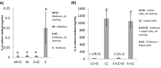
células lúteas não exerceram um efeito luteotrófico nas células; e vii) o uso de óleo mineral na cobertura dos poços de cultura exerceu um efeito embriotrófico, mas absorveu P4 do meio.
Experiências in vivo (dois capítulos experimentais) – novos modelos in vivo - embriões de baixa competência de desenvolvimento (hemi-embriões) e recetoras sub-férteis (vacas leiteiras de alta produção) ou com fertilidade alta (novilhas leiteiras virgens) - foram usados na avaliação do efeito na sobrevivência embrionária e nas concentrações plasmáticas de P4 e
PSPB das recetoras, de tratamentos, na TE, com hCG ou carprofen. Concluiu-se que: i) o tratamento com hCG induziu a formação de CLs secundários, aumentou as concentrações plasmáticas de P4, a taxa de sobrevivência dos hemi-embriões e as taxas de gestação das
recetoras (em vacas). Os embriões foram resgatados para além do reconhecimento materno da gestação (RMG), mas a sobrevivência embrionária posterior, o crescimento até à implantação e a secreção placentária de PSPB até ao Dia 63 de gestação (testados em vacas) não foram afetados; ii) a sobrevivência embrionária após o RMG não está diretamente dependente das concentrações de P4 maternas; iii) o tratamento com o carprofeno não afetou
significativamente as concentrações de P4 ou a sobrevivência embrionária, mas diminuiu o
efeito luteotrófico da hCG.
vii
Thesis title: Embryo-maternal interactions leading to embryonic development and survival in
the bovine – role of progesterone and prostaglandins
Abstract:
The objectives of this thesis were to evaluate steroidogenic and prostanoid embryo-maternal interactions leading to embryonic development and survival in cattle, and to evaluate therapeutic strategies at embryo transfer (ET) designed to enhance embryo survival.
In vitro experiments (three experimental chapters) - bovine early (Day 7) embryos i) had transcription of genes coding for enzymes progesterone (P4) and of prostaglandins (PGs)
synthesis pathways (StAR, P450scc,3β-HSD, PTGS2, PGFS, PGES); ii) produced these
mediators (P4, PGF2α, PGE2) into culture medium; iii) had a significant increase in
transcription levels of the above genes (except StAR) associated to first embryonic cellular differentiation; iv) derived from pre-pubertal oocyte donors had transcription levels of the above genes similar to those of embryos derived from post-pubertal cyclic heifers (except for 3β-HSD, which tended to be higher in embryos from cyclic heifers). Additionally, v) in a developed luteal cells (LC) co-culture model, LC induced an embryotrophic effect, significantly increasing blastocyst yield and quality; however, this embryotrophic effect was not associated with an increase in embryonic gene transcription or production of P4, PGF2α,
PGE2; vi) Embryos co-cultured with LC did not exert a luteotrophic effect upon the cells; and
vii) oil overlaying of culture wells exerted an embryotrophic effect, but absorbed P4 from
culture medium.
In vivo experiments (two experimental chapters
)
- novel in-vivo models considering poor developmental competence embryos (demi-embryos) and either sub-normal fertility recipients (lactating high-yielding dairy cow) or high fertility recipients (virgin dairy heifers) were used to evaluate the effect of hCG and carprofen treatment at embryo transfer on embryo survival and on plasma P4 and PSPB concentrations of recipients. Conclusions were that: i) treatmentwith hCG induced formation of secondary CL, increased plasma P4 concentrations, survival
rate of demi-embryos and pregnancy rate of recipients (only in cows). Embryos were rescued beyond maternal recognition of pregnancy (MPR), but later embryonic survival, growth until implantation and placental PSPB secretion until Day 63 of pregnancy (only tested in cows) were not affected; ii) embryonic survival following MRP is not under direct dependency of maternal P4 concentrations; iii) treatment with carprofen had no significant effect on plasma
P4 concentrations and embryonic survival, but decreased the luteotrophic effect of hCG.
ix
TABLE OF CONTENTS
INTRODUCTION – theme presentation, justification and objectives 1
LITERATURE REVIEW
1 - Embryo-maternal interactions in early pregnancy 5
1.1 - Embryonic development 5
1.1.1 - Activation of embryonic genome 6
1.1.2 - In vitro production of bovine embryos 6
1.1.3 - Assessment of embryo quality 9
1.2 - Corpus luteum (CL) 10
1.2.1 – Steroidogenesis 11
1.2.2 – Utero-ovarian countercurrent transport mechanism 12
1.3 – Embryo-oviductal interactions 13
1.4 – Embryo-uterine interactions 14
1.4.1 - Maternal recognition of pregnancy 14
1.4.2- Implantation and placentation 15
1.4.3. - Pregnancy associated glycoproteins 17
1.5. – Progesterone 18
1.5.1. - Progesterone and the early embryo 20
1.6. - Prostaglandins (PGs) 22
1.6.1. - Prostaglandins and the early embryo 26
2. - Embryonic mortality 27
3. - Strategies to improve embryo survival 30
3.1. - Human chorionic gonadotrophin (hCG) 31
3.2. - Inhibition of luteolytic PGF2α 33
EXPERIMENTAL WORK 34
Chapter I - Development of a bovine luteal cell in vitro culture system suitable for
co-culture with early embryos 34
1 – Abstract 34
2 – Introduction 35
3 – Material and methods 37
3.1– Media composition 37
3.2 - Isolation of enriched luteal cell populations 37
3.3 - Experiment 1 38
3.4 - Experiment 2 38
3.5 - Experiment 3 39
3.6 - Experiment 4 40
3.7 - Measurement of P4 concentrations 40
3.8 - Measurement of PGF2α and PGE2 concentrations 40
x 4 – Results 41 4.1 - Experiment 1 41 4.2 - Experiment 2 43 4.3 - Experiment 3 45 4.4 - Experiment 4 46 5 – Discussion 47
Chapter II – Embryo – luteal cells co-culture: an in vitro model to evaluate steroidogenic
and prostanoid bovine early embryo-maternal interactions 49
1. Abstract 49
2. Introduction 50
3. Material and methods 52
3.1– Experimental design 52
3.2 - Reagents and media 52
3.2.1 - LC media preparation 53
3.2.2 –Media for in vitro embryo production 53
3.3 - LC preparation 54
3.4 - In vitro embryo production 54
3.5 - Embryo evaluation 55
3.6 - Measurement of P4 concentrations 55
3.7 - Measurement of PGs concentrations 56
3.8 - RNA extraction and Real Time PCR analysis 56
3.9 - Statistical analysis 58
4 – Results 58
4.1 - Luteal cells exerted an embryotrophic effect upon co-cultured embryos 60 4.2 - Oil overlay of culture medium exerted an embryotrophic effect upon cultured embryos 60 4.3 - The embryotrophic effects of LC and oil overlay of culture medium were not additive 61 4.4 - Pre-eclosion bovine embryos transcribed genes coding for enzymes of the P4 synthesis
pathway and produced P4 into culture medium 61
4.5 - Pre-eclosion bovine embryos transcribed genes coding for enzymes of the PGs synthesis pathways and produced PGE2 and PGF2α into culture medium 62
4.6 –Luteal cells transcribed genes coding for enzymes of the P4 and PGs synthesis pathways
and produced P4, PGE2 and PGF2α into culture medium 62
4.7 – Embryos of similar quality, either cultured alone or co-cultured with LC had similar transcription levels of genes coding for enzymes of the P4 and PGs synthesis pathways 62
4.8 – Luteal cells transcription levels of genes coding for enzymes of the P4 and PGs synthesis
pathways, and production of P4, PGE2 and PGF2α into culture medium were not affected by
co-culture with embryos 65
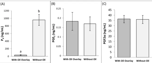
4.9 – Oil overlay of culture medium affected measurement of culture medium concentrations
of P4 but not of PGE2 and PGF2α 65
5 – Discussion 65
5.1 - Embryonic development and quality 65
xi
Chapter III - Effects of oocyte donor age and embryonic stage of development on transcription of genes coding for enzymes of the prostaglandins and progesterone
synthesis pathways in bovine in vitro-produced embryos 70
1 – Abstract 70
2 – Introduction 71
3 - Materials and methods 72
3.1 - Reagents and media 72
3.2 – In vitro embryo production 73
3.3 – RNA extraction and Real Time PCR analysis 74
3.4 – Statistical analysis 76
4 – Results 76
5 – Discussion 79
Chapter IV - Secondary corpora lutea induced by hCG treatment enhanced
demi-embryo survival in lactating high-yielding dairy cows 82
1 – Abstract 82
2 – Introduction 83
3 – Material and Methods 84
3.1 – Experimental design 84
3.2 – Animals and treatments 84
3.3 – Embryo production and bisection 85
3.4 – Blood collection and storage 86
3.5 – Plasma P4 measurement 86
3.6 – Plasma PSPB measurement 86
3.7 – Pregnancy evaluation 87
3.8 – Statistical analysis 87
4 – Results 88
4.1 – Development of secondary CL and pregnancy establishment 88
4.2 – Plasma P4 concentrations 90
4.3 – Plasma PSPB concentrations 92
5 – Discussion 93
5.1 – Pregnancy establishment 93
5.2 – Development of secondary CL and plasma P4 concentrations 94
5.3 – Plasma PSPB concentrations 95
Chapter V - Treatment with human chorionic gonadotrophin and carprofen at embryo transfer: effects on plasma progesterone concentrations of recipient dairy heifers and
preliminary results on demi-embryo survival 97
1 – Abstract 97
2 – Introduction 98
3 – Material and Methods 100
3.1 – Recipients and treatments 100
3.2 – Embryo production, bisection and transfer 100
3.3 – Secondary CL and pregnancy evaluation 101
xii
3.5 – Statistical analysis 102
4 – Results 102
4.1 – Plasma P4 concentrations 102
4.2 – Embryonic size and survival 104
5 – Discussion 105
5.1 – Plasma P4 concentrations 105
5.2 –Embryonic survival 106
GENERAL DISCUSSION AND CONCLUSIONS 109
xiii
LIST OF FIGURES
Figure 1: Effect of CL stage (early, mid, and late) on P4 production by luteal cells in culture
under oil overlaying 42
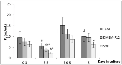
Figure 2: Effect of culture medium (TCM, DMEM-F12, SOF) on P4 production by luteal
cells in culture under oil overlaying 42
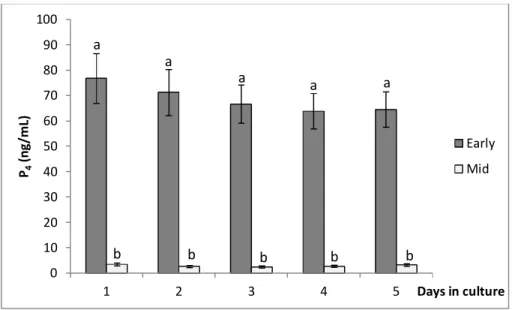
Figure 3: Effect of CL stage (early, mid) on P4 production by luteal cells in culture without
oil overlaying 43
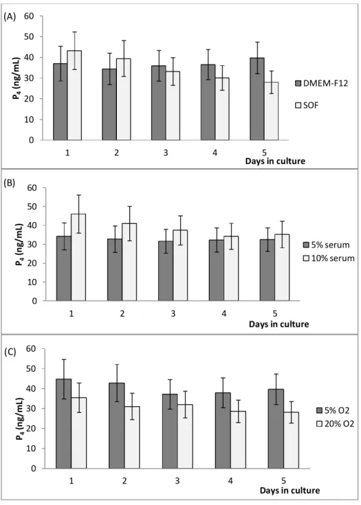
Figure 4: Effects of medium (A), serum concentration in medium (B), and oxygen tension in
culture atmosphere (C) on P4 production by luteal cells in culture without oil overlaying 44
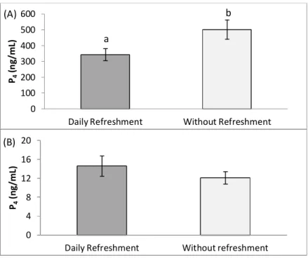
Figure 5: Effect of daily medium refreshment on the production of P4 by early (A) and mid
(B) CL stage luteal cells in culture without oil overlaying 45
Figure 6: Effect of cryopreservation on P4 (A), PGE2 (B), and PGF2α (C) production by early
CL stage luteal cells in culture without oil overlaying 46
Figure 7: Effect of oil overlay on the quantification of P4 (A), PGE2 (B), and PGF2α (C) in the
culture medium of cryopreserved early CL stage luteal cells 46
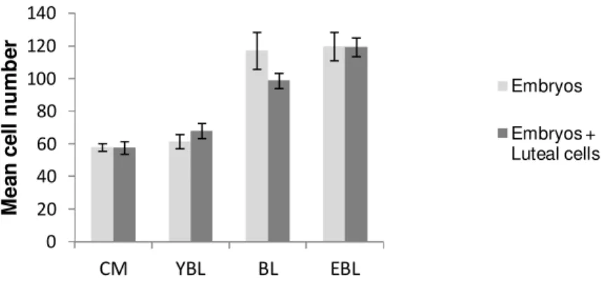
Figure 8: Effect of co-culture with luteal cells (LC) on Day 7 embryonic mean cell number of
different stages of development 60
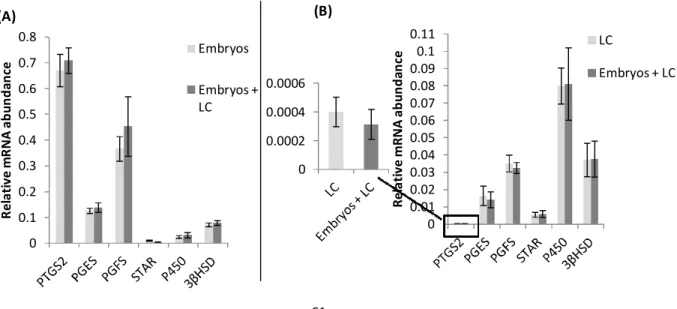
Figure 9: Illustration of the post-hoc LSD analysis of the interaction embryos × luteal cells
(LC) on relative mRNA level for genes coding for enzymes of the prostaglandins (PTGS2, PGES, PGFS) and progesterone (StAR, P450scc and 3β-HSD) synthesis pathways, evaluated
by Real-Time PCR 61
Figure 10: Illustration of the post-hoc LSD analysis of the interaction embryos × luteal cells
(LC) × oil overlaying of culture medium on progesterone (P4) concentrations in Experiment 1
63
Figure 11: Illustration of the post-hoc LSD analysis of the interaction embryos × luteal cells
(LC) on progesterone (P4) concentrations in Experiment 2 63
Figure 12: Illustration of the post-hoc LSD analysis of the interaction interaction embryos ×
luteal cells (LC) × oil overlaying of culture medium on PGE2 concentrations 64
Figure 13: Illustration of the post-hoc LSD analysis of the interaction interaction embryos ×
xiv
Figure 14: Post-hoc analysis of the relative mRNA levels of genes coding for enzymes of the
steroidogenic synthesis pathway, of in vitro produced embryos of different developmental stages of either pre-pubertal or post-pubertal cyclic heifers 77
Figure 15: Post-hoc analysis of the relative mRNA levels of genes coding for enzymes of the
prostaglandins synthesis pathway, of in vitro produced embryos of different developmental stages of either pre-pubertal or post-pubertal cyclic heifers 78
Figure 16: Effect of secondary CL induced by treatment with hCG on Day 7 of the estrous
cycle on plasma P4 concentrations of demi-embryo pregnancies 90
Figure 17: Effects of late embryonic mortality (LEM) and secondary CL induced by
treatment with hCG on Day 7 of the estrous cycle on plasma P4 concentrations of
demi-embryo pregnancies 91
Figure 18: Effect of secondary CL induced by treatment with hCG on Day 7 of the estrous
cycle on plasma PSPB concentrations of demi-embryo pregnancies 92
Figure 19: Plasma PSPB concentrations of single (n = 16) and twin (n =7) demi-embryo
pregnancies 93
Figure 20: Mean plasma P4 concentrations of dairy heifers maintaining pregnancy until Day
xv
LIST OF TABLES
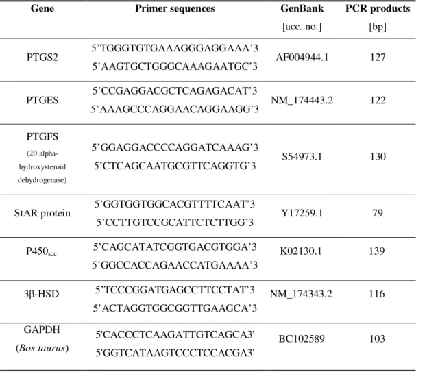
Table 1: Primer sequences, GenBank access number and size of PCR products of target genes
analyzed by Real Time PCR 57
Table 2: Influence of luteal cell co-culture and culture medium mineral oil overlaying on
embryo development and quality in a 4-well dish in vitro culture system (Experiment 1) 59
Table 3: Influence of luteal cell (LC) co-culture on embryo development and quality in a
slide-chamber in vitro culture system (without oil overlay) (Experiment 2) 59
Table 4: Primer sequences, GenBank access number and size of PCR products of target
genes analyzed by Real Time PCR 75
Table 5: Results (P values) of main effects of the factorial ANOVA model applied to the
transcription levels of target genes 76
Table 6: Effect of hCG treatment on Day 7 of the estrous cycle on pregnancy establishment
following demi-embryo bilateral transfer to high-yielding lactating dairy cows 88
Table 7: Effect of secondary CL induced by treatment with hCG on Day 7 of the estrous
cycle on pregnancy establishment following demi-embryo bilateral transfer to high-yielding
lactating dairy cows 89
Table 8: Pregnancy rates of dairy heifers following transfer of one demi-embryo and
treatment with hCG or/and carprofen 104
Table 9: Embryo/fetal losses of dairy heifers following transfer of one demi-embryo and
xvii
LIST OF ABREVIATIONS
3β-HSD – 3 Beta hydroxysteroid dehydrogenase AI – artificial insemination
ANOVA – analysis of variance B2M – beta2-microglobulin BL – blastocyst
BOEC – bovine oviduct epithelial cells BRL cells – Buffalo rat liver cells BSA – bovine serum albumin cDNA – complementary DNA CL – corpus luteum
CM – compact morula
COC – cumulus-oocyte complex COX-2 – cyclooxygenase-2 CRL – crown-rump length
DMEM – Dulbecco’s modified Eagle's medium DNA - deoxyribonucleic acid
E2 – estradiol- 17β
early CL – early luteal phase CL EBL – expanded blastocyst EFM – early fetal mortality
EGA – embryonic genomic control EGF – epidermal growth factor
ELISA – enzyme-linked immunosorbent assay EMM – early embryonic mortality
EP – Prostaglandin E2 receptor
ER – estrogen receptor ET – embryo transfer
FGF – fibroblast growth factor FP – prostaglandin F2α receptor
FSH – Follicle-stimulating hormone
GAPDH – Glyceraldehyde 3-phosphate dehydrogenase GE – uterine glandular epithelia
xviii GnRH – Gonadotrophin-releasing hormone HBSS – Hank’s buffered salt solution hCG – human chorionic gonadotrophin HDL - high density lipoprotein
ICM – inner cell mass
IETS – International Embryo Transfer Society IFN-τ - interferon tau
IGFBP – insulin-like growth factor binding proteins IGF-I – insulin-like growth factor I
IGF-II - insulin-like growth factor II IVP – in vitro production
LC –luteal cells
LDL – low density lipoprotein LE – uterine luminal epithelium LEM – late embryonic mortality LH – luteinizing hormone LIF – leukemia inhibitory factor LLC – large luteal cells
LPS – lipopolysaccharide M – Molar
MET – Maternal-to-embryonic transition MHC – major histocompatibility complex mid CL – mid luteal phase CL
mPR – membrane bound P4 receptor
mRNA – messenger ribonucleic acid MRP – maternal recognition of pregnancy
NSAIDs – non-steroidal anti-inflammatory drugs O2 – oxygen
OA – ovarian artery OXT - oxytocin
OXTR – oxytocin receptor P4 – progesterone
P450scc – cytochrome 450 side-chain cleavage
PAG – pregnancy-associated glycoproteins PBR – Peripheral-type benzodiazepine receptors
xix PBS – phosphate buffered saline
PCR – polymerase chain reaction PDGF – platelet-derived growth factor PG – prostaglandin
PGE2 – prostaglandin E2
PGES – prostaglandin E2 synthase
PGF2α – prostaglandin F2 alpha
PGFS – prostaglandin F2 alpha synthase
PGH2 – prostaglandin H2
PGI2 – prostaciclin
PR – progesterone receptor PKA – protein kinase A PKC – protein kinase C PSPB – Pregnancy-specific protein B PTGS1 – Prostaglandin-endoperoxide synthase 1 PTGS2 – Prostaglandin-endoperoxide synthase 2 RIA – radioimmunoassay RT – reverse transcriptase SEM – standard error mean
sGE – superficial glandular epithelia SLC – small luteal cells
SO – superovulation
SOF – synthetic oviductal fluid medium ß actin – beta actin
StAR – steroidogenic acute regulating protein TCM-199 – Tissue Culture Medium 199 TE –trophectoderm cells
TGF – transforming growth factors TNFα - tumor necrosis factor alpha UOV – utero-ovarian vein
v/v – volume fraction w/v – mass concentration YBL – young blastocyst ZP – zona pellucida
1
INTRODUCTION – theme presentation, justification and objectives
Cattle are an essential worldwide source of animal protein. In Portugal, cattle dairy and meat products represent more than half of the total national animal production (28,1% and 25,0%, respectively; Ministério da Agricultura, do Mar, do Ambiente e do Ordenamento do Território, 2007). In the last decades, dairy cows have been selected to increase their production traits, and this has been associated to a decline in fertility (Rodriguez-Martinez et al., 2008). In Portugal, a steady decline in fertility of dairy cows was also reported (Rocha et al., 2001; Rocha et al., 2010).
Embryonic mortality is the most prevalent component associated to the fertility decrease in the modern high-yielding dairy cow (Leroy et al., 2008). Most embryo losses occur between fertilization and maternal recognition of pregnancy (MRP), around Day 16 of pregnancy (Humblot, 2001; Diskin et al., 2011). During this critical period, embryonic survival depends on a complex crosstalk between the early conceptus and the mother, still poorly understood (Forde et al., 2011b). The presence of a viable conceptus determines the disruption of the luteolytic signal and maintenance of the corpus luteum (CL), providing adequate progesterone (P4) concentrations to induce uterine receptivity (Niswender et al., 2000).
Maternal inadequate circulating concentrations of P4 are thought to be the main cause of early
embryonic loss (Diskin & Morris, 2008). Conversely, high circulating P4 concentrations in the
immediate post-conception period were associated with advancement in embryo development and higher pregnancy rates, both in cattle and in sheep (Lonergan, 2011b). However, the ability of the early embryo to produce P4 and the influence of the early embryo on maternal P4
production remains elusive. It is also unclear whether embryonic development and survival are a result of a direct interaction with the CL or if those events are mediated through uterine effects. Prostaglandins (PGs) are involved in several reproductive events, including luteal regression, ovulation, pregnancy and parturition (Weems et al., 2006). These mediators are produced by the CL, uterus and conceptus, but their precise role in early embryo-maternal interactions is also unclear.
Embryo-maternal crosstalk remains one of the most challenging subjects in reproductive biology. The deciphering of embryo-maternal interactions may allow the development of new therapeutic strategies to enhance embryonic survival. This would have a major impact in cattle reproductive efficiency and profitability of modern cattle industry. Additionally, in a species comparative approach, the investigation of the mechanisms of mammalian early development may provide relevant advancements in our knowledge of the determinants of normal and abnormal deviations of health.
2
The main objective of the work presented in this thesis was to evaluate the embryo-maternal steroidogenic and prostanoid interactions leading to embryonic development and survival in cattle. Additionally, therapeutic strategies to enhance in vivo embryonic survival were also evaluated. This thesis comprises three in vitro studies and two in vivo studies with the following specific objectives:
In vitro studies:
a) To develop a bovine luteal cells in vitro culture system suitable for co-culture with early embryos. This culture system attempts to mimic putative embryo-luteal interactions, ruling out effects of other maternal tissues such as the endometrium; b) To evaluate putative direct steroidogenic and prostanoid interactions between bovine
early embryos and luteal cells, using a novel in vitro embryo – luteal cells co-culture model;
c) To determine the ability of the early embryo to transcribe genes coding for enzymes of the P4 and PGs (prostaglandin E2 - PGE2 and prostaglandin F2 alpha - PGF2α) synthesis
pathways, and to produce these substances. Additionally, the effects of oocyte donor age (prepubertal versus post-pubertal cyclic heifers) and embryonic stage of development on embryonic gene transcription and P4 and PGs production were also
evaluated.
d) To investigate the existence of an embryotrophic effect of luteal origin, and of a luteotrophic effect of embryonic origin, as putative relevant components of the early embryo-maternal crosstalk.
In vivo studies:
a) To develop two novel in vivo models to evaluate embryo-maternal interactions and therapeutic strategies to enhance embryonic survival: the first model considered a low developmental competence embryo plus a high fertility recipient (demi-embryo plus virgin dairy heifer); the second model considered a low developmental embryo plus a poor fertility recipient (demi-embryo plus high-yielding dairy cow). In these models, embryonic growth and survival, and maternal luteal and placental functions were assessed;
3
b) To evaluate treatments with human chorionic gonadotrophin (hCG) and carprofen at embryo transfer (ET), designed to enhance survival of compromised embryos; c) To evaluate treatment with hCG at ET, designed to enhance survival of
compromised embryos in a poor fertility recipient;
d) To investigate the relative effects of presence and location of CL, and maternal plasma P4 concentrations on embryo survival;
e) To describe maternal profiles of plasma P4 and pregnancy-specific protein B
(PSPB) in single and twin demi-embryo pregnancies;
f) To determine the effect of conceptus number on embryonic growth and on maternal luteal and placental functions.
These studies were converted in five manuscripts submitted for publication in international refereed and indexed journals and constitute the five chapters of the experimental work of this thesis, as follows:
Chapter I
Torres, A.*, Batista M.*, Diniz, P., Mateus, L. & Lopes-da-Costa, L. (2012). Development of
a bovine luteal cell in vitro culture system suitable for co-culture with early embryos. In Vitro Cell Dev Biol Anim, 48(9), 583-592. doi: 10.1007/s11626-012-9552-6.
*both authors contributed equally to this work
Chapter II
Torres, A., Batista M., Diniz, P., Mateus, L. & Lopes-da-Costa, L. (2013). Embryo – luteal
cells co-culture: an in vitro model to evaluate steroidogenic and prostanoid bovine early embryo-maternal interactions. In Vitro Cell Dev Biol Anim, Jan 29. doi: 10.1007/s11626-012-9577-x. [Epub ahead of print]
4
Chapter III
Torres, A., Batista, M., Diniz, P., Silva, E., Mateus, L. & Lopes-da-Costa, L. (2012).Effects
of oocyte donor age and embryonic stage of development on transcription of genes coding for enzymes of the prostaglandins and progesterone synthesis pathways in bovine in vitro-produced embryos. Submitted to Zygote.
Chapter IV
Torres, A, Chagas e Silva, J., Deloche, M.C., Humblot, P., Horta, A.E.M., Lopes-da-Costa, L. (2013). Secondary corpora lutea induced by hCG treatment enhanced demi-embryo survival in lactating high-yielding dairy cows. Reprod Domest Anim, Jan 16. doi:
10.1111/rda.12138. [Epub ahead of print]
Chapter V
Torres, A, Chagas e Silva, J., Diniz, P., Lopes-da-Costa, L. (2012). Evaluation of treatments
with hCG and carprofen at embryo transfer in a demi-embryo and recipient virgin heifer. Accepted for publication in Animal.
5
LITERATURE REVIEW
1. Embryo-maternal interactions in early pregnancy
In mammals, and particularly in ruminants, pregnancy establishment is the result of a complex crosstalk between the mother and the developing embryo/fetus. The protagonists of this dialogue and the mechanisms subjacent to their interactions are discussed below.
1.1. Embryonic development
Hartman et al. (1931) first provided a description of bovine ovulated oocytes and two-cell stage embryos. Fifteen years later, a more detailed description of developmental stages, from the unfertilized oocyte to the blastocyst, was reported by Hamilton & Laing (1946). In the oviduct, oocyte and spermatozoa fuse to form the one-cell stage embryo – the zygote. This cell undergoes a series of mitosis, which results in increasing numbers of embryonic cells (blastomeres) without an increase in embryonic size, a process designated by cleavage. The first cell cycle is completed around 30-36 hours post insemination and the second cell cycle 10-12 hours later (Gordon, 1994). At around the 32-cell stage, between Days 5 and 6 post fertilization, compaction of blastomeres occurs, followed by cell polarization and formation of a fluid-filled central cavity, the blastocoel (Van Soom et al., 1992). The latter event, designated blastulation, is the result of fluid transfer across the intercellular connections of outer blastomeres, promoted by ion transport through the Na+/K+ pump (Wrenzycki et al.,
2003). During subsequent cavitation embryonic cells differentiate into trophectoderm (TE) cells, that form a single layer surrounding the blastocoel, and inner cell mass (ICM) cells, resulting in the first cell differentiated embryonic stage – the blastocyst. All embryonic tissues, as well as part of the extra-embryonic membranes, will develop from ICM cells (Rossant & Lis, 1979). Cells of the TE exclusively contribute to the extra-embryonic components of the placenta (Rossant & Croy, 1985). Further increase in blastocoel size induces blastocyst expansion, thinning and rupture of the zona pellucida (ZP), with release of the embryonic mass, a process known as eclosion or hatching.
6
1.1.1. Activation of embryonic genome
The zygote and early cleavage-stage embryo is thought to be transcriptionally quiescent, cleaving under control of cytoplasmic maternally inherited mRNA molecules until genomic activation occurs. The transition from oogenetic to embryonic genomic activation (EGA) is called the maternal-to-embryonic transition (MET; Telford et al., 1990) and allows further embryogenesis to become dependent on the expression of the embryonic genome (Kanka, 2003; Walser & Lipshitz, 2011). In the bovine, onset of MET is thought to occur at the 8- to 16-cell stage. However, it was suggested that the onset of MET may be controlled temporally (i.e., at a time after fertilization) rather than at a developmental stage. Minor transcriptional activity was detected as early as the pronuclear stage after in vitro fertilization, whereas sensitivity to the transcriptional inhibitor α-amanitin was first detected at the 2– to 5–cell stage, and became predominant following the 6– to 8-cell stage (reviewed by Kanka et al., 2012). Using a suppression subtractive hybridization technique, Vigneault et al. (2009) produced a library of newly transcribed genes in bovine 8-cell stage embryos. This allowed the authors to identify more than 300 unique embryonic transcripts at the EGA and some transcripts at the 6– to 8-cell stage. A recent study identified a switch from Cullin 1-like variant 1 transcripts to Cullin 1 variant 3 transcripts at the late 8-cell stage (Vodickova-Kepkova et al., 2011). Authors concluded that Cullin 1-like, variant 1 mRNA represents a maternal transcript that is gradually degraded after fertilization, while Cullin 1 variant 3 transcripts represent new embryonic mRNA synthesized beyond the 8-cell stage.
1.1.2. In vitro production of bovine embryos
The first successful in vitro production of embryos was reported by Shenk (1880 – cited by Gordon, 1994), who inseminated oocytes from rabbit and guinea pig and identified the first cleavage. First attempts of in vitro embryo production in the cow were reported in 1968 (Adams et al., 1968; Sreenan et al., 1968). However, the birth of a live calf following in vitro fertilization and ET at the 4-cell stage was only achieved in 1981 (Brackett et al., 1982). Since the 1960s, several animal species, including rabbits, sheep, cattle and even chicken eggs, were used for in vivo culture of bovine embryos, mainly within the oviduct, which is thought to play an important role in early embryonic development (Gordon, 1994).
Initially, cattle embryos in vitro cultured from the first stages of development did not progress beyond the 8- to 16-cell stage, whereas only embryos beyond this stage could achieve normal
7
compaction; blastulation and hatching in culture (Wright & Bondioli, 1981). This blockage in in vitro development was later shown to be caused by inadequate culture conditions, and could be reversed by changes in the medium composition and co-culture with somatic cells (Gordon, 1994). The use of oviduct organ cultures to support mammalian in vitro embryo development was first reported in the mouse (Biggers et al., 1962). In cattle, successful in vitro culture until the blastocyst stage of development was first achieved through the co-culture with trophoblastic vesicles and oviduct epithelial cells co-co-culture (Camous et al., 1984; Eyestone & First, 1989). A variety of somatic cells, including primary cultures of reproductive tissues like granulosa (Fabbri et al., 2000), cumulus (Goto et al., 1988) and endometrial cells (Soong et al., 1998), and established cell lines like Vero (Huang et al., 1997) or BRL cells (Kubisch et al., 2001), were also shown to allow successful embryo culture.
Although the mechanisms by which somatic cells improve early embryo development remain elusive, several embryotrophic effects were described during or following co-culture with somatic cells: faster cleavage, higher blastocyst rate, reduced cell fragmentation, increased blastocyst cell numbers, improved morphological quality grade, reduced apoptosis, enhanced hatching, maintenance of viability prior to transfer, increased hCG secretion, “rescue” of poor quality embryos or subjected to manipulation procedures, improved pregnancy rates and live births (reviewed by Orsi & Reischl, 2007). Putative mechanisms of somatic cells action include removal of deleterious components from the culture medium, protection against oxidative stress and modulation of medium physico-chemical properties, altogether termed ‘negative conditioning’. Somatic cells may also secrete embryotrophic factors (termed ‘positive conditioning’), including specific proteins and various growth factors, like leukaemia inhibitory factor (LIF), colony stimulating factors (e.g. granulocyte-macrophage-colony stimulating factor–GM-CSF), fibroblast growth factor (FGF), epidermal growth factor (EGF), platelet-derived growth factor (PDGF), haematopoietic stem cell factor, transforming growth factors (TGF)-α, β1 and β2, insulin-like growth factors (IGF)-I and -II, and their binding proteins IGFBP-1, -2, -3, -4 and -5, as well as polyamines (reviewed by Orsi & Reischl, 2007). These beneficial effects of co-culture with somatic cell acquire more relevance with increasing duration of co-culture (Wiemer et al., 1989; Lai et al., 1996). Co-cultures can be used to study embryo-maternal interactions (Lee & Yeung, 2006; Pereira et al., 2006, 2009). However, co-culture systems should address the different and changing requirements of somatic cells and embryos throughout the culture period (Gardner, 1998). Media designed for embryo culture may not provide the specific requirements of somatic cells, with consequential loss of their in vivo morphological and functional properties (Reischl
8
et al., 1999). Conversely, media designed for somatic cells culture may not support normal embryonic development. Altogether, this may invalidate the use of co-culture systems to study physiologic early embryo-maternal interactions. Therefore, an in vitro maternal reproductive tissue (luteal cells, oviductal cells, uterine cells) co-culture system suitable for embryo culture needs to be developed.
Sanitary concerns and deleterious epigenetic effects induced by in vitro culture conditions (see below), led scientists to develop new media formulations capable of supporting in vitro embryo development in the absence of somatic cells co-culture. New media should mimic the histotroph produced by oviductal and uterine cells. Formulation of the synthetic oviductal fluid (SOF; Tervit et al., 1972), based on the composition of sheep oviductal fluid, allowed development until the blastocyst stage, without somatic cell support. Since then, media have been improved in order to satisfy embryo metabolic needs and avoid genic expression disturbances associated with the use of complex media (Purpera et al., 2009).
Supplementation of SOF with serum enhanced embryo development (McLaughlin et al., 1990; Lim et al., 1994). Serum contains proteins, vitamins, energy sources, lipids and growth factors, promoting compaction, blastulation and high embryo cell numbers (Gordon, 1994). However, media high serum content was linked to disturbed embryonic morphology, metabolism and gene expression, and was also associated to the large offspring syndrome (Van Wagtendonk-de Leeuw et al., 1998; Niemann &Wrenzycki, 2000; Purpera et al., 2009). These effects, as well as risks of contamination, led scientists to develop chemically, protein free, defined media. Unfortunately, a consensually reliable medium suitable for bovine in vitro embryo culture has not been fully established.
Oviductal and uterine intraluminal oxygen tensions were shown to be lower than the atmospheric in many species (Mastroianni & Jones, 1965; Mitchell & Yochim, 1968; Fischer & Bavister, 1993). Low oxygen (O2) tensions (5% instead of the 20% typical of air) are
beneficial to in vitro embryo culture (Thompson et al., 1990; Liu & Foote, 1995; Lonergan et al., 1999). Culture under a low O2 tension atmosphere diminishes the need for co-culture,
since one of the benefits of co-culture is the protection against oxidative stress (Watson et al., 1994, Rizos et al., 2001). However, somatic cells, particularly those from well vascularized tissues like luteal cells, usually require an atmospheric oxygen tension in co-culture systems (Orsi & Reischl, 2007; Corrêa et al., 2008; Vajta et al., 2010). It is a common practice in embryo cultures the overlaying of the culture medium with paraffin or mineral oil, in order to prevent pH alterations and dehydration. Oil overlaying of culture medium also allows culture in micro drops, which enhances development, compared to large volume medium culture (Fukui et al., 1996). However, oil can absorb lipid-soluble components of culture media
9
(Miller & Pursel, 1987) eventually restricting their effects on embryonic and somatic cells metabolism. Additionally, movement of these lipophilic substances towards the oil fraction results in erroneous measurements in culture medium.
Despite decades devoted to improvement of culture conditions, in vitro production still leads to lower embryonic developmental competence, compared to that achieved in vivo (Lonergan et al., 2006). This difference in developmental competence is translated in terms of morphology, cryotolerance, gene expression patterns, and pregnancy rate following transfer (Rizos et al., 2002a, 2002b). Transfer of in vivo derived zygotes (i.e., matured and fertilized in vivo) to in vitro culture results in a high blastocyst rate, but the resulting blastocysts tend to be of poor quality. In contrast, culturing in vitro derived zygotes (i.e., maturated and fertilized in vitro) in vivo, in the oviducts of the same or of different species, although not affecting the blastocyst rate, substantially improves blastocyst quality. These observations indicate that developmental competence, at least to the blastocyst stage, is controlled to a large extent by oocyte quality, while the post-fertilization environment mainly affects embryo quality (Lonergan, 2011a).
.
1.1.3. Assessment of embryo quality
The ultimate test of embryo quality is the ability to establish a pregnancy, resulting in the birth of a healthy offspring. As this represents a very expensive and time consuming methodology, researchers have been trying to identify markers of embryonic quality. Morphology assessment is widely used for embryo selection prior to transfer. In the bovine, blastomeres are opaque because of lipid droplet accumulation, making it difficult to assess nuclear and nucleolar morphology. Linder and Wright (1983) developed a morphological classification system with four grades (excellent, good, fair and poor), which were associated to pregnancy rates. The International Embryo Transfer Society (IETS) standardized an embryo quality classification, based on morphological assessment (Stringfellow & Seidel, 1998). Features such as color of the blastomeres, extent of compaction, timing of blastocyst formation and expansion, and embryo diameter at hatching can be linked with embryo quality (Van Soom et al., 2003). The timing of the first cleavage division is indicative of the developmental potential of the early embryo (Van Soom et al., 1992, 1997; Grisart et al., 1994; Holm et al., 1998; Lonergan et al., 2000), the faster cleaving embryos being those that develop to blastocyst at a higher rate. Embryo cell numbers have also been used as an indicator of bovine embryo quality. Data clearly shows that total cell number and ICM
10
number are higher in good than in poor quality embryos (reviewed by Gordon, 1994). Differential staining of the ICM and TE cells allows the calculation of the ICM/TE cells ratio (Van Soom et al., 2001).
More recently, development of molecular biology tools, such as the polimerase chain reaction (PCR), Real Time-PCR and microarray analysis, significantly widened our knowledge on embryo developmental competence. Microarray technology allowed the comparison between the transcriptional profiles of in vivo and in vitro produced embryos (Vodickova-Kepkova et al., 2011). These powerful tools identified a plethora of genes that may potentially be regarded as markers of oocyte and embryo quality. Oocyte and embryo developmental competence is probably a quantitative trait, dependent on small transcriptional changes of many individual genes in a well-orchestrated pattern (Kanka et al., 2012). Certainly, many of these genes are still not identified. In a future scenario, we can envisage the construction of a micro-array like tool that gathers the complete assemble of genes predictive markers of oocyte and embryo quality. Nevertheless, some genes, like gpx1 (Bermejo-Alvarez, et al., 2010), igf2r (Liu et al., 1997; Young et al., 2001), slc2a1 (Balasubramian et al., 2007), ptgs2 (El-Sayed et al., 2006; Saint-Dizier et al., 2011), plac8 and akr1b1 (El-(El-Sayed et al., 2006), tp53 (Favetta et al., 2004) and shc1shc (Leroy et al., 2010), were already suggested to be linked to embryo quality.
1.2. Corpus luteum (CL)
The CL is a transient endocrine gland that develops from the follicle wall after ovulation (Reynolds & Redmer, 1999). Granulosa cells (inner layer of the follicle wall) differentiate into large luteal cells (LLC), which represent 30% of the steroidogenic cells of the mature CL. These LLC secrete about 70% of the P4 in a constitutive way (unresponsive to LH or cAMP
stimulation). Theca cells (outer layer of the follicle wall) differentiate into small luteal cells (SLC), which comprise 70% of the steroidogenic cells of the mature CL. These SLC secrete only 30% of the P4 and, unlike LLC, require LH stimulation for maximal production (Farin et
al., 1989; Niswender et al., 1994). The CL consists of steroidogenic luteal cells and non-steroidogenic cells, i.e. vascular endothelial cells, fibroblasts, pericytes and immune cells such as lymphocytes, leucocytes and macrophages (Lei et al., 1991).
The initial growth rate of the CL is similar to that of a developing tumor, however growth ceases at mid-cycle and CL size remains relatively constant if pregnancy is established (Reynolds et al., 1994). Increase in size is dependent on an increase in SLC number and on
11
growth of LLC (Niswender, 1994). Neo-angiogenesis occurring within the developing CL leads to a highly dense and perfused vascular network, so that nearly every steroidogenic cell is in contact with a capillary (Redmer & Reynolds, 1996). Growth of the CL is stimulated by the autocrine and paracrine action of steroids, eicosanoids, cytokines and other growth factors (Reynolds & Redmer 1999; Berisha & Schams 2005). In the absence of pregnancy, the CL undergoes functional and morphological regression, a process termed luteolysis, which allows for the resumption of a new ovarian cycle. Functional regression occurs before morphological regression (O’Shea & McCoy, 1988; McCracken et al., 1999). At the 16th day of the cycle, PGF2α from uterine origin reaches the CL, induces a sharp decrease in P4 production and
involution of luteal tissue, resulting in the formation of the corpus albicans, rich in collagen and connective tissue.
1.2.1 Steroidogenesis
Cholesterol, the common precursor of all steroids, is mainly diet derived and transported to the ovaries by circulating lipoproteins (low density lipoproteins – LDL – and high density lipoproteins – HDL; Ohashi et al., 1982). Under lipid deprivation, LC are also able to synthesize cholesterol from acetate (Cook et al., 1967; Cook & Nalbandov, 1968). The uptake of LDL by LC occurs through receptor mediated endocitosis (Brown & Goldstein, 1986), while HDL uptake is typically accomplished by membrane-bound HDL binding proteins (Lestavel & Fruchart, 1994). There appears to be some species differences in their preference for LDL or HDL, but either source can be used by LC of most species (Niswender et al., 2000).
Once inside the cell, cholesterol can be metabolized for steroidogenesis or sterified with long-chain fatty acids and stored as cholesterol esters inside lipid droplets. In the cytoplasm, free cholesterol is transported to the mitochondria through a mechanism that involves cytoskeletal elements and sterol carrier proteins (Niswender, 2002), and then from the outer to the inner mitochondrial membrane (Stone & Hechter, 1954). This transport of cholesterol appears to be the rate-limiting step in the biosynthesis of P4, as well as the primary site of acute hormonal
regulation (Wiltbank et al., 1993). Steroidogenic acute regulatory protein (StAR) may bind cholesterol in the cytosol and transport it to the mitochondrial membrane. Phosphorylation of StAR by protein kinase A (PKA) stimulates cholesterol transport, whereas phosphorylation by protein kinase C (PKC) may inhibit this process. Peripheral-type benzodiazepine receptors (PBR) are involved in the transport from the outer to the inner mitochondrial membrane. The
12
natural ligand of PBR, endozepine, also appears to be critical in cholesterol transport regulation, as targeted deletion of this molecule reduces basal steroidogenesis (Niswender, 2002). Once in the mitochondrial matrix, cytochrome P450 complex (P450scc), adrenodoxin
and adrenodoxin reductase cleave the side chain of cholesterol to form pregnenolone (P5)
(Stone & Hechter, 1954). This steroid is then transported to the smooth endoplasmatic rethiculum, where 3-β-hydroxysteroid dehydrogenase/∆5, ∆4 isomerase (3β-HSD) converts P
5
to P4 (Hanukoglu, 1992), which is then thought to leave the cell by diffusion (Niswender et
al., 2002). Because LC store minimal amounts of steroids, the regulation of circulating concentrations of steroids is accomplished primarily by the level of steroid synthesis and metabolism (Bose et al., 2002).
1.2.2. Utero-ovarian countercurrent transport mechanism
The anatomical connections between the arterial, venous and lymphatic circulations of the ovary, oviduct and uterus allow for the passive transfer of energy (heat) and substances from one to another, without entering the systemic circulation and organ metabolization (at the liver, kidneys or lungs). Early studies in sheep demonstrated that ligation or surgical separation of the main uterine vein ipsilateral to the CL-bearing ovary inhibited the cyclic regression of the CL (Barrett et al., 1971; Baird & Land, 1973). Later on, Ginther (1974) demonstrated that PGF2α of endometrial origin reaches the CL through direct passive transfer
from the uterine vein to the ovarian artery, a process named utero-ovarian countercurrent transport mechanism. Transfer of steroid hormones and peptides through this mechanism was demonstrated in several species (Einer-Jensen, 1988), and significant differences between the concentrations of steroids in ovarian arterial blood and systemic blood was reported (Hunter et al. 1983).
On the other hand, substances produced in the ovary and released through the ovarian venous flow may reach ovarian arterial blood (rebound), the oviduct and the tubaric portion of the uterus. In ruminants, an ovarian-uterine countercurrent transport mechanism is critical for P4
of luteal origin reach high concentrations in the uterine horn ipsilateral to the CL-bearing ovary (Weems et al. 1988). A preferential transfer of ovarian steroids to the uterus was also demonstrated in women, during both the follicular and luteal phases (Cicinelli et al., 2004). This ovarian-uterine countercurrent transport mechanism is regarded as a local (ipsilateral) regulatory mechanism, involving mainly steroids and peptides (Einer-Jensen & Hunter, 2005). This is supported by early experiments involving anastomosis of the uterine vein or the
13
ovarian artery from the pregnant to the nonpregnant uterine horn, which indicated that both luteolytic and luteoprotective mediators only act locally (Mapletoft et al., 1975, 1976). Additionally, embryo/conceptus transfer and hysterectomy experiments in sheep indicated that the luteolytic and luteoprotective mechanisms do not act systemically, but are locally established between the uterus and the CL (Moor et al., 1969, 1970).
1.3. Embryo-oviductal interactions
The oviduct is the siege of critical reproductive events, including oocyte maturation, sperm capacitation, fertilization and early embryonic development (Hunter, 1988; Gandolfi et al., 1989; Ellington, 1991). The proximal part of the oviduct, referred to as the ampulla, is implicated in the transport of the oocyte-cumulus complex and in fertilisation, whereas the distal part, named the isthmus, is involved in sperm storage and embryo transport towards the uterus (Kolle et al., 2009; Hunter, 2012). Disturbances in events occurring in the oviduct may lead to infertility (Hunter, 2012).
The transport of oocytes and embryos is thought to be mainly modulated by endocrine and autocrine/paracrine signals locally produced by the oocyte/embryo. In rats and hamsters, fertilized eggs reach the uterus a little earlier than unfertilized eggs, suggesting an influence of embryos on oviduct activity, in which platelet-activating factor may be involved (Velasquez et al., 1995). In the mare, unfertilized oocytes remain and degenerate within the oviduct, while viable embryos progress to the uterus (Van Niekerk & Gerneke, 1966). In this species, the embryo enters the uterus 5 to 6 days following ovulation, when the morula or young blastocyst can secrete prostaglandins to stimulate local myosalpingeal activity (Weber et al., 1991b). Estrogens and PGs such as PGF2α, increase oviductal smooth muscle contractility and
the speed of embryonic transport (Lindblom et al., 1978; Weber et al., 1991a; Kissler et al., 2004), whereas P4 induces oviductal smooth muscle relaxation and reduces the speed of
embryonic transport (Lindblom & Hamberger, 1980). Oocyte and embryo transport is also driven by ciliary beating at the oviductal epithelium (Lyons et al., 2006). Digital videomicroscopic analysis established that, in cattle, the mechanisms of transport are different in the ampulla and the isthmus (Kölle et al., 2009). In the ampulla, transport occur depth between the folds, while in the isthmus transport is straight and fast. Interestingly, at the ampulla, viable oocytes attach to the oviductal epithelium, while degenerated oocytes float in the oviductal lumen. These observations indicate that the oviduct is able to select viable oocytes. Also, the early embryo (24–48 h after fertilization) was able to modify local
14
oviductal vascularization, induce the formation of secretory cells, and regulate ciliary beating. Altogether, the above studies indicate that the early embryo has the ability to change the oviductal environment, which prompts for an early embryo-oviductal crosstalk.
1.4. Embryo-uterine interactions
1.4.1. Maternal recognition of pregnancy
Ruminants developed a unique mechanism to rescue the CL from luteolysis and maintain pregnancy (Wooding, 1992). In sheep, Martal et al. (1979) first demonstrated that CL maintenance resulted from the production of a proteinaceous factor present in the pre-implantation elongating conceptus. This factor, initially called trophoblastin and later ovine trophoblast protein 1 (oTP-1) (Godkin et al., 1982), is now known as ovine interferon-tau (oIFN-τ) (Roberts et al., 1999). In the bovine, a similar secretory product of the trophoblast cells of the bovine conceptus is known as bovine interferon-tau (bIFN-τ; Helmer et al., 1987; Thatcher et al., 1989; Roberts et al., 1992). These trophoblastins were shown to be produced by the elongating blastocyst from Day 10 to 21-25, peaking around Days 14 to 16 (Spencer & Bazer, 2004). Around Day 16 of pregnancy, IFN-τ indirectly maintains the CL by attenuating (in cows; Meyer et al., 1995) or altering (in ewes; Zarco et al., 1988) the luteolytic pulses of uterine PGF2α. In the ewe, IFN-τ suppresses transcription of genes coding for oestrogen
receptors (ER) and oxytocin receptors (OTXR) in the uterine epithelium. In the cow, IFN-τ suppresses transcription of genes coding for OTXR but has no effect in uterine ER (Mann et al., 1999). Generation of uterine luteolytic episodes of PGF2α requires luteal secretion of
oxytocin (OTX) and the interaction of OTX of pituitary and ovarian origin at their receptors, located mainly on endometrial epithelial cells (Demmers et al., 2001). Thus, the luteolytic mechanism requires a sequence of events involving positive feedback loops between the endometrium and the CL (McCracken et al., 1999). The presence of a viable conceptus within the uterus diminishes, and in most cases, totally abolishes the pulsatile release of PGF2α
(Roberts et al., 1999). It is still controversial whether IFN-τ acts only on the uterus or also at the systemic level. In sheep, Oliveira et al. (2008) and Bott et al. (2010) reported high levels of IFN-τ in the uterine venous blood on Day 15 of pregnancy, as well as an increase in transcription of IFN-stimulated genes in peripheral tissues (leukocytes, liver, and CL) of pregnant sheep and cows. However, others (Gotkin et al., 1987; Lee et al., 2012) reported that
15
was retained within the uterus, since IFN-τ is a relatively large protein (19 kDa) and it cannot be transferred to the ovary through the utero-ovarian pathway. Additionally, the above teams did not detect the presence of IFN-τ, analysed by Western-blott and enzyme-linked immunosorbent assay (ELISA), in the uterine vein, ovarian artery and CL of Days 12, 14 and 16 of both cyclic and pregnant animals.
In addition to its antiluteolytic actions, bIFN-τ alters the transcriptome of the endometrium, in order to enhance conceptus elongation and to establish uterine receptivity to implantation (reviewed by Spencer et al., 2008). A recent study using RNAseq (Forde et al., 2012) showed that, although IFN-τ is critical for MRP, there may be other relevant mechanisms in the embryo-maternal crosstalk leading to pregnancy establishment. Comparing the endometrium of pregnant and cyclic heifers on Day 16, several differentially expressed genes regulated by IFN-τ were identified. Expression of some of these genes in blood cells may be predictive of the pregnancy status of dairy cows as early as Day 18 following insemination. However, several other differentially transcribed genes are not directly regulated by IFN-τ in vivo and may therefore be regulated by yet unknown conceptus-derived factors.
Secretion of IFN-τ during early pregnancy is also relevant for maternal tolerance against the fetal allograft. Major histocompatibility complex (MHC) class I molecules, consisting of an alpha chain and a beta2-microglobulin (B2M), regulate immune rejection responses by discriminating self and non-self-proteins (Choi et al., 2003). These molecules are increased by type I IFNs stimulation. In the ovine uterus, MHC class I alpha chain and B2M, although expressed primarily in endometrial luminal epithelium and glandular epithelium on Days 10 and 12 of the estrous cycle and pregnancy, were shown to have an increased expression restricted to endometrial stromal cells and glandular epithelium, on Days 14 to 20 of pregnancy. Also, intrauterine infusion of IFN-τ increased MHC class I and B2M expression in endometrial stromal cells and glandular epithelium, but not in luminal epithelium and superficial glandular epithelium. Silencing MHC class I alpha chain and B2M genes in endometrial luminal epithelium and superficial glandular epithelium during pregnancy may be critical in preventing immune rejection of the conceptus allograft (Bazer et al., 2008).
1.4.2. Implantation and Placentation
During early pregnancy, the endometrium produces a complex mix of compounds, known as the histotroph, which is released into the uterine lumen. The histotroph contains nutrients such as amino acids and glucose, cytokines and other growth factors, fatty acids, transport proteins
16
for vitamins and minerals and enzymes (Bazer et al., 2012). Studies using the sheep uterine gland knockout model demonstrated that functional uterine glands are essential for ewes to experience normal estrous cycles and to support conceptus development (Spencer et al., 2004). The endometrium plays a central role in the context of early embryo-maternal crosstalk. This dialogue induces dynamic changes in the uterine epithelia, tightly regulated by steroid hormones, cytokines and growth factors, which establish uterine receptivity towards the developing conceptus (Spencer et al., 2008).
Implantation may be invasive (interstitial or eccentric) or non-invasive (central) depending on whether the TE invades through the uterine luminal epithelia and superficial glandular epithelia into the stroma or not. Implantation in domestic animals is protracted and superficial, with conceptus TE only attaching to uterine luminal epithelia and superficial glandular epithelia. This protracted pre-implantation period allows large spherical conceptuses to migrate (horse) and hatched blastocysts to elongate (swine and ruminant species) before apposition and attachment to the endometrium, prior to implantation (Bazer et al., 2009). In ruminants, a dialogue between the free-floating blastocyst and the receptive endometrium, initiates the process of implantation (Tabibzadeh & Babaknia, 1995). After hatching from the ZP, blastocysts start to elongate on Day 12 (sheep) or Day 15 (cattle) to filamentous conceptuses that occupy the entire length of the uterine horn (Spencer et al., 2008). This elongation, which is not observed in vitro (Heyman et al., 1984), is critical for production of IFN-τ, MRP and implantation (Spencer et al., 2008).
In the cow, implantation is preceded by blastocyst elongation, and apposition, attachment, and adhesion of the TE to the endometrial luminal epithelium (Chavatte-Palmer & Guillomot, 2007). TE-derived cells form a single layer that covers the external surface of the placenta, in contact with the endometrium. Together with the somatic (parietal) mesoderm, they will constitute the chorion. The allantois, which is a sac-like structure that arises as an out-pocketing of the fetal hindgut, ultimately expands to fuse with the chorion, forming the chorioallantois, a richly vascularized membrane (Schlafer et al., 2000). TE-derived cells are referred to as trophoblast cells, which include two morphologically and functionally distinct types: mononucleate and binucleate trophoblast cells (Igwebuike, 2006). Mononucleate cells comprise the majority of the interface and are primarily involved in nutrient exchange, although they are responsible for the production of the pregnancy recognition signal, IFN-τ (Farin et al., 1990). Binucleate cells secrete into the maternal circulation hormones such as placental lactogen, P4 and pregnancy-specific (-associated) proteins (PAGs) (Becker set al.,
1998). These cells migrate across the fetal-maternal interface to fuse with endometrial epithelial columnar cells forming trinucleate hybrid cells. Continued binucleate cell migration