Universidade de Lisboa
Faculdade de Medicina
Early markers of heart and kidney
damage in Fabry disease
Patrício Ricardo da Terra Aguiar
Orientadores: Professor Doutor José Luís Bliebernicht Ducla Soares
Professora Doutora Derralynn Arlene Hughes
Tese especialmente elaborada para obtenção do grau de Doutor em
Medicina, especialidade de Medicina Interna
2018
Universidade de Lisboa
Faculdade de Medicina
Early markers of heart and kidney damage in Fabry
disease
Patrício Ricardo da Terra Aguiar Orientadores: Professor Doutor José Luís Bliebernicht Ducla Soares Professora Doutora Derralynn Arlene Hughes Tese especialmente elaborada para obtenção do grau de Doutor em Medicina, especialidade de Medicina Interna Júri:Presidente: Doutor José Augusto Gamito Melo Cristino, Professor Catedrático, Presidente do Conselho Científico da Faculdade de Medicina da Universidade de Lisboa.
Vogais:
- Univ. Prof. Dr. Med. Christoph Kampmann, Universitätsprofessor und Leiter der Sektion Pädiatrische Kardiologie, Universitätsmedizin Mainz;
- Dr. Derralynn Hughes, Senior Lecture – Institute of Immunity and Transplantation – Royal Free Hospital and Faculty of Medical Sciences – University College London (co-orientadora);
- Doutor António José Murinello de Sousa Guerreiro, Professor Catedrático da Faculdade de Ciências Médicas da Universidade Nova de Lisboa;
- Doutor Rui Manuel Martins Victorino, Professor Catedrático da Faculdade de Medicina da Universidade de Lisboa; - Doutora Dulce Alves Brito, Professora Auxiliar Convidada da Faculdade de Medicina da Universidade de Lisboa; - Doutor Edgar Avito Fernandes Almeida, Professor Auxiliar Convidado da Faculdade de Medicina da Universidade de Lisboa. 2018
As opiniões expressas nesta publicação são da exclusiva responsabilidade do seu autor.
“Põe quanto És no Mínimo que Fazes Para ser grande, sê inteiro: nada Teu exagera ou exclui. Sê todo em cada coisa. Põe quanto és No mínimo que fazes. Assim em cada lago a lua toda Brilha, porque alta vive.” Ricardo Reis, in "Odes"
Declaration of work and integrity
I confirm that the work presented in this thesis is my own and I did not resort to the practice of plagiarism or any form of falsification of results. Where information has been derived from other sources, I confirm that this has been indicated in the thesis.
Moreover, I state that I was the main responsible for the research idea and design, as well as the data acquisition, the data analysis and interpretation and the statistical analysis. I also participated in laboratorial tasks, but under the supervision and with help of other members of the research team.
I also declare that I acted according to the code of ethical conduct of the University of Lisbon and of the University College London. Lisbon, 17th of December, 2018 Patrício Ricardo da Terra Aguiar ________________________________________________
Acknowledgements
When I left Lisbon, five years ago and arrived in London on the 12th of February 2012, I was far from imagining my future. All hopes, with a willingness to learn and “brilliant” ideas, which I now realise were so naïve, but no experience or knowledge about this world of research. However, someone believed in me and held my hand while I developed my thoughts and beliefs. Returning back to Portugal, I had to take the great decision – would I be able to put my life on hold for 4 years? Was my family or I prepared to plunge headfirst into a PhD? I think that no-one knows the right answer when diving deep into an unknown reality. But now, 5 years and many achievements later, I can say that we, my family and I, were brave and now the world is really different. To my wife Lígia and my sons Diogo and Miguel I owe you all the most heartfelt gratitude! You always supported, encouraged and believed in me, and your never-ending faith in my abilities and ideas were crucial in keeping me on the right path. Every time I said good-bye to you, one more journey to London, your confidence made the distance become shorter and sublimated my homesickness into willingness to work… just few days and I will be back home! I know that you gave your best and your hard work and love were the strength I needed and the reason to continue… I will always love you! To my parents, I need to say that you have always been an example of strength and tenacity… you taught me the values and love for knowledge… you gave me the wings, without which I could never have flown through these utopian paths! To my brother and my parents-in law thank you for your trust and confidence in my skills and for all the support when I was abroad.
To Dr Derralynn Hughes all my gratitude, you believed me and in my ideas… you always heaped praise on my work, respected my opinion and set me in the right direction… I will always admire your knowledge and intelligence! Professor Ducla Soares, you were unbelievable, you exceeded yourself so that I could have the best... you always recognized my abilities and, pointing out the dangers, you helped me to walk a path of great accomplishments! To Dr Francisco Araújo, you mentored me as a doctor and also in Fabry Disease, and taught me much more that you could ever imagine, your brilliant mind was always an example for me. My deepest thanks for all the ideas that you shared with me, that turned
all these projects into a reality. To Diogo Cruz, my mate since the beginning of this journey, you gave me all the time and support that I needed; thank you for believing in me!
To everyone at the Lysosomal Disorders Unit at Royal Free Hospital, who welcomed me with arms wide open since the very first day, you made me feel at home and treated me as one of yours! Professor Atul Mehta and Dr Uma Ramaswami for your welcome and availability. Linda, Niamh, Allan, Juniebel, Allison, Ming, June, Shannon, Mark, June, Tracey, Rob and Hatim – I will never forget you and how you helped me throughout this entire adventure, but I will always be around giving you more work! My special thanks to Pat, you always knew everything that I required, and Jacira, because without you none of this would have been possible, you gave your best and helped me with everything… I will never forget your friendship! To Professor Perry Elliot, Vimal, Oliver Guttman and Oliver Watkinson, thank you for helping me with all cardiac data and echocardiograms. To Olga Azevedo, your friendship, availability and trust were crucial, you always helped me with everything… your hard work, intelligence and passion for your project are an example of resilience and tenacity… just keep on and you will have everything that you want, because you deserve it! To Rita, thank you… your organization and help were invaluable.
To Dr Rui Pinto and Dr Carlos Cardoso – your availability and confidence in me were outstanding. You did not know me, but you trusted that we could start this project together, and without you none of this could have happened; I knew nothing about any laboratorial work, but you cast aside the stones from my path. André Domingues, all my gratitude for your hard work during this project.
At my own Hospital, Professor Manuela Fiúza, Professor Dulce Brito, Professor Ana Almeida and Sofia Jorge, thank you for your help with the timely Cardiology and Nephrology follow-up of these patients and all the data. A special thank you to Susana Gonçalves for your consistent availability and help with every echocardiogram appointment and data. Dr Vitor Ramalinho, I must also express all my gratitude for your willingness to evaluate all the controls. Professor Braz Nogueira, Dr Brito Avô and Dr Anabela Oliveira, thank you for your support and your confidence in my skills.
A special thank you to Francisco Nunes, Fernando Canhão and Carlos Cardoso, you started this adventure with me and from day one trusted that I would get what I had proposed to… you always defended my ideas and me! Rafael Fernandes and Nuno Cobrado, thank you for your continuous confidence. Ana Amorim, all my gratitude for the great support over these last years – you always sorted out any problem that I posed you without any complaint, you were always available for any question and, not least, you really believed in this project!
To everyone working at Hospital Ortopédico de Sant’Iago do Outão, but especially Dr Isabel Bragança, Dr Mário Alcatrão and Cristina, for all the care, attention and support given … I will never forget the great moments that we had together!
My final acknowledgment goes to all the patients and controls that accepted to participate in these studies – you are the reason for this never-ceasing search for knowledge and I will always do my best for you.
Introductory note
This thesis focuses in the identification of new biomarkers of heart and kidney involvement in Fabry disease, not only of the very early pre-clinical stages, but also their prognostic value.
In the introduction section I present a brief overview of Fabry disease epidemiology, pathophysiology, clinical manifestations and treatment, with a special attention to the rational underlying the urgent need for better biomarkers in Fabry disease. Furthermore, I also review the biomarker concept and its usefulness in this disorder, as well as the current state of the art in terms of the available biomarkers in Fabry disease. Moreover, an overview about searching new biomarkers and the available evidence supporting the studied biomarkers related to myocardial fibrosis and glomerular and tubular injury is also presented. The global goals of this thesis are enumerated at the end of the introduction in the aims section and the subsequent sections are the result of the original work performed during the PhD:
- The “cardiomyopathy study” was designed to understand the role of the biomarkers related to collagen metabolism, not only in the identification of the pre-clinical stage of Fabry disease cardiomyopathy, but also their prognostic value in terms of the identification of patients at greater risk of a progressive disorder (increase in left ventricular mass). Part of the results have been accepted for publication in the Journal of
American Heart Association and another paper is being prepared with the remaining
results.
- In the “nephropathy study” I have studied several biomarkers of glomerular and tubular injury in terms of the identification of patients with incipient Fabry disease nephropathy and patients prone to a progressive decline in glomerular filtarion rate. Some results have been published in the Molecular, Genetics and Metabolism and the remaining are being prepared for publication.
Finally, I present the ongoing projects of biomarkers research and the future perspectives.
All the publications are included as appendices.
Table of Contents
Abbreviations ... 10 List of figures ... 13 List of tables ... 16 Abstract ... 20 Resumo ... 21 Introduction ... 23 1. Anderson-Fabry disease ... 23 1.1 Aetiology, genotype-phenotype correlations and epidemiology ... 23 1.2 Pathology and pathophysiology ... 25 1.2.1 Cardiomyopathy ... 25 1.2.2 Nephropathy ... 30 1.3 Clinical manifestations and natural history ... 36 1.3.1 Cardiomyopathy ... 36 1.3.2 Nephropathy ... 41 1.4 Treatment ... 45 1.4.1 Cardiomyopathy ... 45 1.4.2 Nephropathy ... 51 2. Biomarkers ... 57 2.1 Definition and application in Fabry disease ... 57 2.2 Fabry disease biomarkers used in the clinical practice ... 59 2.2.1 Total disease burden ... 59 2.2.2 Fabry disease cardiomyopathy ... 63 2.2.3 Fabry disease nephropathy ... 70 2.3 Searching for new biomarkers ... 72 2.3.1 Experimental biomarkers in Fabry disease ... 73 3. Circulating biomarkers of myocardial fibrosis ... 77 3.1 Myocardial fibrosis: relevance and available circulating candidate biomarkers ... 77 3.1.1 Circulating candidate biomarkers related to collagen metabolism ... 80 3.2 Biomarkers related to collagen metabolism in cardiac diseases: state of the art ... 87 3.2.1 Fabry disease ... 87 3.2.2 Hypertrophic cardiomyopathy due sarcomere protein gene mutations ... 88 3.2.3 Dilated cardiomyopathy and hypertensive heart disease ... 90 4. Urinary biomarkers of glomerular injury ... 934.1 Glomerular physiology: rational for the categorization of biomarkers ... 93 4.2 Urinary transferrin excretion ... 95 4.2.1 Rational and pathophysiology ... 95 4.2.2 Urinary transferrin excretion in renal diseases: state of the art ... 97 4.3 Urinary collagen type IV excretion ... 100 4.3.1 Rational and pathophysiology ... 100 4.3.2 Urinary collagen type IV excretion in renal diseases: state of the art ... 102 5. Urinary biomarkers of tubular injury ... 105 5.1 Tubular physiology and pathology: rational for the categorization of biomarkers ... 105 5.2 Urinary α1-microglobulin excretion ... 107 5.2.1 Rational and pathophysiology ... 107 5.2.2 Urinary α1-microglobulin excretion in renal diseases: state of the art ... 108 5.3 Urinary N-acetyl-β-D-glucosaminidase excretion ... 111 5.3.1 Rational and pathophysiology ... 111 5.3.2 Urinary N-acetyl-glucosaminidase excretion in renal diseases: state of the art ... 112 5.4 Urinary alanine aminopeptidase excretion ... 115 5.4.1 Rational and pathophysiology ... 115 5.4.2 Urinary alanine aminopeptidase excretion in renal diseases: state of the art ... 115 Aims ... 117 Materials and methods ... 119 1. Research design ... 119 2. Population ... 121 2.1 Cardiomyopathy study ... 121 2.2 Nephropathy study ... 122 3. Recruitment and compliance ... 124 4. Clinical data ... 124 4.1 General clinical data ... 124 4.2 Cardiomyopathy study ... 125 4.2.1 Echocardiogram ... 126 4.2.2 Cardiac magnetic resonance imaging ... 127 4.2.3 Outcomes ... 127 4.3 Nephropathy study ... 128 4.3.1 Albuminuria ... 128 4.3.2 Serum creatinine and glomerular filtration rate ... 129 4.3.3 Outcomes ... 129 4.4 Data handling and archiving ... 130
5. Biomarkers assessment ... 130 5.1 Cardiomyopathy study ... 130 5.1.1 Biological samples ... 131 5.1.2 Serological tests ... 132 5.2 Nephropathy study ... 133 5.2.1 Biological samples ... 134 5.2.2 Serological tests ... 134 6. Statistical analysis ... 135 Results ... 138 1. Cardiomyopathy study ... 138 1.1 Pilot study ... 138 1.1.1 Population characteristics ... 138 1.1.2 Biomarkers related to collagen metabolism ... 138 1.2 Population characteristics ... 140 1.3 Biomarkers related to collagen metabolism at baseline ... 143 1.3.1 Concentration in healthy controls and Fabry disease patients ... 143 1.3.2 Correlation with echocardiographic variables ... 146 1.3.3 Correlation with late gadolinium enhancement ... 147 1.3.4 Correlation with other variables ... 148 1.4 Association between predictive variables and clinical endpoints ... 149 1.4.1 Predictors of left ventricular mass ... 149 1.4.2 Diagnostic accuracy to identify Fabry Disease ... 150 1.4.3 Diagnostic accuracy to identify tissue Doppler imaging abnormalities ... 151 1.4.4 Diagnostic accuracy to identify left ventricular hypertrophy ... 152 1.4.5 Diagnostic accuracy to identify late gadolinium enhancement ... 153 1.5 Biomarkers related to collagen metabolism in the longitudinal study: ... 156 1.5.1 Compliance and population characteristics ... 156 1.5.2 Variation in controls and Fabry disease patients ... 158 1.5.3 Baseline biomarkers concentration and risk of progression ... 161 1.5.4 Correlation between variations of biomarkers and left ventricular mass ... 163 1.5.5 Longitudinal variation and risk of progression ... 164 1.5.6 Longitudinal variation: influence of gender and treatment ... 167 1.6 Discussion ... 169 1.6.1 Increased collagen type I synthesis ... 169 1.6.2 Collagen type I degradation: matrix metalloproteinases down-regulation ... 173 1.6.3 Prognostic value of biomarkers: correlation with cardiac imaging ... 175
1.6.4 Biomarkers related to collagen metabolism: influence of therapeutics ... 178 1.6.5 Collagen type I biomarkers: previous findings in FD cardiomyopathy ... 180 1.6.4 Difficulties and limitations ... 181 2. Nephropathy study ... 182 2.1 Pilot study ... 182 2.1.1 Population characteristics ... 182 2.1.2 Novel biomarkers of glomerular or tubular damage ... 182 2.2 Population characteristics ... 183 2.3 Glomerular and tubular damage biomarkers at baseline ... 186 2.3.1 Concentration in healthy controls and Fabry disease patients ... 186 2.3.2 Correlation with urinary albumin excretion ... 188 2.3.3 Correlation with estimated glomerular filtration rate ... 189 2.3.4 Correlation with other variables ... 191 2.4 Association between predictive variables and clinical endpoints ... 193 2.4.1 Predictors of estimated glomerular filtration rate ... 193 2.4.2 Diagnostic accuracy to identify Fabry Disease ... 193 2.4.3 Diagnostic accuracy to identify chronic kidney disease stage ≥2 ... 194 2.5 Glomerular and tubular damage biomarkers in the longitudinal study ... 195 2.5.1 Compliance and population characteristics ... 195 2.5.2 Variation in controls and Fabry disease patients ... 198 2.5.3 Baseline concentration and risk of progression ... 199 2.5.4 Correlation between variations of biomarkers and glomerular filtration rate ... 205 2.5.5 Longitudinal variation and risk of progression ... 206 2.5.6 Longitudinal variation: influence of gender and treatment ... 208 2.6 Discussion ... 211 2.6.1 Glomerular damage biomarkers ... 214 2.6.2 Tubular damage biomarkers ... 218 2.6.3 Difficulties and limitations ... 222 Final discussion and future perspectives ... 224 Scientific output ... 232 1. Publications ... 232 2. Communications in scientific meetings ... 233 References ... 234 Appendices ... 276
Abbreviations
A: atrial contraction transmitral peak velocity A1MG: α1-microglobulin AAP: alanine aminopeptidase ACEi: angiotensin converting enzyme inhibitors ACR: urinary albumin to creatinine ratio ADAMST: a disintegrin-like and metalloproteinase domain with thrombospodin type motif A dur: duration of transmitral forward wave ARB: angiotensin receptor blockers Ar dur: duration of the pulmonary venous retrograde wave Ar vel: velocity of the pulmonary venous retrograde wave ATP: adenosine triphosphate AUC: area under the curve AV: atrioventricular B-AP: bone-specific alkaline phosphatase BP: blood pressure C: carboxy CITP: collagen type I carboxy-terminal telopeptide CI: confidence interval CKD: chronic kidney disease CKD-EPI: Chronic Kidney Disease Epidemiology Collaboration ColIV: collagen type IV DC: dilated cardiomyopathy DMT: divalent metal transporter DNA: deoxyribonucleic acid DOR: diagnostic odds ratio E: rapid filling transmitral peak velocity E’: early diastolic mitral annular velocity ECLIA: electro-chemiluminescence immunoassay EDTA: ethylenediamine tetra-acetic acid ELISA enzyme-linked immunosorbent assayEOW: every other week ERT: enzyme replacement therapy ESRD: end-stage renal disease FD: Anderson-Fabry disease FIPI: Fabry International Prognostic Index FLC: free light chains Gb3: globotriaosylceramide Gb4: globoside GBM: glomerular basement membrane GFR: glomerular filtration rate GHL: tripeptide glycyl-histidyl-lysine HbA1C: glycated haemoglobin HBP: arterial hypertension HCM: hypertrophic cardiomyopathy due sarcomere protein gene mutations IL-6: interleukin 6 IQR: interquartile range IVRT: isovolumic relaxation time IVS: interventricular septum KDIGO: Kidney Disease Improving Global Outcomes LA: left atrial LGE: late gadolinium-contrast enhancement LR-: likelihood ratio for a negative test result LSD: lysosomal storage disorder LV: left ventricular LVEDP: left ventricular end-diastolic pressure LVPW: left ventricular posterior wall MDRD: modification of diet in renal disease MMP: matrix metalloproteinase MRI: magnetic resonance imaging MSSI: Mainz severity score index mTOR: mammalian target of rapamycin N: amino
NAG: N-acetyl-β-D-glucosaminidase NO: nitric oxide NT-proBNP: amino-terminal fragment of the pro-hormone of brain natriuretic peptide NYHA: New York Heart Association OR: odds ratio PICP: procollagen type I carboxy-terminal propeptide PINP: procollagen type I amino-terminal propeptide PIIINP: procollagen type III amino-terminal propeptide PCr: creatine phosphate PPV: positive predictive value PW: posterior wall RBP: retinol-binding protein RNA: ribonucleic acid ROC: receiver operating characteristic ROS: reactive oxygen species RV: right ventricle S: sensitivity S’: systolic mitral annular velocity SD: standard deviation Sp: specificity SPARC: secreted protein acidic and rich in cysteine SR: strain-rate sVCAM-1: soluble vascular adhesion molecule-1 T1DM: type 1 diabetes mellitus T2DM: type 2 diabetes mellitus TDI: tissue Doppler imaging TGF-β1: transforming growth factor-β1 TIMP: tissue inhibitor of metalloproteinases TM: thrombomodulin UTE: urinary transferrin excretion VEGF: vascular endothelial growth factor
List of figures
Figure 1: Pathophysiology of Fabry disease cardiomyopathy. Glycosphingolipid storage is probably the main trigger to the activation of several pathways, ending in cellular dysfunction and increased cell stress; this will promote increased collagen synthesis and cardiomyocyte hypertrophy. Page 29
Figure 2: Pathophysiology of Fabry disease nephropathy. Glycosphingolipids storage in several kidney compartments is the main trigger to the activation of several pathways, ending in glomerular sclerosis and tubulointerstitial fibrosis; these are the main findings leading to progressive chronic kidney disease. Page 34
Figure 3: Steps involved in the synthesis and degradation of collagen type I fibrils. Several peptides released during this process reach the blood-stream and may be measured in serum. Page 82
Figure 4: Research design. Two parallel studies were developed with the main objective of searching for new biomarkers of FD cardiomyopathy and nephropathy, where each FD patient or healthy control could be enrolled in any study. Page 120
Figure 5: Cardiomyopathy pilot study: balance between collagen synthesis and degradation in healthy controls and FD patients. Left image: non-adjusted for bone turnover; right image: adjusted for bone turnover. Page 140
Figure 6: Cardiomyopathy study: study flow diagram. Page 141
Figure 7: Biomarkers related to collagen metabolism with significant differences in controls and FD patient subgroups. Upper left image: PICP; upper right image: PICP to B-AP ratio; lower left image: CITP adjusted for bone turnover; lower right image: MMP-1. Page 144
Figure 8: Balance between collagen synthesis and degradation in healthy controls and FD patient subgroups. Left image: non-adjusted for bone turnover; right image: adjusted for bone turnover. Page 145
Figure 9: Correlation between balance collagen synthesis and degradation and MMP-1 activity. Left image: non-adjusted for bone turnover; right image: adjusted for bone turnover. Page 145 Figure 10: Correlation various biomarkers related to collagen metabolism and LV mass. Left upper image: PICP; right upper image: PICP adjusted for bone turnover; left lower image: MMP-1; right lower image: PICP to CITP ratio. Page 147
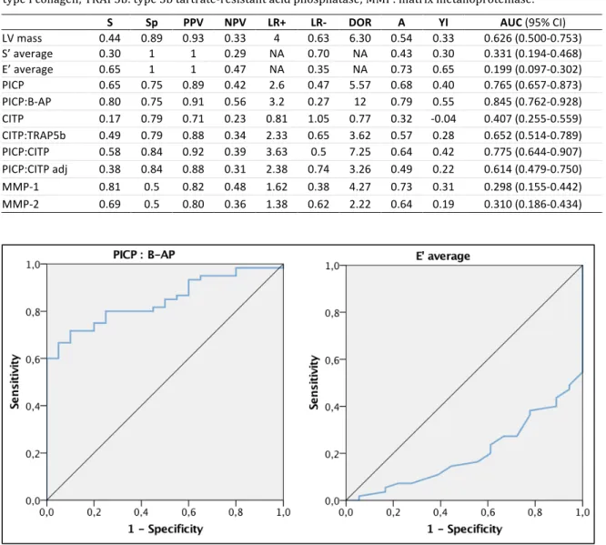
in cardiac MRI. Left image: PICP to B-AP ratio; right image: PICP to CITP ratio adjusted for bone turnover. Page 148
Figure 12: Global diagnostic accuracy to identify Fabry disease. Left image: PICP to B-AP ratio; right image: E’ average. Page 151 Figure 13: Global diagnostic accuracy to identify LV hypertrophy. Left upper image: PICP; right upper image: NT-proBNP; left lower image: MMP-1; right lower image: E’. Page 154
Figure 14: Global diagnostic accuracy to identify LGE. Left image: PICP to CITP ratio; right image: LV mass. Page 155 Figure 15: Cardiomyopathy study: flow diagram of longitudinal evaluation. Page 156 Figure 16: Cardiomyopathy study: longitudinal variation of indexed LVM and E/E’ average by FD subgroups. Left image: LVM; right image: E/E’ average. Page 158
Figure 17: Cardiomyopathy study: longitudinal variation of biomarkers by subgroups of FD patients. Page 160
Figure 18: Cardiomyopathy study: longitudinal variation of balance between collagen type I synthesis and degradation by subgroups of FD patients. Left image: non-adjusted for bone turnover; right image: adjusted for bone turnover. Page 161
Figure 19: Correlation between LVM variation and PICP (left image) or CITP/TRAP5b (right image) variation. Page 164
Figure 20: Longitudinal variation of LVM according to PICP variation quartiles (left image) and comparison of PICP concentration variation (right image) between subgroups of progression. Right image - from left to right: non-progressors, only LVEDP progressor patients, only LV mass progressor patients and patients presenting LVEDP and LV mass progression. Page 166
Figure 21: Main findings of cardiomyopathy study. A significant increase in PICP (biomarker of collagen type I synthesis) and a significant decrease in MMP-1 (biomarker related to collagen type I degradation) was found; when collagen type I build up, in patients with left ventricular hypertrophy, a significant increase in PICP to CITP ratio was identified. Page 170
Figure 22: Nephropathy study: study flow diagram. Page 184
Figure 23: Glomerular and tubular damage biomarkers in controls and FD patient subgroups. Page 187
Figure 24: Correlation between NAG and ACR. Left image: entire FD cohort; right image: subgroup 1 of FD patients. Page 188
Figure 25: Subgroup 1: correlation between ACR and novel biomarkers. Left upper image: UTE; right upper image: ColIV; left lower image: A1MG; right lower image: AAP. Page 189 Figure 26: Correlation between eGFR and ACR or urinary NAG excretion. Left upper image: ACR (entire FD cohort); right upper image: NAG (entire FD cohort); left lower image: ACR (subgroup 1 of FD patients); right lower image: NAG (subgroup 1 of FD patients). Page 190 Figure 27: Boxplot comparing ACR and urinary NAG excretion between CKD stages. Page 191 Figure 28: Global diagnostic accuracy to identify Fabry disease. Left image: ACR; right image: NAG. Page 194
Figure 29: Global diagnostic accuracy to identify CKD stage ≥2. Left image: ACR; right image: NAG. Page 195 Figure 30: Nephropathy study: flow diagram of longitudinal evaluation. Page 196 Figure 31: Nephropathy study: longitudinal variation of kidney function by subgroups and CKD stage in FD patients. Left image: FD subgroups; right image: CKD stage. Page 197 Figure 32: Nephropathy study: longitudinal variation of biomarkers by subgroups of FD patients. Page 200 Figure 33: Nephropathy study: longitudinal variation of biomarkers according to CKD stage at baseline. Page 201
Figure 34: Longitudinal variation of GFR according to ACR (left image) or NAG (right image) quartiles at baseline. Page 202
Figure 35: Comparison of ACR (left image) or NAG (right image) baseline concentration between subgroups of progression. From left to right: non-progressor, only ACR progressor patients, only GFR progressor patients and patients presenting ACR and GFR progression. Page 204
Figure 36: Correlation between GFR variation and ACR (left image) or NAG (right image) variation. Page 205
Figure 37: Comparison of ACR (left image) or NAG (right image) variation in concentration between subgroups of progression. From left to right: non-progressors, only ACR progressor patients, only GFR progressor patients and patients presenting ACR and GFR progression. Page 208
List of tables
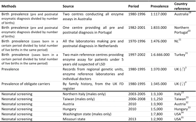
Table 1: studies of prevalence of Fabry disease (adapted from Germain DP30). Page 26 Table 2: Fabry disease biomarkers used in clinical practice. Page 60 Table 3: Potential circulating biomarkers for assessment of cardiac fibrosis (adapted from López B333). Page 79 Table 4: Cardiomyopathy pilot study: population characteristics. Page 138 Table 5: Cardiomyopathy pilot study: echocardiographic characteristics of the study population. Page 139 Table 6: Cardiomyopathy pilot study: biomarkers related to collagen metabolism. Page 139 Table 7: Cardiomyopathy study: clinical characteristics of the study population. Page 142 Table 8: Cardiomyopathy study: mutation frequency. Page 142 Table 9: Cardiomyopathy study: echocardiographic characteristics of the study population. Page 143 Table 10: Biomarkers related to collagen metabolism in controls and FD subgroups. Page 144 Table 11: Correlation between biomarkers related to collagen metabolism and cardiac imaging parameters. Page 146 Table 12: Influence of other variables in biomarkers of collagen type I turnover. Page 149 Table 13: Multivariate regression analysis of predictive variables affecting PICP or MMP-1. Page 149 Table 14: Predictive model of LV mass by univariate and multivariate regression analysis. Page 150Table 15: Diagnostic accuracy of serum and imaging biomarkers to identify Fabry Disease. Page 151
Table 16: Diagnostic accuracy of blood and imaging biomarkers to identify decreased S’ in patients with FD. Page 152
Table 17: Diagnostic accuracy of blood and imaging biomarkers to identify decreased E’ in patients with FD. Page 153
Table 18: Diagnostic accuracy of blood and imaging biomarkers to identify LV hypertrophy in patients with FD. Page 154
Table 19: Diagnostic accuracy of blood and imaging biomarkers to identify LGE in patients with FD. Page 155
Table 20: Cardiomyopathy study: longitudinal variation of disease severity, kidney function and treatment. Page 157
Table 21: Cardiomyopathy study: longitudinal variation of echocardiographic variables. Page 157
Table 22: Patients progressing according to LVM and LVEDP criteria in the entire FD cohort and by subgroups. LVM and LVEDP progressors were defined in the methods sections. Page 158 Table 23: Cardiomyopathy study: longitudinal variation of biomarkers in control group. Page 159
Table 24: Cardiomyopathy study: longitudinal variation of biomarkers in the entire FD cohort. Page 159
Table 25: Distribution of LVM or LVEDP progressors according to quartiles of the baseline concentration of the biomarkers. Page 162
Table 26: Comparison of the baseline concentration of the biomarkers between LVM or LVEDP non-progressors and progressors. Page 163
Table 27: Correlation between the variation of the biomarkers and LVM or LVEDP variation. Page 163 Table 28: Comparison of the variation in the concentration of the biomarkers between LVM or LVEDP non-progressors and progressors. Page 164 Table 29: Distribution of LVM or LVEDP progressors according to quartiles of the variation in the concentration of the biomarkers. Page 165 Table 30: Distribution of progressors (LV mass and/or LVEDP) and non-progressors in the first and fourth quartiles of biomarkers variation. Page 166 Table 31: Progressing and non-progressing Fabry disease patients: analysis by gender. Page 167 Table 32: Comparison of the baseline concentration of the biomarkers between LVM or LVEDP non-progressors and progressors: stratification by gender. Page 167 Table 33: Comparison of the variation in the concentration of the biomarkers between LVM or LVEDP non-progressors and progressors: stratification by gender. Page 168
Table 34: Cardiomyopathy study: comparison of the variation in the concentration of the biomarkers between patients under or not under ERT or ACEi / ARB. Page 169
Table 35: Cardiomyopathy study: comparison of the variation in the concentration of the biomarkers between patients starting ERT or ACEi / ARB, or not, during the study. Page 169 Table 36: Nephropathy pilot study: population characteristics. Page 182
Table 37: Nephropathy pilot study: kidney function in study population. Page 183
Table 38: Nephropathy pilot study: novel biomarkers of glomerular or tubular damage. Page 183 Table 39: Nephropathy study: clinical characteristics of the study population. Page 185 Table 40: Nephropathy study: mutation frequency. Page 185 Table 41: Nephropathy study: kidney function in the population of the study. Page 186
Table 42: Studied biomarkers of kidney injury in controls and FD subgroups. Page 186 Table 43: Correlation between novel biomarkers of kidney injury and ACR for all FD cohort and subgroup 1. Page 188 Table 44: Correlation between novel biomarkers of kidney injury and estimated GFR for all FD cohort and subgroup 1. Page 190 Table 45: Biomarkers in patients with FD according to CKD stage. Page 191 Table 46: Influence of other variables in biomarkers of kidney injury. Page 192 Table 47: Correlation of biomarkers with ACR and eGFR stratified by gender. Page 192 Table 48: Predictive model of eGFR by univariate and multivariate regression analysis. Page 193 Table 49: Diagnostic accuracy of eGFR and urinary biomarkers to identify Fabry Disease. Page 194 Table 50: Diagnostic accuracy of biomarkers to identify CKD stage ≥2. Page 195 Table 51: Nephropathy study: longitudinal variation of kidney function, severity and treatment. Page 197 Table 52: Patients progressing according to ACR and GFR criteria in the entire FD cohort and by subgroups. ACR and GFR progressors were defined in the methods sections. Page 198 Table 53: Nephropathy study: longitudinal variation of biomarkers in control group. Page 198
Table 54: Nephropathy study: longitudinal variation of biomarkers in the entire FD cohort. Page 198 Table 55: ACR variation according to quartiles of the baseline concentration of the biomarkers. Page 202 Table 56: GFR variation according to quartiles of the baseline concentration of the biomarkers. Page 202
Table 57: Distribution of ACR or GFR progressors according to quartiles of the baseline concentration of the biomarkers. Page 203
Table 58: Comparison of the baseline concentration of the biomarkers between ACR or GFR non-progressors and progressors. Page 204 Table 59: Correlation between the variation of the biomarkers and ACR or GFR variation. Page 205 Table 60: Comparison of the variation in the concentration of the biomarkers between ACR or GFR non-progressors and progressors. Page 206 Table 61: Distribution of ACR or GFR progressors according to quartiles of the variation in the concentration of the biomarkers. Page 207
Table 62: Distribution of progressors (ACR and/or GFR) and non-progressors in the first and fourth quartile of the biomarkers’ variation. Page 208
Table 63: Progressing and non-progressing Fabry disease patients: analysis by gender. Page 208 Table 64: Comparison of the baseline concentration of the biomarkers between ACR or GFR non-progressors and progressors: stratification by gender. Page 209
Table 65: Comparison of the variation in the concentration of the biomarkers between ACR or GFR non-progressors and progressors: stratification by gender. Page 210
Table 66: Nephropathy study: comparison of the variation in the concentration of the biomarkers between patients under or not under ERT or ACEi / ARB. Page 210
Table 67: Nephropathy study: comparison of the variation in the concentration of the biomarkers between patients starting ERT or ACEi / ARB, or not, during the study. Page 211
Abstract
Anderson-Fabry disease is an X-linked lysosomal storage disorder, causing significant morbidity and premature death; cardiac and renal involvements are the major determinants of overall disease prognosis. Fabry cardiomyopathy is characterized by a hypertrophic phenotype, in the setting of histological cardiomyocyte hypertrophy and myocardial fibrosis; myocardial fibrosis is an irreversible event and affects the prognosis. A progressive chronic kidney disease characterizes Fabry disease nephropathy; however it is clinically silent for a long period, because heavy storage may occur in renal cells with minimal or no changes on standard renal tests. Furthermore, accumulating evidence suggests that early enzyme replacement therapy is effective in preventing progression of both cardiomyopathy and nephropathy. Therefore, in this thesis I have studied two linked research questions in a multicenter, prospective, longitudinal (evaluation at baseline, 12 months and 24 months) and diagnostic test study:
1) Role of biomarkers related to collagen type I metabolism in the diagnosis of incipient and prognosis of Fabry cardiomyopathy, in a cohort of 60 patients with Fabry disease and 20 healthy controls, according to subgroups of increasing disease severity. I found that collagen type I synthesis is increased in Fabry disease cardiomyopathy, even in the earlier stages of the disease, and this profibrotic state has good prognostic value for and is likely to be critical to the development of overt left ventricular hypertrophy. Moreover, inhibition of enzymes involved in collagen type I cleavage also seems crucial to myocardial collagen deposition and is related to risk of progressive diastolic dysfunction.
2) Identification of early and prognostic biomarkers of Fabry nephropathy in a cohort of 78 patients with Fabry disease and 25 healthy controls, according to subgroups of increasing disease severity. I have shown that two biomarkers of glomerular damage (urinary transferrin and collagen type IV excretion) and three biomarkers of tubular injury (urinary α1-microglobulin, N-acetyl-β-D-glucosaminidase and alanine aminopeptidase excretion) may overcome the limitations of albuminuria as a sensitive marker of early renal dysfunction; furthermore N-acetyl-β-D-glucosaminidase presented the better prognostic value in the identification of patients at risk for chronic kidney disease progression. These biomarkers may also define novel early stages of nephropathy characterized by mesangial expansion and/or tubular damage.
Resumo
A Doença de Fabry é uma doença lisossomal de sobrecarga com um padrão de hereditariedade ligado ao cromossoma X, que condiciona morbilidade significativa e mortalidade precoce; o envolvimento cardíaco e renal são os principais determinantes do prognóstico global. A cardiomiopatia da Doença de Fabry é caracterizada por um fenótipo hipertrófico, condicionado pela hipertrofia dos cardiomiócitos e fibrose miocárdica; esta última é irreversível e afecta o prognóstico. A doença renal crónica progressiva caracteriza a nefropatia da Doença de Fabry; contudo, esta é clinicamente silenciosa por um longo período, na medida em que pode ocorrer extensiva acumulação de substractos nas células renais na ausência de alterações nos testes standard de avaliação da função renal. Mais ainda, evidência crescente sugere que a instituição precoce de terapêutica de substituição enzimática é efectiva na prevenção da progressão quer da cardiomiopatia quer da nefropatia. Por conseguinte, na presente tese eu abordei duas questões interrelacionadas, num estudo de testes de diagnóstico, multicêntrico, prospectivo e longitudinal (avaliação basal e aos 12 e 24 meses):
1) A função dos biomarcadores relacionados com o metabolismo do colagénio tipo I no diagnóstico precoce e na determinação do prognóstico na cardiomiopatia da Doença de Fabry, num coorte de 60 doentes com Doença de Fabry e 20 controlos saudáveis, de acordo com subgrupos de gravidade crescente. Demonstrei que a síntese de colagénio tipo I encontra-se aumentada na cardiomiopatia da Doença de Fabry, mesmo nos estádios mais precoces da doença e que este estado profibrótico apresenta bom valor prognóstico para e parece ser essencial ao desenvolvimento de hipertrofia ventricular esquerda. Constei ainda que a inibição das enzimas envolvidas na degradação do colagénio tipo I também afigura-se como crucial à deposição de colagénio no miocárdio e encontra-se relacionada com o risco de disfunção diastólica progressiva.
2) Identificação de biomarcadores precoces e de prognóstico na nefropatia da Doença de Fabry num coorte de 78 doentes com Doença de Fabry e 25 controlos saudáveis, de acordo com subgrupos de gravidade crescente. Evidenciei que dois biomarcadores urinários de lesão glomerular (transferrina e colagénio tipo IV) e três biomarcadores urinários de dano tubular (α1-microglobulina, N-acetil-β-D-glucosaminidase e alanina aminopeptidase) podem ultrapassar as limitações da
albuminuria como marcador sensível de disfunção renal precoce; mais ainda, a excreção de N-acetil-β-D-glucosaminidase apresentou melhor valor prognóstico na identificação de doentes em risco de doença renal crónica progressiva. Estes biomarcadores também poderão definir novos estádios precoces da nefropatia da Doença de Fabry caracterizados por expansão do mesângio e/ou lesão tubular.
Introduction
1. Anderson-Fabry disease
1.1 Aetiology, genotype-phenotype correlations and epidemiology
Anderson-Fabry disease (FD) is an X-linked lysosomal storage disorder (LSD), independently first described by Johannes Fabry and William Anderson in 1898,1 who reported patients with “angiokeratoma corporis diffusum”, the red-purple maculopapular skin lesions that are characteristic of the disease. It was only in 1967 that FD was found to be caused by mutations in the GLA gene (that encodes the enzyme α-galactosidase A).2 The deficiency of this enzyme leads to the lysosomal accumulation of neutral glycosphingolipids (mainly globotriaosylceramide [Gb3]) in several cells, causing organ failure. Involvement of the heart, kidney and brain in FD causes significant morbidity and premature death.3, 4
The GLA gene was mapped to the q22.1 region of the X chromosome. It contains seven exons and the coding part of the gene consists of 1290 base pairs, encoding a polypeptide of 429 amino acids.5 A total of 672 mutations / variants have been described,
most of them (455) missense / nonsense.6 The great majority of the GLA mutations are
private (each family has its own mutation) and theoretical considerations point to 3-10% of patients with de novo mutations.7
GLA gene encodes the lysosomal enzyme α-galactosidase A (a homodimeric molecule), responsible for the degradation of neutral glycosphingolipids with terminal α-galactosyl moieties in cells throughout the body. Its main substrate is Gb3, but there are also other minor substrates, including digalactosylceramide, P1 antigen and blood group B glycolipids. Gb3 originate from metabolic turnover of membrane glycosphingolipids (like globoside [Gb4]) present in large amounts in the kidneys, liver, vascular endothelium, lungs and erythrocytes.5
Despite its “monogenic” origin, FD is clinically heterogeneous, with some patients presenting a classical phenotype (full-blown clinical picture with “acroparesthesias”, angiokeratomas, hypohydrosis, corneal opacity and renal, cardiac and cerebrovascular involvement) and others a late-onset / attenuated form (mainly affecting the heart).8 Genetic factors certainly contribute to this heterogeneity, with patients exhibiting out-of-frame short length rearrangements and splice-site and nonsense mutations, leading to a
premature stop codon and a truncated and most likely non-functional enzyme, presenting a classical phenotype.5 In patients with missense mutations, enzymatic activity may be variably affected: mutations in functionally important regions or leading to large structural changes show the classical phenotype, whereas late-onset forms are associated with mutations located apart from the active site, conditioning small structural changes.9 However, in patients within the same family or with the same mutation, the clinical picture may vary widely, thus other genetic, epigenetic and environmental factors also contribute to clinical heterogeneity.
Furthermore, unlike many other X-chromosomal conditions, female FD patients may show signs and symptoms commonly seen in male patients. However, disease onset occurs later and the phenotype tends to be milder (though clinically heterogeneous with several patients with classic FD).10, 11 Thus, with this state of intermediate disease penetrance in female FD patients, the terms “X-linked recessive” or “X-linked dominant” may not capture the wide spectrum of variable expression in heterozygotes and FD may be referred to as “X-linked”.12
Phenotype and penetrance in FD female patients may be explained according to the principles of random X chromosome inactivation (lionization), resulting in a mosaic with some cells expressing “wild type” and others “mutant” GLA gene.13 The celular and tissue levels of α-galactosidase A activity will depend on the balance between “wild type” and “mutant” X-inactivation, varying from organ to organ and with female FD patients exhibiting skewed X-inactivation favouring “mutant” GLA gene, presenting a more severe phenotype.14 However, there is no correlation between plasma or leucocyte α-galactosidase A activity and disease severity, and cross correction of the metabolic defect by uptake of the “wild type” enzyme by the cells with the “mutant” GLA gene would prevent females from developing clinically significant disease.15, 16 Thus, these findings suggest that cross complementation is impaired or insufficient in FD, possibly due to defective uptake, for reasons as yet unclear, of “wild type” enzyme by the “mutant” cells.16 Moreover, “cross-induction” of the defect may be another pathophysiological link
explaining the clinical manifestations in female FD patients. Lyso-Gb3, a deacylated form of Gb3, is increased in the great majority of female FD patients; it is widely diffusible and has the capacity to inhibit α-galactosidase A activity in “wild type” cells.17
smooth muscle cells proliferation (directly contributing to FD vasculopathy) and production of extracellular matrix proteins by podocytes (leading to glomerular damage).17, 18
FD is pan-ethnic, but due to its rarity, it is difficult to determine its true prevalence. Moreover, in most countries, there are various diagnostic centres, making it difficult to collect the diagnoses. Three main studies addressing the birth prevalence of FD reported incidences ranging from 1:117,000 in Australia, to 1:833,000 in Northern Portugal.19-21 In the Australian epidemiological study, no data was obtained on
heterozygote females, but the incidence determined in hemizygote males could be extrapolated to give a combined incidence of 1 in 58,000. However, the true prevalence of FD is likely to be much higher, due to the existence of late-onset forms / attenuated phenotypes of the disease as identified by the screening of high-risk populations and newborns. Several newborn screening initiatives of FD raised the incidence of FD up to 1:3,100 male births in Italy or even 1:1,250 male births in Taiwan, with most of the identified mutations associated with attenuated phenotypes with predominant cardiac involvement, giving an estimated ratio as high as 11:1 of attenuated to classic phenotypes.22, 23 A summary of prevalence and newborn screening studies is shown in table 1.
1.2 Pathology and pathophysiology 1.2.1 Cardiomyopathy
In FD cardiomyopathy, glycosphingolipid storage (mainly Gb3) occurs in all cardiac cell types, namely cardiomyocytes, conduction system cells, valvular fibroblasts, endothelial cells, vascular smooth muscle cells and cardiac nerves (including autonomic nervous system).24-26 In female FD patients there is a mosaic pattern of storage, due to the random X-inactivation.27 However, autopsy studies in extremely hypertrophied hearts showed that Gb3 infiltration of the myocardium accounts only for 1-3% of the wet weight of the hypertrophic heart and cannot explain its degree, which indicates that activation of several pathological pathways and other histological features are also important.28 Hence, Gb3 storage is probably a primer for lysosomal and cellular malfunctioning, triggering intracellular common signalling pathways leading, ultimately, to
cardiomyocytes hypertrophy, apoptosis and necrosis, followed by replacement fibrosis. These events are responsible for the diastolic dysfunction and contractility impairment seen in the clinical setting.28, 29
Table 1: Studies on prevalence of Fabry disease. LSD: lysosomal storage disorder; FD: Fabry disease; UK: United Kingdom; NL: Netherlands; USA: United States of America (adapted from Germain DP30).
Methods Source Period Prevalence Country reference
Birth prevalence (pre and postnatal enzymatic diagnosis divided by number of births)
Two centres conducting all enzyme assays in Australia
1980-1996 1:117.000 Australia19 Birth prevalence (pre and postnatal
enzymatic diagnosis divided by number of births)
One centre providing all pre and
postnatal diagnosis in Portugal 1982-2001 1:833.000 Northern Portugal20 Birth prevalence (cases born in a
certain period divided by total number of live births in the same period)
All the laboratories making pre and postnatal diagnosis in Netherlands
1970-1996 1:476.000 NL21
Birth prevalence (cases born in a certain period divided by total number of live births in the same period)
Two main reference centres providing enzyme assay for patients under 5 years old suspected of LSD
1997-2002 1:6.666.000 Turkey31 Prevalence Records from regional genetic units,
enzyme reference laboratories and individual doctors
1980-1995 1:370.000 UK (♂)3 Prevalence of obligate carriers By family history, from the UK FD
register 1980-1995 1:345.000 UK (♀)
4
Neonatal screening Northern Italy (males only) 2003-2005 1:3,100 Italy22 Neonatal screening Taiwan (males only) 2006-2008 1:1,250 Taiwan23
Neonatal screening Austria 2010 1:3,900 Austria32
Neonatal screening Hungary 2010 1:5,000 Hungary33
Neonatal screening Washington state (males only) 1:7,800 USA34
Neonatal screening Missouri state 2013 1:2,900 USA35
The link between lysosomal storage and cellular dysfunction is not well understood, but energy depletion (related to respiratory chain enzymes dysfunction), abnormal production of reactive oxygen species (ROS), ischaemia and circulating growth-promoting factors are certainly involved.
Cellular energy depletion, due to inefficient cardiomyocytic energy utilization, is one of the most accepted hypotheses. It has been proposed as a common mechanism for several types of metabolic and even sarcomeric hypertrophic cardiomyopathy.36 A study
using cultured skin fibroblasts from four FD patients and ten controls has shown, in FD patients, a significant reduction of the activity of respiratory chain complexes I+III, IV and V, as well as a significant decrease of cellular content of high-energy phosphate compounds (creatine phosphate [PCr] and adenosine diphosphate) and adenosine monophosphate.37 Moreover, studies with cardiac phosphorus-31 magnetic resonance spectroscopy (a non-invasive technique enabling in vivo determination of PCr and
adenosine triphosphate [ATP]) in FD have shown a significant decrease in PCr, ATP and the PCr to ATP ratio, as well as a significant inverse correlation between the PCr to ATP ratio and the annual increase in left ventricular (LV) mass, indicating mitochondrial energy depletion.38, 39 This mitochondrial dysfunction will lead to inefficient sarcomeric ATP
utilization and impaired contractility, resulting in hypertrophy. The reason for the reduction of respiratory chain enzyme activities in FD remains unclear. However, in vitro, lysosphingolipids may have direct deleterious effects on mitochondrial function, by direct binding to mitochondrial membranes and inhibition of oxidative phosphorylation.40
Several studies suggest that oxidative stress is also implicated in the link between lysosomal storage and cellular dysfunction in FD cardiomyopathy and vasculopathy. In cultured FD endothelial cells, incubation with increasing doses of Gb3 increased intracellular ROS generation in a dose-dependent manner, which was significantly lowered (even in the cells loaded with Gb3) by addition of an antioxidant (vitamin C). In the same study, incubation of the cell line with plasma from FD patients also led to increased ROS production, even if the endothelial cells were pre-treated with a glycosphingolipid synthase inhibitor or recombinant enzyme replacement therapy (ERT) to decrease Gb3 loading, suggesting that other factors in FD patient plasma may also contribute to oxidative stress in vascular endothelial cells.41 This data was confirmed in
vivo by demonstration, in FD patients under ERT, of reduced glutathione and glutathione
peroxidase activity and increased superoxide dismutase / catalase ratio in erythrocytes (leading to decreased levels of antioxidant defences), as well as oxidative damage of lipid and proteins (increased levels of malondialdehyde [a product of lipid peroxidation] and carbronyl group and dityrosine [both resulting from oxidative damage to proteins]).42 Moreover, oxidative damage to deoxyribonucleic acid (DNA) purines is significantly higher in FD patients, despite efficient DNA repair.43
Gb3 loading may also cause a deregulation of nitric oxide (NO) pathway, with down-regulation of endothelial NO synthase, up-regulation of inducible NO synthase and enhanced cyclooxygenase-2 expression in microvascular (but not in macrovascular) cardiac endothelial cells.44 Moreover, FD causes increased nitrotyrosine expression in
brain tissue from FD patients45 and elevated levels of nitrotyrosine in an endothelial FD
cell line and in the plasma of FD patients,46 attributable to endothelial NO synthase
Regarding cardiomyocytes oxidative damage, in a study involving 18 FD patients performing endomiocardial biopsies, immunohistochemistry has shown that inducible NO synthase and nitrotyrosine expression was increased in FD patients compared with hypertrophic cardiomyopathy due sarcomere protein gene mutations (HCM) and normal controls. Oxidative damage to DNA, assessed by immunostaining for 8-hydroxydeoxyguanosine, was also only identifiable in FD patients (25% of cardiomyocyte nuclei) and not in controls; cardiomyocytes apoptotic cell death was 187-fold greater in FD patients than in controls and only detected in nuclei with oxidative DNA damage.47
The disease pathophysiology is potentiated by ischaemia, which may occur even in the absence of significant epicardial coronary artery disease. Elliott P et al., in a study including 10 FD male patients (none had angiographically significant coronary artery disease), showed that resting and hyperaemic myocardial blood flow and coronary flow reserve were significantly reduced in FD patients, with comparable resting coronary reserve, attributable to abnormal microvascular function.48 This might be due to
increased susceptibility, for unknown reasons, of microvascular endothelial myocardial cells to deregulation of NO pathway,44 increased oxygen demand of the hypertrophied
muscle cells, decreased capillary density, increased diastolic filling pressures (impairing blood flow throughout the subendocardium) and sphingolipid storage within endothelial and vascular smooth muscle cells of small arterioles and capillaries.5
Furthermore, incubation of FD patients plasma with mouse vascular smooth muscle cells and neonatal cardiomyocytes, suggests that there are circulating growth-promoting factors inducing cell hypertrophy and proliferation, correlating with LV mass and carotid common artery intima-media thickness.49
Cellular dysfunction affects myofilaments and cardiomyocyte contractility; there is evidence of myofilament proteolysis (revealed by presence of degradation products of troponin I and desmin) and dislodgement and disarray (partly due to intracellular glycosphingolipids storage) and myofibrillolysis. In this context, it was reported that the active tension of cardiomyocytes was four times lower in Fabry cardiomyocytes compared to controls, and this correlated well with the extent of myofibrinolysis. Furthermore, the resting tension was six times higher than in controls, which was shown to correlate with tissue Doppler imaging (TDI) data, suggesting that increased stiffness of cardiomyocytes contributes to diastolic dysfunction in FD cardiomyopathy.29
Myofilament dysfunction, with impaired relaxation and contractility and increased cell stress may trigger, as hypothesized for HCM, expression of stress-responsive trophic and mitotic factors (in an attempt of structural and functional cell repair), a possible direct cause of myocyte hypertrophy and disarray and increased interstitial collagen synthesis (figure 1).50
Figure 1: Pathophysiology of Fabry disease cardiomyopathy. Glycosphingolipid storage is probably the main trigger to the activation of several pathways, ending in cellular dysfunction and increased cell stress; this will promote increased collagen synthesis and cardiomyocyte hypertrophy. ROS: reactive oxygen species. energy deple*on Glycophingolipid storage Other factors (?) ROS growth
factors ischemia pathways other
cellular dysfunc*on ê cardiomyocyte relaxa*on ê cardiomyocyte contrac*lity é cell stress cardiomyocyte (hypertrophy) fibroblasts (é collagen synthesis) é growth factors
Histologically, the most prominent feature in patients with advanced FD cardiomyopathy is the severe cardiomyocyte hypertrophy, mainly attributable to intracytoplasmic glycosphingolipids storage, appearing as perinuclear vacuoles in light microscopy and as electron-dense deposits consisting of parallel or concentric lamellae in electron microscopy.24, 29, 51-53 There is disarray and peripheral displacement of the myofibrils, with some of the fibres displaying signs of disintegration (being surrounded by small collections of foamy histiocytes).24, 28, 29
Myocardial fibrosis is also one of the hallmarks of FD cardiomyopathy. Fibrosis is unequally distributed along the LV wall, being more prominent within the mid-myocardial layers and the posterolateral segments of the LV29, 51 and ranging from mild fibrosis (with
fibres embedded in loose fibrous tissues) in the interventricular septum (IVS),28, 53 to
marked fibrosis (with almost no myocardial cells observed) in the base of the LV posterior wall (PW).54 Moreover, fibrosis seems to be predominantly interstitial diffuse (with only
focal areas of replacement fibrosis) and, in one study, it did not correlate with TDI measurements of systolic or diastolic LV function.29, 54
Furthermore, massive accumulation of lipids is also present in the conduction tissue, tricuspid and mitral valves and endothelium cells of the myocardial capillaries.24, 55 In FD patients with attenuated phenotypes with predominant cardiac involvement, no lysosomal inclusions were observed in cardiac capillary endothelial cells.54
Concluding, Gb3 storage is probably the trigger for the activation of several signalling pathways leading to cellular dysfunction and, ultimately, to cardiomyocyte hypertrophy and fibrosis, in direct relationship with the clinical manifestations of LV hypertrophy and diastolic dysfunction.
1.2.2 Nephropathy
As mentioned above for cardiomyopathy, Gb3 storage also occurs in all renal cells, namely the endothelial, glomerular, interstitial and tubular cells, and the disease progression results in the development of glomerulosclerosis, tubular atrophy and interstitial fibrosis.56, 57 However, the pathophysiology of FD nephropathy and the
relationship between Gb3 storage and the aforesaid findings are not completely understood. Podocyte and endothelial damage, as well as other mechanisms may play an important role in FD nephropathy pathogenesis.
Though present in all glomerular cells, Gb3 storage is most abundant in podocytes.57-60 Moreover, podocytes are unique neuron-like glomerular cells that are highly differentiated, with only limited potential of self-renewal, a fact that could make them particularly susceptible to damage by lysosomal storage disorders. Podocyte injury leads to loss of integrity of the glomerular filtration barrier and progression to chronic kidney disease.61 Several pathways are certainly involved in podocytes damage, namely
autophagy dysregulation, cytoskeleton disorganization (and podocyturia) and activation of fibrotic and inflammatory pathways.
Dysregulation of the autophagic pathway is one of the contributors to podocytes damage. In vitro studies in a human podocytes cell line, a FD phenotype was established using small hairpin ribonucleic acids (RNA) to reduce α-galactosidase A expression and activity. In these α-galactosidase A deficient podocytes, Gb3 accumulation was accompanied by an increase in autophagosomes and a down-regulation of mammalian target of rapamycin (mTOR) signalling cascade (a well-known inhibitor of autophagy).62
This data was confirmed in kidney biopsies of 5 patients, showing increased number of autophagic vacuoles in FD kidney tissue (notably in podocytes), which decreased in renal biopsies performed after three years of ERT.63 Sphingolipid storage may have a direct role in the dysregulation of the autophagic pathway, since ceramide is an activator of autophagy.64
Gb3 overload in podocytes may also interfere, by mechanical stress, with the distribution of synaptopodin, an actin-associated protein highly expressed in podocyte foot processes that is involved in cytoskeletal reorganization.65 Moreover, Gb3 may interact with F-actin causing cell contraction, slit diaphragm widening and the coupling with integrins.65, 66 Integrins, mainly αvβ3 (also known as vitronectin receptor), are
essential to anchor podocytes to the basement membrane and their activation triggers podocyte contraction and migration and, ultimately, detachment from the glomerulus. In FD patients, increased urinary excretion of αvβ3 integrin was found in both classical and
attenuated phenotypes; also its increased expression was observed in podocytes of the kidney tissue from a patient with a classical phenotype.67 Therefore, integrin activation
seems to be actively involved in podocytes detachment from the glomerular basement membrane in FD. This phenomenon will lead to higher levels of podocyturia, as previously shown in two studies with FD patients, one of them reporting a significant correlation
between podocyturia and albuminuria.66, 68 Podocytopenia is the main consequence, and
it is believed that when each glomerulus loses more than 40% of its 500 podocytes, it undergoes obliteration.69
In a human glomerular podocytes cell line, as lyso-Gb3 increases, there is activation of fibrotic pathways, leading to the expression and production of extracellular matrix proteins (fibronectin and collagen type IV [ColIV]), mediated by the transforming growth factor-β1 (TGF-β1) and Notch1 pathways. Paricalcitriol and calcitriol, vitamin D receptor activators, prevented the up-regulation of these mediators. This expansion of extracellular matrix is certainly involved in the glomerulosclerosis pathogenesis.18, 70
Inflammatory response is also up-regulated in FD glomerular podocytes, as shown by higher urinary excretion of CD80 in FD patients, as well as increased CD80 messenger RNA expression in podocytes cultured with lyso-Gb3.71 CD80 (lymphocyte activation antigen 7-1) is normally located in antigen presenting cells (not expressed by normal podocytes) and modulates T helper and T cytotoxic cells activity.72 Increased expression
of CD74, a macrophage migration inhibitory factor receptor that regulates the expression of lethal cytokines, had also been shown.18 Furthermore, Notch1 pathway, a key mediator
of proinflammatory response, was up-regulated in a podocytes cell line cultured with lyso-Gb3, with increase expression of chemokines MCP-1 (Monocyte Chemoattractant Protein-1) and RANTES (Regulated on Activation, Normal T Cell Expressed and Secreted), mediated by the recruitment of the transcription of NFkB (nuclear factor kappa B). This data was confirmed in kidney biopsies from FD patients, with immunohistochemistry confirming Notch1 expression in glomerular podocytes and tubular cells.70
Vasculopathy is also certainly involved in FD nephropathy pathogenesis. As mentioned above for FD cardiomyopathy, production of ROS and dysregulation of NO pathway are major contributors to endothelial dysfunction.41, 42, 44-46 The classical theory emphasized and suggested that kidney fibrosis and advanced kidney lesions were a consequence of ischaemic tissue damage, derived from microvascular endothelial disease and/or necrosis of vascular smooth muscle cells.57, 60 This hypothesis was supported by
pathological studies showing universal lipid deposits at a very early age, unlike the nonspecific changes (like glomerulosclerosis, tubular atrophy and interstitial fibrosis) which were age-related and firstly involving the vessels.57 However, these are nonspecific
patients younger than 25 years old, suggesting that they are secondary features and not the early pathogenic event. Therefore, the current knowledge on the importance of podocyte injury in glomerulosclerosis pathogenesis indicates that vasculopathy is certainly involved in FD pathogenesis, but it is not the sole or even the main pathogenic mechanism.
Tubulointerstitial injury is also involved in FD nephropathy pathogenesis. Gb3 may have direct toxic effects in tubular cells, causing focal tubular atrophy and interstitial fibrosis. Although there is weak evidence supporting this mechanism, the finding of glomerular enlargement in the early stages of glomerular and tubular injury could corroborate that the glomeruli upstream of affected tubules may function poorly and that other glomeruli may undergo hypertrophy to compensate. Hyperfiltration in these glomeruli may trigger a secondary form of focal segmental glomerulosclerosis.60 Diffuse involvement of interstitial cells, with electron dense inclusions, has also been reported.57, 73 By causing tubular injury, proteinuria may have, as in other nephropathy models, a
direct role in chronic kidney disease (CKD) progression and may provide a link between the aforesaid pathological processes and the development of tubulointerstitial disease.74, 75
These abovementioned effects of storage in all renal cells will give rise to progressive nephropathy, characterized by nonspecific degenerative lesions, namely mesangial widening, segmental and/or global glomerular sclerosis, tubular atrophy and interstitial fibrosis (figure 2).56, 57, 60, 73, 76 These well known histological findings in advanced FD nephropathy contrast with the limited knowledge about the histology of patient with incipient nephropathy. Lipid deposition related lesions are characteristic of FD nephropathy. As ceramide tails of glycosphingolipids are dissolved during paraffin embedding of kidney tissue, the most characteristic finding in routine light microscopy of kidney biopsies is vacuolization. This is particularly prominent in podocytes, parietal epithelial cells of Bowman’s capsule and Henle’s loop and distal tubular cells.56, 57, 60, 76 In light microscopy, glycosphingolipids deposits are best assessed in toluidine blue stained semi-thin sections of glutaraldehyde / formaldehyde fixed and epoxy-embedded tissue. In the glomeruli the largest amount of lipid material is seen in podocytes followed by parietal epithelial, mesangial and glomerular endothelial cells.57, 59, 77 Tubular inclusions are more prominent in distal