Microsatellite markers: what they mean and why they are so useful
Maria Lucia Carneiro Vieira
1, Luciane Santini
1, Augusto Lima Diniz
1and Carla de Freitas Munhoz
1 1Departamento de Genética, Escola Superior de Agricultura “Luiz de Queiroz” (ESALQ), Universidade de
São Paulo (USP), Piracicaba, SP, Brazil.
Abstract
Microsatellites or Single Sequence Repeats (SSRs) are extensively employed in plant genetics studies, using both low and high throughput genotyping approaches. Motivated by the importance of these sequences over the last de-cades this review aims to address some theoretical aspects of SSRs, including definition, characterization and bio-logical function. The methodologies for the development of SSR loci, genotyping and their applications as molecular markers are also reviewed. Finally, two data surveys are presented. The first was conducted using the main data-base of Web of Science, prospecting for articles published over the period from 2010 to 2015, resulting in approxi-mately 930 records. The second survey was focused on papers that aimed at SSR marker development, published in the American Journal of Botany’s Primer Notes and Protocols in Plant Sciences (over 2013 up to 2015), resulting in a total of 87 publications. This scenario confirms the current relevance of SSRs and indicates their continuous utiliza-tion in plant science.
Keywords: SSR biological function, genomic distribution, genotyping approaches, molecular marker, practical utility.
Received: February 29, 2016; Accepted: May 13, 2016.
Brief introduction
Ongoing technological advances in all fields of knowledge mean that we cannot be sure which technolo-gies will survive the impact of innovation, and for how long. Over the years, advances in molecular genetics meth-odology have lead to widespread use of codominant molec-ular markers, especially Simple Sequence Repeats (SSRs) and, more recently, Single Nucleotide Polymorphisms (SNPs). This paper attempts to present an overview of how the concept of SSRs has evolved and how their biological functions were discovered. We also address the develop-ment of methods for identifying polymorphic SSRs, and the application of these markers in genetic analysis. It reveals that much remains to be explored regarding these se-quences, particularly in relation to cultivated and wild plants.
Definition and genome occurrence of
microsatellites and their use as genetic markers
Microsatellites (1 to 10 nucleotides) and mini-satellites (> 10 nucleotides) are subcategories of tandem re-peats (TRs) that, together with the predominant inter-spersed repeats (or remnants of transposable elements), make up genomic repetitive regions. TRs are evolutionarily
relevant due to their instability. They mutate at rates be-tween 103and 106per cell generation i.e., up to 10 orders of
magnitude greater than point mutations (Gemayel et al.,
2012).
Microsatellites, Simple Sequence Repeats (SSR), Short Tandem Repeats (STR) and Simple Sequence Length Polymorphisms (SSLP) are found in prokaryotes and eukaryotes. They are widely distributed throughout the ge-nome, especially in the euchromatin of eukaryotes, and coding and non-coding nuclear and organellar DNA (Pérez-Jiménezet al., 2013; Phumichaiet al., 2015).
There is a lot of evidence to back up the hypothesis that SSRs are not randomly distributed along the genome. In a comparative study, SSR distribution was found to be highly non-random and to vary a great deal in different
re-gions of the genes ofArabidopsis thalianaand rice
(Law-son and Zhang, 2006). In the major cereals, for instance, authors have tended to categorize microsatellites based on different criteria. In barley andAvenaspecies, SSRs were
classified in two types: those with unique sequences on ei-ther flank and those intimately associated with retrotrans-posons and other dispersed repetitive elements. The second type was found to be less polymorphic in oat cultivars (Ramsayet al., 1999; Liet al., 2000). Using publicly
avail-able DNA sequence information on the rice genome, Temnykhet al.(2001) categorized microsatellites based on
length and noticed that longer perfect repeats (³20 nucleo-tides) were highly polymorphic. Microsatellites with SSRs shorter than 12 bp were found to have a mutation potential DOI: http://dx.doi.org/10.1590/1678-4685-GMB-2016-0027
Send correspondence to Maria Lucia Carneiro Vieira. USP/ESALQ, P.O. Box 83, 13400-970, Piracicaba, SP, Brazil. E-mail: mlcvieir@usp.br
no different from that of most unique sequences. Moreover, authors reported that ~80% of GC-rich trinucleotides oc-curred in exons, whereas AT-rich trinucleotides were distributed roughly evenly throughout all genomic compo-nents (coding sequences, untranslated regions, introns and intergenic spaces). Tetranucleotide SSRs were predomi-nantly situated in non-coding, mainly intergenic regions of the rice genome. It was later established that the SSR distri-butions in different regions of the maize genome were non-random, and that density was highest in untranslated regions (UTR), gradually falling off in the promotor, intron, intergenic, and coding sequence regions, in that or-der (Qu and Liu, 2013).
On the other hand, comparisons of microsatellite
dis-tributions inRumex acetosaandSilene latifolia
chromo-somes showed that some motifs (e.g. CAA or TAA) are strongly accumulated in non-recombining regions of the
sex chromosome (Y) in both plant species (Kejnovskyet
al., 2009). Similarly, a very large accumulation consisting
mainly of microsatellites on the heterochromatic W chro-mosome was reported in a group of fish species (Leporinus
spp.) that share a ZW sex system, showing an interconnec-tion between heterochromatinizainterconnec-tion and the accumulainterconnec-tion of repetitive sequences, which has been proposed as the ba-sis of sex chromosome evolution (Poltronieriet al.,2014).
Generally speaking, it can be affirmed that the occur-rence of SSRs is lower in gene regions, due to the fact that SSRs have a high mutation rate that could compromise gene expression. Studies indicate that in coding regions there is a predominance of SSRs with gene motifs of the tri-and hexanucleotide type, the result of selection pressure against mutations that alter the reading frame (Zhanget al.,
2004; Xuet al., 2013b). In humans, the consensus is that
SSRs can also originate in coding regions, leading to the appearance of repetitive patterns in protein sequences. In protein sequence database studies, it was reported that tan-dem repeats are common in many proteins, and the mecha-nisms involved in their genesis may contribute to the rapid evolution of proteins (Kattiet al., 2000; Huntley and
Gol-ding, 2000).
Repeat polymorphisms usually result from the addi-tion or deleaddi-tion of the entire repeat units or motifs. There-fore, different individuals exhibit variations as differences in repeat numbers. In other words, the polymorphisms ob-served in SSRs are the result of differences in the number of repeats of the motif caused by polymerase strand-slippage in DNA replication or by recombination errors. Strand-slippage replication is a DNA replication error in which the template and nascent strands are mismatched. This means that the template strand can loop out, causing contraction. The nascent strand can also loop out, leading to repeat expansion. Recombination events, such as unequal crossing over and gene conversion, may additionally lead to SSR sequence contractions and expansions. According to several authors, the longer and purer the repeat, the
higher the mutation frequency, whereas shorter repeats with lower purity have a lower mutation frequency.
Mutations that have evaded correction by the DNA mismatch repair system form new alleles at SSR loci. For this reason, different alleles may exist at a given SSR locus, which means that SSRs are more informative than other molecular markers, including SNPs.
As for their composition, SSRs can be classified ac-cording to motif as:i)perfect if composed entirely of
re-peats of a single motif; ii) imperfect if a base pair not
belonging to the motif occurs between repeats;iii)
inter-rupted if a sequence of a few base pairs is inserted into the motif; oriv)composite if formed by multiple, adjacent,
re-petitive motifs (reviewed in Oliveiraet al., 2006; revisited
by Mason, 2015).
SSRs have been the most widely used markers for genotyping plants over the past 20 years because they are highly informative, codominant, multi-allele genetic mark-ers that are experimentally reproducible and transferable among related species (Mason, 2015). In particular, SSRs are useful for wild species (i) in studies of diversity
mea-sured on the basis of genetic distance; (ii) to estimate gene
flow and crossing over rates; and (iii) in evolutionary
stud-ies, above all to infer infraspecific genetic relations. On the other hand, for cultivated plants SSRs are commonly used for (i) constructing linkage maps; (ii) mapping loci
in-volved in quantitative traits (QTL); (iii) estimating the
degree of kinship between genotypes; (iv) using
marker-assisted selection; and (v) defining cultivar DNA
finger-prints (Jonahet al., 2011; Kalia et al., 2011). SSRs have
been particularly useful for generating integrated maps for plant species in which full-sib families are used for con-structing linkage maps (Garciaet al., 2006; Souzaet al.,
2013; Pereira et al., 2013), and for combining genetic,
physical, and sequence-based maps (Temnykh, 2001), pro-viding breeders and geneticists with a tool to link pheno-typic and genopheno-typic variation (see Mammadovet al., 2012;
Haywardet al., 2015 for review articles).
These markers are enormously useful in studies of population structure, genetic mapping, and evolutionary processes. SSRs with core repeats 3 to 5 nucleotides long are preferred in forensics and parentage analysis. It is worth noting that a number of SSR search algorithms have been developed, including TRF (Benson, 1999), SSRIT
(Temnykh, 2001), MISA (Thielet al., 2003), SSRFinder
(Gao et al., 2003), TROLL (Castelo et al., 2002) and SciRoKo (Kofleret al., 2007).
Detailing the biological functions of SSRs
Despite the wide applicability of SSRs as genetic markers since their discovery in the 1980s, little is known about the biological importance of microsatellites (Tautz
and Renz, 1984), especially in plants. Morgante et al.
(2002) estimated the density of SSRs in Arabidopsis
maize (Zea mays) and wheat (Triticum aestivum) and
ob-served a high frequency of SSRs in transcribed regions, es-pecially in untranslated regions (UTRs). Interestingly, there are substantial data indicating that SSR expansions or contractions in protein-coding regions can lead to a gain or loss of gene function via frameshift mutation or expanded toxic mRNAs. SSR variations in 5’-UTRs could regulate gene expression by affecting transcription and translation, but expansions in the 3’-UTRs cause transcription slippage and produce expanded mRNA, which can disrupt splicing and may disrupt other cellular functions. Intronic SSRs can affect gene transcription, mRNA splicing, or export to cy-toplasm. Triplet SSRs located in UTRs or introns can also induce heterochromatin-mediated-like gene silencing. All these effects can eventually lead to phenotypic changes (Li
et al., 2004; Nalavadeet al., 2013).
In fact, variation in the length of DNA triplet repeats has been linked to phenotypic variability in microbes and to several human disorders, including Huntington’s disease
which is caused mainly by (CAG)nexpansions. Moreover,
the frequencies of different codon repeats vary consider-ably depending on the type of encoded amino acid. In plants, a triplet repeat-associated genetic defect was identi-fied in a wild variety ofA. thalianathat carries a
dramati-cally expanded TTC/GAA repeat in the intron of the gene encoding the large subunit 1 of the isopropyl malate iso-merase. Expansion of the repeat causes an environment-dependent reduction in the enzyme’s activity and severely impairs plant growth, whereas contraction of the expanded repeat can reverse the detrimental effect on the phenotype (Sureshkumaret al., 2009).
Historically, tandem repeats have been designated as nonfunctional DNA, mainly because they are highly unsta-ble. With the exception of tandem repeats involved in hu-man neurodegenerative diseases, repeat variation was often believed to be neutral with no phenotypic consequences (see Gemayelet al., 2012).
The detection of microsatellites in transcripts and reg-ulatory regions of the genome encouraged scientific inter-est in discovering their possible biological functions. More and more publications have presented evidence that micro-satellites play a role in relevant processes, such as the regu-lation of transcription and transregu-lation, organization of chromatin, genome size and the cell cycle (Nevo, 2001; Li
et al., 2004; Gaoet al., 2013).
As mentioned above, most of the knowledge acquired on microsatellites occurring in genes was obtained by studying humans and animals, indicating their relationship with the manifestation of disease. In bacteria, maintaining numerous microsatellite variants provides a source of highly mutable sequences that enable prompt generation of novel variations, ensuring the survival of the bacterial pop-ulation in widely varying environments, and adaptation to pathogenesis and virulence. Nevertheless, few studies have focused on whether the typical instability of microsatellites
is linked to phenotypic effects in plants (Liet al., 2004; Gao et al., 2013). However, thanks to whole genome
sequenc-ing the important role repeats might play in genomes is be-ing elucidated.
The consensus is that the biological function of a microsatellite is related to its position in the genome. For instance, SSRs in 5’-UTRs serve as protein binding sites, thereby regulating gene translation and protein component and function, as classically demonstrated for the human
genes for thymidylate synthase (Horie et al., 1995) and
calmodulin-1 (Toutenhoofdet al., 1998). Ten years later,
SSR densities in different regions (5’-UTRs, introns, cod-ing exons, 3’-UTRs, and upstream regions) in housekeep-ing and tissue-specific genes in human and mouse were compared. Specifically, SSRs in the 5’-UTRs of house-keeping genes are more abundant than in tissue-specific genes. Additionally, it was suggested that SSRs may have an effect on gene expression and may play an important role in contributing to the different expression profiles of housekeeping and tissue-specific genes (Lawson and Zhang, 2008).
In plants, despite the fact that a high density of SSRs has been detected in 5’-UTR regions (Fujimoriet al., 2003;
Tranbargeret al., 2012; Zhaoet al., 2014), there are few
studies verifying their effect on the regulation of gene ex-pression. Additionally, tri- and hexanucleotide coding re-peats appear to be controlled by stronger mutation pressure in coding regions than in other gene regions. Consequently, in plants there is less allele variability in exonic SSRs than in intronic SSRs. The biased distribution of microsatellites and microsatellite motifs also suggests that microsatellites of different types play different roles in different gene re-gions, such as within promoters, introns and exons in plants (Liet al., 2004; Gemayelet al., 2012; Gaoet al., 2013).
Comparison among SSRs located in CDS, 5’ UTR
and 3’ UTR in the transcriptome ofSargassum thunbergii,
an economically important brown macroalgae has con-firmed that UTR regions harbored more microsatellite compared to the CDS, and the length variation of microsatellite was significantly affected by repeat motif size. Remarkably were the results relative to the function of microsatellite-containing transcripts. After an enrichment analysis, four pathways, i.e. ubiquitin-mediated proteoly-sis, RNA degradation, spliceosome and terpenoid back-bone biosynthesis were obtained, providing new insights into the function and evolution of microsatellite in tran-script sequences (Liuet al., 2016).
Microsatellites located in introns can play a role in the transport and alternative splicing of mRNA and in gene si-lencing, as well as in the regulation of transcription, acting independently or in combination with SSRs present in 5’-UTR regions (Kaliaet al., 2011). A number of examples
of the effects of intronic SSRs in humans were reviewed by Liet al.(2004), including an increase in the expression of
(CA)nrepeats in the 5’-UTR region and (GT)nrepeats in the
first intron.
The 3’-UTR region is also subject to alterations due to the presence of SSRs which cause slippage during the transcription or modification of target regions whose trans-lation is controlled by miRNAs (Liet al., 2004; Gaoet al.,
2013). An example of the effect of polymerase slippage in 3’-UTR regions is the multisystem disorder myotonic dys-trophy type 1, caused by expansion of a CTG trinucleotide repeat. Normal alleles have 5 to 34 CTG repeats, but alleles with > 50 CTG repeats are associated with disease
manifes-tations (see Ranum and Day, 2002; Liet al., 2004; Bird,
2015).
Finally, microsatellites are known to affect expres-sion if present in gene promoters and intergenic regions. In the promotor, SSRs render gene expression vulnerable to possible alterations caused by expansion or contraction of repeat sequences. These alterations result in an increase or reduction in the level of gene expression caused by changes in transcription factor linkage sites and can even culminate
in gene silencing. Tandem repeats in intergenic regions can cause changes in the secondary structure of the DNA by forming loops and altering the chromatin, which indirectly results in alterations in the expression of nearby genes (Gao
et al., 2013).
In spite of the scarcity of studies on the functional changes brought about by SSRs in plants, their effects are believed to be similar to those found in humans. For in-stance, the occurrence of trinucleotide repeats in
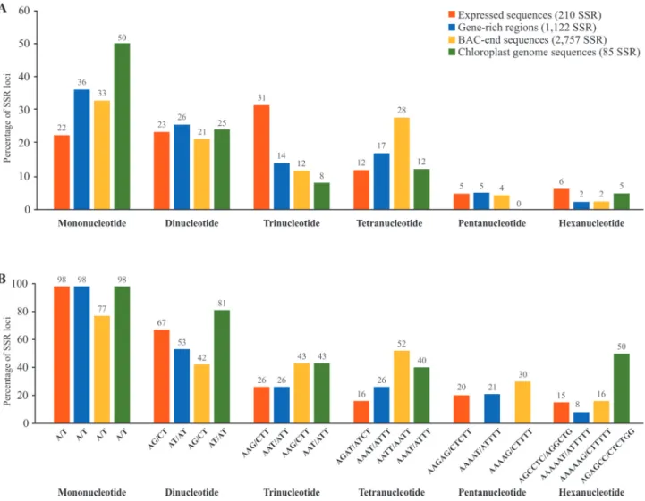
Arabidopsisgenome was found to be twice as frequent in coding regions, suggesting selection for certain stretches of amino acids (Morganteet al., 2002). Using data generated in our laboratory, we have compared the percentage of SSRs having mono-, di-, tri, tetra-, penta and hexanucleotide motifs in expressed sequences, gene-rich regions, BAC-end sequences and chloroplast genome se-quences ofPassiflora edulis, and identified the prevalent motif in each case. We also noticed the prevalence of tri-and hexanucleotide motifs in expressed sequences (Figu-re 1).
Recently, based on the genomes available in the Phytozome database, Zhaoet al.(2014) analyzed the
distri-bution of tandem repeats in 29 species of terrestrial plants and two species of algae, in which the density of repeat se-quences was higher in introns and coding sese-quences; in plants, 5’-UTR and upstream intergenic 200 nucleotide re-gions had the first and second highest densities.
In cDNA libraries constructed using plant and repro-ductive tissues ofElaeis guineensis, SSRs were observed in
both coding regions and UTRs (Tranbargeret al., 2012).
The majority were identified in open reading frames, indi-cating a possible effect on the gene product and conse-quently on gene function.On the other hand, mutations in
SSRs located in UTRs could affect transcription, transla-tion or transcript splicing (Tranbargeret al., 2012).
An important example of the functioning of SSRs in
plants was reported by Liu et al. (2014b) using a
high-throughput sequencing approach to characterize miRNAs and their targeted transcripts in different tissues of sweet orange. These miRNAs were evenly distributed across the genome in several small clusters, and 69 pre-miRNAs were co-localized with SSRs. Noticeably, the loop size of a par-ticular pre-miRNA was influenced by the repeat number of the CUU codon. Another important aspect is the instability of microsatellites. Studies conducted on transgenic plants ofA. thalianashowed that this instability increases as the
plant ages, mainly due to a drop in the efficiency of DNA repair mechanisms (Golubovet al., 2010). This peculiarity
means that SSR markers can be used to assess the impacts
of mutagenic contaminants. Mutagenesis induced inPisum
sativumby high doses of lead was detected based on the
in-stability of microsatellites at a locus involved in metabolizing glutamine (Rodriguezet al., 2013).
Microsatellite alterations associated with diseases in humans are widely known and can give the false impres-sion that the effects of these mutations are predominantly adverse. On the contrary, some examples provide evidence that SSR alleles can offer potential selective advantages (Kashi and King, 2006). It was therefore time to abandon the presumption that SSRs are junk DNA. SSRs are cur-rently qualified as relevant to population adaptation and phenotypic plasticity within and across generations and gene-associated tandem repeats act as evolutionary facilita-tors, providing abundant, robust variation and thus en-abling rapid development of new forms (Nevo, 2001; Kashi and King, 2006).
Development of SSR markers, including
de
novo nucleotide sequences for finding SSRs
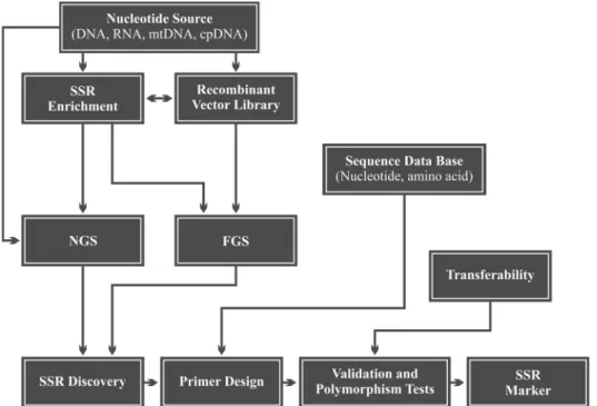
The development of SSR markers can basically be di-vided into the following stages: (i) prior knowledge of
nu-cleotide sequences in which SSRs occur; (ii) design of
oligonucleotides (or primers) complementary to the regions flanking the SSR; (iii) validation of primers by PCR and
electrophoresis of the product of the reaction, and (iv)
de-tection of polymorphisms among individuals (Mason, 2015). A schematic workflow showing how an SSR marker can be obtained is given in Figure 2. Interestingly, the effi-ciency of SSR marker development was found to be associ-ated with the microsatellite class. In rice, for instance, the rate of successful amplification varied from 31.7% (AT re-peats) up to 87% (GAA rere-peats). The following figures
were observed for other SSR classes: GA, 83.8%; CA, 71.8%; GC-rich trinucleotides, 64.45%; ATT, 78,3%; CAT
and CAA, 83,3% and tetranucleotides, 71.4% (Temnykhet
al., 2001).
Microsatellites were originally developed from both coding and non-coding regions of plant genomes, and sev-eral sources were used to search for SSRs, including a vari-ety of DNA libraries (genomic, genomic-enriched for SSR, bacterial artificial chromosome and cDNA libraries), as well as public databases, including expressed sequence tag (EST) databases (see Hanaiet al., 2007).
In prospecting for SSRs, the first step consists of con-structing enriched genomic libraries and various enrich-ment methods have been successfully developed (Billotte
et al., 1999; Maio and Castro, 2013). To construct and
se-quence genomic libraries, the DNA is fragmented, ligated to adaptors and inserted into vectors for transforming Esch-erichia coli. Most protocols involve a stage of enrichment
for repetitive sequences that can be achieved using selec-tive hybridization, PCR or both techniques (Senanet al.,
2014). In enrichment by hybridization, positive clones are detected using radioactively or chemically labeled SSR probes. Finally, these clones are selected by PCR amplifi-cation and sequencing (Semagnet al., 2006; Blair et al.,
2009). Another way of enriching a library is to use bio-tinized SSR probes that are captured by streptavidin-coated
beads (Nunomeet al., 2006). The captured DNA is eluted,
amplified, cloned and sequenced. The enriched libraries are screened to identify clones containing SSRs, producing the subsample of repetitive sequences that is intrinsic to this approach. PCR-based methods can bias the sampling of re-petitive sequences in non-enriched libraries, since fragment selection and amplification are dependent on complemen-tarity with specific primers for the SSR and cloning vector. However, non-enriched libraries and alternative methods derived from other molecular markers (e.g. RAPD and AFLP) have also been used to find SSRs (see Senanet al.,
2014).
The advances made in Next Generation Sequencing (NGS) have provided a new scenario for detecting micro-satellites. Various NGS-based projects have been devel-oped over the last few decades, generating an enormous quantity of sequences made available in public databases and widely used for prospecting for microsatellites. Auto-mation of the original sequencing method proposed by Sanger and Coulson (1975) has made it possible to
se-quence the complete genome ofA. thaliana(Arabidopsis
Genome Initiative, 2000). However, because of the high cost of the Sanger method when sequencing complete genomes, it has been replaced by NGS platforms or a com-bination of both methods (Schnableet al., 2009).
NGS has been very useful for various studies, includ-ing prospectinclud-ing for new SSR markers. Successors of the Sanger sequencing method include the 454 FLX (Roche), Solexa (Illumina), SOLiD (Applied Biosystems) and
HeliScope True Single Molecule Sequencing (Helicos) platforms. Third generation platforms are also currently available, including a platform developed by Pacific Bio-sciences (PacBio), based on a new sequencing technology, SMRT sequencing, which has the advantage of producing longer DNA reads.
Each platform has specific characteristics in terms of the number and size of reads generated, run time, as well as the accuracy and cost of each base read, with both advan-tages and disadvanadvan-tages compared to other platforms (Egan
et al., 2012). In order to advice researchers in sequencing
technology choice, Alicet al.(2016) published a review
about different high-throughput sequencing methods and 50 stand-alone softwares used to control errors. Control er-ror analysis is one of the most important steps in
sequenc-ing data analysis, mainly inde novosequencing projects,
that lack a reference genome. Furthermore, sequences that contain repetitive regions are challenges to be overcome by error correction methods, due to their vulnerability to er-rors. Initiatives for sequencing the complete genomes of various species use combinations of different platforms with the aim of incorporating the best features of each and extracting the maximum amount of information.
Currently, 454 and Illumina are the NGS platforms most widely used for developing SSR markers. However, the PacBio SMRT sequencing technology is being consid-ered an economically viable alternative for discovering microsatellites (Grohmeet al., 2013).
In-silico prospecting and transferability of SSR
markers
With the advent of NGS, it was necessary to create databases for storing the information generated. In addition to genomic sequences, a large quantity of expressed se-quence tags (EST) derived from cDNA libraries (i.e. origi-nating from mRNA) were also generated. The online database platforms for nucleotide, protein and transcript data available for the majority of plant species are relatively small when compared to model species, such asA. thaliana, Glycine max, O. sativaandZ. mays. Since the protocols for
obtaining and isolatingde novoSSR loci can be expensive
and not viable in some cases, the investigation of these ele-mentsin silico(i.e. in the actual databases) is a promising
strategy. This approach is possible only because SSR loci primers are transferable among different, phylogenetically matching species (Kuleunget al., 2004).
The possibility of interchanging this genetic informa-tion is ascribed to the synteny between matching species. Although there are some exceptions resulting from struc-tural rearrangements, synteny is an import attribute of plant genomes and is inversely proportional to the phylogenetic distance between species (Kalóet al., 2004). The
evolutionary advantages, and are therefore subject to low selection pressure (Zhu, 2005).
Microsatellites found in the chloroplast genome of higher plants (cpSSRs) consist basically of mononucleotide repeats (A and T) (Bryanet al., 1999). Contrarily, we have found 50, 25, 8, 12 and 5% of mono-, di-, tri-, tetra- and hexanucleotides respectively in the microsatellites of the
chloroplast genome ofPassiflora edulis(Figure 1A), but
we have confirmed that mononucleotide repeats consisted predominantly of A/T repeats (98%, Figure 1B). In terms of transferability, cpSSRs are particularly promising for the study of phylogenetically distant species, since the regions flanking them are strongly conserved, so that universal primers can be developed (Weising and Gardner, 1999; Ebert and Peakall, 2009).
Genotyping
After identifying the sequences containing SSRs, specific primers must be synthesized (18 and 25 bp in length), complementary to the flanking regions, followed by amplification and polymorphism testing. According to
Guichouxet al.(2011), a number of experimental problems
can arise during SSR amplification, which can compromise allele calling and binning, resulting in increased error rates or the need for extensive manual corrections. These authors itemized possible solutions for aiding researchers to solve these problems, such as stuttering or shadow bands,
non-template addition of a nucleotide by theTaq
polymer-ase, primer mispriming, etc.
Once the SSR markers have been produced, genotyp-ing can begin. It is a relatively easy and low-cost procedure. The allele variants of a given SSR locus can be identified by agarose gel electrophoresis (AGE) or polyacrylamide gel electrophoresis (PAGE), low-complexity methods used routinely in molecular genetics laboratories. PAGE geno-typing is more labor intensive but provides better resolu-tion, allowing identification of given polymorphisms for a single base pair (Penhaet al., 2013; Mason, 2015).
Alterna-tively, marked SSR primers can be synthesized with fluo-rescent markers for genotyping by capillary electrophoresis
using conventional sequencers (Araújo et al., 2007;
Csencsicset al., 2010; Agarwalet al., 2015). In this case,
each DNA sample is loaded into a capillary containing a polyacrylamide matrix in which the electrophoresis is per-formed. The fluorescence emitted by the marked primer is captured and the molecular mass of the amplified fragment is determined. The result is an electropherogram showing luminescence peaks corresponding to each amplified allele. Lastly, the genotyping stage consists of comparing the
electropherograms of different individuals (see Culley et
al., 2013; Mason, 2015), a technique that is particularly
widely used when working with complex genome species, such as sugarcane and other polyploids (Morais TBR de, 2012, Doctoral Thesis. Escola Superior de Agricultura
“Luiz de Queiroz, University of São Paulo, Piracicaba, SP, Brazil).
The most appropriate genotyping method for each project is defined according to the species under investiga-tion, the sensitivity required in determining allele varia-tions, the availability of the equipment and cost effectiveness. The amplification and genotyping stages can be perfected to multiplex different SSR loci, cutting costs and saving time, and allowing large scale analysis (Brown
et al., 1996; Guichoux et al., 2011; Lepais and Bacles,
2011). There are two ways of performing multiplexed anal-ysis of microsatellite loci. The first is by multiplexed PCR, in which different SSR primers are placed in the same reac-tion tube. The following stages are essential:i)determining
the length (in bp) of the alleles at each SSR locus;ii)
select-ing loci whose allele lengths are not superimposed;iii) in silicotesting at melting temperature (Tm) and the possible
formation of secondary structures between the primers of the SSR loci selected. The second multiplexed SSR loci analysis method entails multiplexed genotyping. In this case, amplifications are performed separately, but the am-plified products of a biological sample are mixed and loaded into the same electrophoresis gel channel or se-quencing capillary.
Guichouxet al.(2011) have published an outstanding
analysis of current trends in microsatellite genotyping. Sev-eral aspects are reviewed, including the ovSev-erall cost of SSR genotyping as a function of the degree of multiplexing and the number of genotyped samples. For instance, the most widely cited commercial kit has a cost per sample of 1.88. The authors then suggest solutions to cut the final cost per sample. According to these authors, most of the work done to develop and optimize SSR multiplexing actually consists of phases common to all SSR development projects.
In the past, alternative methods have been developed to facilitate genotyped PCR multiplexing by capillary elec-trophoresis, such as the M13 tailed primer method (Oetting
et al., 1995). In this method, the sequencing reaction is per-formed as a multiplexed PCR using the M13 (reverse) primer, conjugated with a fluorescent colorant and various modified SSR (forward) primers. The SSR primers are modified by a 19-bp extension at the 5’ end, identical to the M13 nucleotide sequence. In the first PCR cycle, amplifi-cation is based on the SSR primers, forming an M13 an-nealing site at the 3’ end, used in the second amplification cycle. A variant of this technique (Multiplex-Ready PCR) was subsequently published with the aim of cutting the cost of primer marking, which is usually 5 to 10 times that of conventional primer synthesis (Haydenet al., 2008).
Current overview
Liet al.(2002); Buschiazzo and Gemmell (2006); Oliveira et al.(2006); Sun et al. (2009); Guichoux et al.(2011);
Gemayelet al.(2012); Senanet al.(2014); Mason (2015).
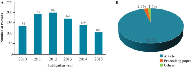
With the aim of investigating the use of microsatellite markers over the period from 2010 to 2015 in the genetic analysis of cultivated plants, we conducted a search in the
main database of Web of Science (Web of ScienceTMCore
Collection). We entered “microsatellite” or “SSR marker” in the title field and “crop*” in the topic field. To avoid se-lecting records related to plant pathogens and insect pests, the following terms were excluded from the topic field: bacteria (bacter*), fungi (fung*), insect (insect*) and pathogen (pathogen*). Finally, the search was refined by selecting the field of Plant Science, and all resulting hits were manually checked. We found 933 unique records (Figure 3, Supplementary Material Table S1) showing that microsatellites continue to be used as high-relevance mo-lecular markers in the genetic analysis of cultivated plants. The number of publications rose steadily until 2012, and then fell back, possibly due to the ease with which genetic studies could be carried out using SNPs.
Recent studies have shown that the easiest way of identifying SSR loci is by using NGS to sequence the
ge-nome or transcriptome. Zalapaet al.(2012) reviewed
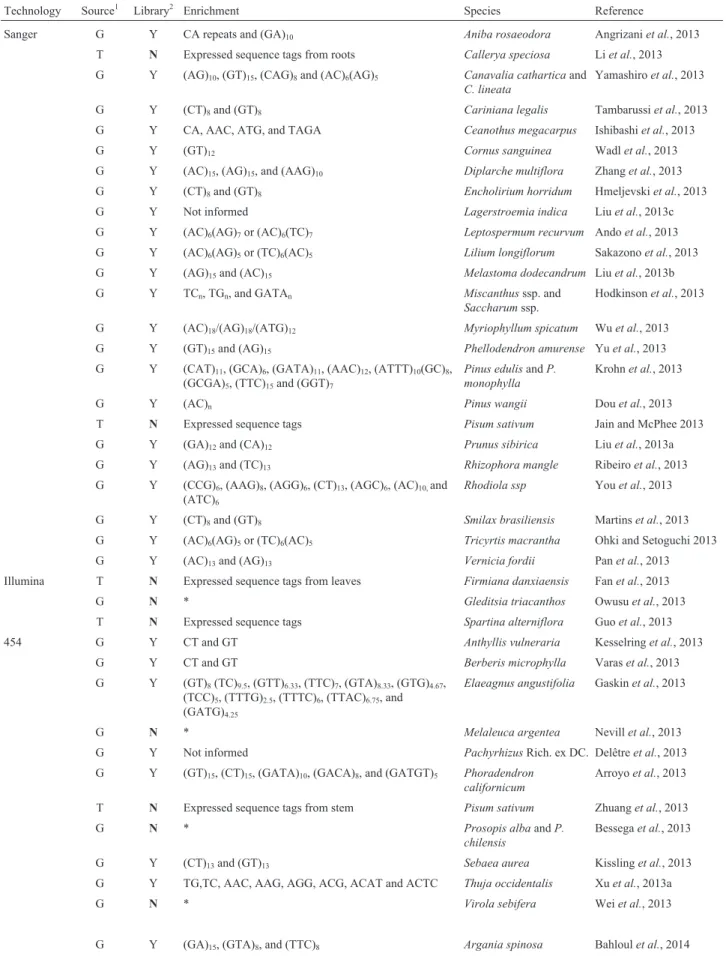
pa-pers published in the American Journal of Botany’s Primer Notes and Protocols in Plant Sciences, an important monthly journal that centralizes a significant number of publications related to the discovery and use of SSRs in plants. Note that the use of the Sanger method was predom-inant up to this time, as well as the use of genomic libraries enriched with sequences containing microsatellites. Since then, there is a tendency to replace this method by NGS ge-nome or transcriptome sequencing.
With the aim of comparing this scenario with the cur-rent situation, we conducted a similar survey based on pa-pers published in the AJB from January 2013 to December 2015, selecting only those papers in which sequences were
generated by developing SSR markers. A total of 87 papers were published during this period, the majority of which in-volved using the Sanger method to sequence genomic li-braries enriched with SSRs. It is worth noting that the use of NGS for prospecting for and generating SSR markers has been on the increase, surpassing the Sanger method in 2015 (Table 1). We also realized that the enrichment stage might no longer be advantageous, due to the number of sequences generated by NGS. On the contrary, since the composition of the nucleotide base and the frequency of SSR motifs can actually vary among plant genomes (Liet al., 2002), the
en-richment stage with a small number of motifs should allow curtailment or subsampling of the population of SSRs to be discovered.
Another interesting trend is that the Illumina platform is being routinely used for transcriptome sequencing. The advantage of developing SSR markers from transcribed se-quences includes the possibility of finding associations with genes and phenotypes (Liet al., 2002). As observed by
Zalapaet al.(2012), a common factor of all the papers,
irre-spective of the sequencing method, is that only a small frac-tion of the SSR loci discovered have been assessed. As mentioned earlier, obtaining a sequence is only the first stage in the marker development process. Primer design and PCR optimization still represent a bottleneck. Further-more, there is always the possibility that the locus is mono-morphic, i.e. non-informative.
One strategy for working around this limitation is to
track loci polymorphisms in silico, during the stage at
which regions that contain SSRs are identified. This can be done using two or more genetically contrasting individuals or their progeny (F1) for performing NGS, increasing the
possibility of sampling alleles based on the alignment of the sequences obtained, and thereby avoiding the synthesis and
testing of primers for monomorphic loci (Iorizzo et al.,
2011).
Table 1- Recent studies involved in the detection and development of SSR markers in plants, using different sequencing technologies.
Technology Source1 Library2 Enrichment Species Reference
Sanger G Y CA repeats and (GA)10 Aniba rosaeodora Angrizaniet al., 2013
T N Expressed sequence tags from roots Callerya speciosa Liet al., 2013 G Y (AG)10, (GT)15, (CAG)8and (AC)6(AG)5 Canavalia catharticaand
C. lineata
Yamashiroet al., 2013
G Y (CT)8and (GT)8 Cariniana legalis Tambarussiet al., 2013
G Y CA, AAC, ATG, and TAGA Ceanothus megacarpus Ishibashiet al., 2013
G Y (GT)12 Cornus sanguinea Wadlet al., 2013
G Y (AC)15, (AG)15, and (AAG)10 Diplarche multiflora Zhanget al., 2013
G Y (CT)8and (GT)8 Encholirium horridum Hmeljevskiet al., 2013
G Y Not informed Lagerstroemia indica Liuet al., 2013c G Y (AC)6(AG)7or (AC)6(TC)7 Leptospermum recurvum Andoet al., 2013
G Y (AC)6(AG)5or (TC)6(AC)5 Lilium longiflorum Sakazonoet al., 2013
G Y (AG)15and (AC)15 Melastoma dodecandrum Liuet al., 2013b
G Y TCn, TGn, and GATAn Miscanthusssp. and Saccharumssp.
Hodkinsonet al., 2013
G Y (AC)18/(AG)18/(ATG)12 Myriophyllum spicatum Wuet al., 2013
G Y (GT)15and (AG)15 Phellodendron amurense Yuet al., 2013
G Y (CAT)11, (GCA)6, (GATA)11, (AAC)12, (ATTT)10(GC)8,
(GCGA)5, (TTC)15and (GGT)7
Pinus edulisandP. monophylla
Krohnet al., 2013
G Y (AC)n Pinus wangii Douet al., 2013
T N Expressed sequence tags Pisum sativum Jain and McPhee 2013 G Y (GA)12and (CA)12 Prunus sibirica Liuet al., 2013a
G Y (AG)13and (TC)13 Rhizophora mangle Ribeiroet al., 2013
G Y (CCG)6, (AAG)8, (AGG)6, (CT)13, (AGC)6, (AC)10,and
(ATC)6
Rhodiola ssp Youet al., 2013
G Y (CT)8and (GT)8 Smilax brasiliensis Martinset al., 2013
G Y (AC)6(AG)5or (TC)6(AC)5 Tricyrtis macrantha Ohki and Setoguchi 2013
G Y (AC)13and (AG)13 Vernicia fordii Panet al., 2013
Illumina T N Expressed sequence tags from leaves Firmiana danxiaensis Fanet al., 2013
G N * Gleditsia triacanthos Owusuet al., 2013
T N Expressed sequence tags Spartina alterniflora Guoet al., 2013 454 G Y CT and GT Anthyllis vulneraria Kesselringet al., 2013
G Y CT and GT Berberis microphylla Varaset al., 2013 G Y (GT)8(TC)9.5, (GTT)6.33, (TTC)7, (GTA)8.33, (GTG)4.67,
(TCC)5, (TTTG)2.5, (TTTC)6, (TTAC)6.75, and
(GATG)4.25
Elaeagnus angustifolia Gaskinet al., 2013
G N * Melaleuca argentea Nevillet al., 2013
G Y Not informed PachyrhizusRich. ex DC. Delêtreet al., 2013 G Y (GT)15, (CT)15, (GATA)10, (GACA)8, and (GATGT)5 Phoradendron
californicum
Arroyoet al., 2013
T N Expressed sequence tags from stem Pisum sativum Zhuanget al., 2013
G N * Prosopis albaandP.
chilensis
Bessegaet al., 2013
G Y (CT)13and (GT)13 Sebaea aurea Kisslinget al., 2013
G Y TG,TC, AAC, AAG, AGG, ACG, ACAT and ACTC Thuja occidentalis Xuet al., 2013a
G N * Virola sebifera Weiet al., 2013
Technology Source1 Library2 Enrichment Species Reference
Sanger G Y (CT)8, (GT)8 Byrsonima cydoniifolia Bernardeset al., 2014
Cp N * Lemna minor Waniet al., 2014
G Y CT Lobelia inflata Hugheset al., 2014
G Y (GT)8and (CT)8 Passiflorassp. Cerqueira-Silvaet al., 2014
G Y (CT)8and (GT)8 Piper solmsianum Yoshidaet al., 2014
G Y (AC)6(AG)5or (TC)6(AC)5 Scrophularia incisa Wanget al., 2014
G Y (AC)15and (AG)15 Spiraeassp Khanet al., 2014
G Y (AC)6(AG)5, (TC)6(AC)5 Vitex rotundifolia Ohtsukiet al., 2014
G Y (GA)nand (GT)n Xanthosoma sagittifolium Cathebraset al., 2014
Illumina T N Expressed sequence tags from roots Buxusspp. Thamminaet al., 2014
G N * Macadamiassp. Nocket al., 2014
T N Expressed sequence tags from leaves Ostryopsisssp. Liuet al., 2014a
G N In silicomining Phoenix dactylifera Aberlenc-Bertossiet
al., 2014
G N * SolidagoL. Becket al., 2014
G N * Saxifraga granulata Meeret al., 2014
454 G Y (GA)15, (GTA)8, and (TTC)8 Argania spinosa Bahloulet al., 2014
G N * Agave utahensis Byerset al., 2014
G N * Bidens alba Luet al., 2014
G Y CT and GT Nephromassp. Belinchónet al., 2014 G Y TG, TC, AAC, AAG, AGG, ACG, ACAT, and ACTC Parietaria judaica Bossuet al., 2014
Sanger G Y (CT)8and (GT)8 Cabomba aquatica Barbosaet al., 2015
G Y (AT)8, (GA)8, and (GAA)8 Calibrachoa heterophylla Silva-Ariaset al., 2015
G Y GA, GT, AGA, ACT, and ATC Campanula pyramidalis Radosavljeviæet al., 2015 G Y (AC)15and (AG)15 Commelina communis Liet al., 2015
G Y (AG)10 Ilex chinensis Chenet al., 2015
G Y Not informed Fothergilla intermedia Hatmakeret al., 2015 G Y (AC)6(AG)5or (GA)5(CA)5 Hepatica nobilis var.
ja-ponica
Kameokaet al., 2015
G Y (CT)8and (GT)8 Philcoxia minensis Scatignaet al., 2015
G Y (AG)17, (AC)17, (AAC)10, (CCG)10, (CTG)10, and
(AAT)10
Psittacanthus schiedeanus
Gonzálezet al., 2015
G Y (AG)17, (AC)17, (AAC)10, (CCG)10, (CTG)10, and
(AAT)10
Quillaja saponaria Letelieret al., 2015
G Y (AC)15and (AG)15 Saxifraga egregia Zhanget al., 2015
G Y (TTC)10, (CG)10, and (GT)10 Vellozia squamata Duarte-Barbosaet al., 2015
Illumina T/Cp N Expressed sequence tags from leaves Artocarpus moraceae Gardneret al., 2015 T N Expressed sequence tags from leaves Bombax ceiba Juet al., 2015 T N Expressed sequence tags from leaves Carallia brachiata Qianget al., 2015
G N * Dendrobium calamiforme Trapnellet al., 2015
T N Expressed sequence tags from leaves Lablab purpureus, Lathyrus sativus
Chapman 2015
Psophocarpus
tetragonolobusandVigna subterranea
Finally, visiting the website of the 24thedition of the
Plant and Animal Genome (PAG) Conference (San Diego, CA) held in January 2016, we were able to find 63 work-shops, abstracts and posters in which the term SSR was em-ployed. We categorized these studies according to the groups of species analyzed and found the great majority of them (~90%) related to plants. We also checked references to SNPs and found about 150 studies, two-thirds related to plants and a third to domesticated animals (cattle, chicken, horse, pig, sheep and fish). A few (1.5% for SSRs and 5% for SNPs) proposed advances in experimental approaches
or novel bioinformatics tools
(https://pag.confex.com/pag/xxiv/meetingapp.cgi). Are
SNPs destined to replace SSRs as the preferred marker? It seems clear that this will occur, but we do believe that SSRs will still be applicable in future plant genetic and genomic studies.
References
Aberlenc-Bertossi F, Castillo K, Tranchant-Dubreuil C, Chérif E, Ballardini M, Abdoulkader S, Gros-Balthazard M, Chabril-lange N, Santoni S, Mercuri Aet al.(2014)In silicomining
of microsatellites in coding sequences of the date palm (Arecaceae) genome, characterization, and transferability. Appl Plant Sci 2:1300058.
Agarwal G, Sabbavarapu MM, Singh VK, Thudi M, Sheelamary S, Gaur PM and Varshney RK (2015) Identification of a non-redundant set of 202in silicoSSR markers and applica-bility of a select set in chickpea (Cicer arietinum L.). Euphytica 205:381-394.
Alic AS, Ruzafa D, Dopazo J and Blanquer I (2016) Objective re-view of de novostand-alone error correction methods for NGS data: Objective review of de novo stand-alone NGS
correctors. Wiley Interdiscip Rev Comput Mol Sci 6:111-1146.
Ando S, Kaneko S, Isagi Y, Repin R and Kitayama K (2013) De-velopment of SSR markers for the tropical alpine tree spe-ciesLeptospermum recurvum(Myrtaceae) on Mount
Kina-balu in Borneo. Appl Plant Sci 1:1200010.
Angrizani RC, Contim LAS and Lemes MR (2013) Development and characterization of microsatellite markers for the endan-gered Amazonian treeAniba rosaeodora(Lauraceae). Appl Plant Sci 1:1200516.
Arabidopsis Genome Initiative (2000) Analysis of the genome se-quence of the flowering plantArabidopsis thaliana. Nature 408:796-815.
Araújo IS, Intorne AC, Pereira MG, Lopes UV and Filho GA de S (2007) Development and characterization of novel tetra-, tri-and di-nucleotide microsatellite markers in cacao (Theobroma cacaoL.). Mol Breed 20:73-81.
Arroyo JM, Munguia-Vega A, Rodríguez-Estrella R and Bas-compte J (2013) Isolation of 18 microsatellite loci in the desert mistletoe Phoradendron californicum(Santalaceae) via 454 pyrosequencing. Appl Plant Sci 1:1300048. Bahloul YE, Dauchot N, Machtoun I, Gaboun F and Cutsem PV
(2014) Development and characterization of microsatellite loci for the Moroccan endemic endangered speciesArgania spinosa(Sapotaceae). Appl Plant Sci 2:1300071.
Barbosa TDM, Trad RJ, Bajay MM and Amaral MCE (2015) Microsatellite markers isolated fromCabomba aquaticas.l. (Cabombaceae) from an enriched genomic library. Appl Plant Sci 3:1500076.
Beck JB, Semple JC, Brull JM, Lance SL, Phillips MM, Hoot SB and Meyer GA (2014) Genus-wide microsatellite primers for the goldenrods (Solidago; Asteraceae). Appl Plant Sci 2:1300093.
Belinchón R, Ellis CJ and Yahr R (2014) Microsatellite loci in two epiphytic lichens with contrasting dispersal modes:
Technology Source1 Library2 Enrichment Species Reference
T N Expressed sequence tags from leaves and cambium Thujopsis dolabrata var. hondae
Satoet al., 2015
454 G N * Cyperus fuscus Böckelmannet al., 2015
G N In silicomining Metasequoia
glyptostroboides
Jinet al., 2015
G N * Pilosella alpicola Vítet al., 2015
G N * Pulsatilla vulgaris DiLeoet al., 2015
G Y (AG)10, (AC)10, (AAC)8, (ACG)8, (AAG)8, (AGG)8,
(ACAT)6, and (ATCT)6
Quercus variabilis Wanget al., 2015
G Y CT and GT Salix humboldtiana Bozziet al., 2015
G N * Silene acaulis Mülleret al., 2015
G Y TG, TC, AAC, AAG, AGG, ACG, ACAT, and ACTC Veronicasubsect. Pentasepalae
López-Gonzálezet al., 2015
G N * Vinca minor Moelleret al., 2015
1G = genome, T = transcriptome, Cp = chloroplast DNA 2SSR enrichment library: Y, yes;
N, no
* Total genomic DNA sequencing
Nephroma laevigatum and N. parile (Nephromataceae). Appl Plant Sci 2:1400080.
Benson G (1999) Tandem repeats finder: a program to analyze DNA sequences. Nucleic Acids Res 27:573-580.
Bernardes V, Anjos DE dos, Gondim SG de CA, Murakami DM, Bizão N and Telles MP de C (2014) Isolation and character-ization of microsatellite loci in Byrsonima cydoniifolia
(Malpighiaceae) and cross-amplification in B. crassifolia. Appl Plant Sci 2:1400016. d
Bessega CF, Pometti CL, Miller JT, Watts R, Saidman BO and Vilardi JC (2013) New microsatellite loci forProsopis alba
andP. chilensis(Fabaceae). Appl Plant Sci 1:1200324. Billotte N, Lagoda PJL, Risterucci A-M and Baurens F-C (1999)
Microsatellite-enriched libraries: applied methodology for the development of SSR markers in tropical crops. Fruits 54:277-288.
Bird TD (2015) Myotonic dystrophy type 1. 1999 Sep 17 [Up-dated 2015 Oct 22]. In: Pagon RA, Adam MP, Ardinger HH, Wallace SE, Amemiya A, Bean LJ, Bird TD, Fong C-T, Mefford HC, Smith RJ,et al.(eds) GeneReviews,
Univer-sity of Washington, Seattle (WA),
http://www.ncbi.nlm.nih.gov/books/NBK1165/.
Blair MW, Torres MM, Pedraza F, Giraldo MC, Buendía HF and Hurtado N (2009) Development of microsatellite markers for common bean (Phaseolus vulgarisL.) based on screen-ing of non-enriched, small-insert genomic libraries. Genome 52:772-782.
Böckelmann J, Wieser D, Tremetsberger K, Šumberová K and Bernhardt K-G (2015) Isolation of nuclear microsatellite markers for Cyperus fuscus(Cyperaceae). Appl Plant Sci
3:1500071.
Bossu A, Bertaudière-Montès V, Dubut V and Manel S (2014) Microsatellite primers inParietaria judaica(Urticaceae) to
assess genetic diversity and structure in urban landscapes. Appl Plant Sci 2:1400036.
Bozzi JA, Liepelt S, Ohneiser S, Gallo LA, Marchelli P, Leyer I, Ziegenhagen B and Mengel C (2015) Characterization of 23
polymorphic SSR markers in Salix humboldtiana
(Salicaceae) using next-generation sequencing and cross-amplification from related species. Appl Plant Sci 3:1400120.
Brown SM, Hopkins MS, Mitchell SE, Senior ML, Wang TY, Duncan RR, Gonzalez-Candelas F and Kresovich S (1996) Multiple methods for the identification of polymorphic sim-ple sequence repeats (SSRs) in sorghum [Sorghum bicolor
(L.) Moench]. Theor Appl Genet 93:190-198.
Bryan GJ, McNicoll J, Ramsay G, Meyer RC and De Jong WS (1999) Polymorphic simple sequence repeat markers in chloroplast genomes of Solanaceous plants. Theor Appl Genet 99:859-867.
Buschiazzo E and Gemmell NJ (2006) The rise, fall and renais-sance of microsatellites in eukaryotic genomes. Bioessays 28:1040-1050.
Byers C, Maughan PJ, Clouse J and Stewart JR (2014) Micro-satellite primers inAgave utahensis(Asparagaceae), a key-stone species in the Mojave Desert and Colorado Plateau. Appl Plant Sci 2:1400047.
Castelo AT, Martins W and Gao GR (2002) TROLL—Tandem Repeat Occurrence Locator. Bioinformatics 18:634-636.
Cathebras C, Traore R, Malapa R, Risterucci A-M and Chaïr H (2014) Characterization of microsatellites in Xanthosoma sagittifolium (Araceae) and cross-amplification in related species. Appl Plant Sci 2:1400027.
Cerqueira-Silva CBM, Santos ESL, Vieira JGP, Mori GM, Jesus ON, Corrêa RX and Souza AP (2014) New microsatellite markers for wild and commercial species of Passiflora
(Passifloraceae) and cross-amplification. Appl Plant Sci 2:1300061.
Chapman MA (2015) Transcriptome sequencing and marker de-velopment for four underutilized legumes. Appl Plant Sci 3:1400111.
Chen W-W, Xiao Z-Z, Tong X, Liu Y-P and Li Y-Y (2015) De-velopment and characterization of 25 microsatellite primers for Ilex chinensis (Aquifoliaceae). Appl Plant Sci 3:1500057.
Csencsics D, Brodbeck S and Holderegger R (2010) Cost-effec-tive, species-specific microsatellite development for the
en-dangered dwarf bulrush (Typha minima) using
next-generation sequencing technology. J Hered 101:789-793. Culley TM, Stamper TI, Stokes RL, Brzyski JR, Hardiman NA,
Klooster MR and Merritt BJ (2013) An efficient technique for primer development and application that integrates fluo-rescent labeling and multiplex PCR. Appl. Plant Sci. 1:1300027.
Delêtre M, Soengas B, Utge J, Lambourdière J and Sørensen M (2013) Microsatellite markers for the yam beanPachyrhizus
(Fabaceae). Appl Plant Sci 1:1200551.
DiLeo MF, Graf R, Holderegger R, Rico Y and Wagner HH (2015) Highly polymorphic microsatellite markers in
Pulsatilla vulgaris(Ranunculaceae) using next-generation sequencing. Appl Plant Sci 3:1500031.
Dou J-J, Zhou R-C, Tang A-J, Ge X-J and Wu W (2013) Develop-ment and characterization of nine microsatellites for an en-dangered tree, Pinus wangii (Pinaceae). Appl Plant Sci 1:1200134.
Duarte-Barbosa M, Bajay MM, Zucchi MI and Pivello VR (2015) Development and characterization of 47 novel microsatellite markers forVellozia squamata(Velloziaceae). Appl Plant Sci 3:1400087.
Ebert D and Peakall R (2009) A new set of universal de novo se-quencing primers for extensive coverage of noncoding chloroplast DNA: new opportunities for phylogenetic stud-ies and cpSSR discovery. Mol Ecol Resour 9:777-783. Egan AN, Schlueter J and Spooner DM (2012) Applications of
next-generation sequencing in plant biology. Am J Bot 99:175-185.
Fan Q, Chen S, Li M, He S, Zhou R and Liao W (2013) Develop-ment and characterization of microsatellite markers from the transcriptome of Firmiana danxiaensis (Malvaceae s.l.).
Appl Plant Sci 1:1300047.
Fujimori S, Washio T, Higo K, Ohtomo Y, Murakami K, Matsubara K, Kawai J, Carninci P, Hayashizaki Y, Kikuchi Set al.(2003) A novel feature of microsatellites in plants: a distribution gradient along the direction of transcription. FEBS Lett 554:17-22.
Gao L, Tang J, Li H and Jia J (2003) Analysis of microsatellites in major crops assessed by computational and experimental ap-proaches. Mol Breed 12:245-261.
Garcia FAA, Kido AE, Meza NA, Souza BHM, Pinto RL, Pastina MM, Leite SC, Silva GJA da, Ulian CE, Figueira Aet al.
(2006) Development of an integrated genetic map of a sug-arcane (Saccharumspp.) commercial cross, based on a max-imum-likelihood approach for estimation of linkage and linkage phases. Theor Appl Genet 112:298-314.
Gardner EM, Laricchia KM, Murphy M, Ragone D, Scheffler BE, Simpson S, Williams EW and Zerega NJC (2015) Chloro-plast microsatellite markers forArtocarpus(Moraceae) de-veloped from transcriptome sequences. Appl Plant Sci 3:1500049.
Gaskin JF, Hufbauer RA and Bogdanowicz SM (2013) Micro-satellite markers for Russian olive (Elaeagnus angustifolia; Elaeagnaceae). Appl Plant Sci 1:1300013.
Gemayel R, Cho J, Boeynaems S and Verstrepen KJ (2012) Be-yond Junk-Variable Tandem repeats as facilitators of rapid evolution of regulatory and coding sequences. Genes 3:461-480.
Golubov A, Yao Y, Maheshwari P, Bilichak A, Boyko A, Belzile F and Kovalchuk I (2010) Microsatellite instability in
Arabidopsis increases with plant Development. Plant Physiol 154:1415-1427.
González C, Harvey N and Ornelas JF (2015) Development and characterization of microsatellite loci in the mistletoe
Psittacanthus schiedeanus (Loranthaceae). Appl Plant Sci
3:1400099.
Grohme MA, Soler RF, Wink M and Frohme M (2013) Micro-satellite marker discovery using single molecule real-time circular consensus sequencing on the Pacific Biosciences RS. BioTechniques 55:253-256.
Guichoux E, Lagache L, Wagner S, Chaumeil P, LéGer P, Lepais O, Lepoittevin C, Malausa T, Revardel E, Salin F et al.
(2011) Current trends in microsatellite genotyping. Mol Ecol Resour 11:591-611.
Guo W, Huang Y, He Z, Yan Y, Zhou R and Shi S (2013) Devel-opment and characterization of microsatellite loci for smooth cordgrass, Spartina alterniflora (Poaceae). Appl
Plant Sci 1:1200211.
Hanai LR, de Campos T, Camargo LEA, Benchimol LL, de Souza AP, Melotto M, Carbonell SAM, Chioratto AF, Consoli L, Formighieri EFet al.(2007) Development, characterization,
and comparative analysis of polymorphism at common bean SSR loci isolated from genic and genomic sources. Genome 50:266-277.
Hatmaker EA, Wadl PA, Mantooth K, Scheffler BE, Ownley BH and Trigiano RN (2015) Development of microsatellites from Fothergilla intermedia(Hamamelidaceae) and cross
transfer to four other genera within Hamamelidaceae. Appl Plant Sci 3:1400123.
Hayden MJ, Nguyen TM, Waterman A and Chalmers KJ (2008) Multiplex-ready PCR: A new method for multiplexed SSR and SNP genotyping. BMC Genomics 9:80.
Hayward AC, Tollenaere R, Dalton-Morgan J and Batley J (2015) Molecular marker applications in plants. In: Batley J (ed) Plant Genotyping. Springer, New York, NY, pp 13-27
Hmeljevski KV, Ciampi MB, Baldauf C, Reis MSD and Forzza RC (2013) Development of SSR markers forEncholirium horridum (Bromeliaceae) and transferability to other Pitcairnioideae. Appl Plant Sci 1:1200445.
Hodkinson TR, Cesare M de and Barth S (2013) Nuclear SSR markers for Miscanthus, Saccharum, and related grasses (Saccharinae, Poaceae). Appl Plant Sci 1:1300042. Horie N, Aiba H, Oguro K, Hojo H and Takeishi K (1995)
Func-tional analysis and DNA polymorphism of the tandemly re-peated sequences in the 5’-terminal regulatory region of the human gene for thymidylate synthase. Cell Struct Funct 20:191-197.
Hughes PW, Jaworski AF, Davis CS, Aitken SM and Simons AM (2014) Development of polymorphic microsatellite markers for Indian tobacco,Lobelia inflata(Campanulaceae). Appl Plant Sci 2:1300096.
Huntley M and Golding GB (2000) Evolution of simple sequence in proteins. J Mol Evol 51:131-140.
Iorizzo M, Senalik DA, Grzebelus D, Bowman M, Cavagnaro PF, Matvienko M, Ashrafi H, Van Deynze A and Simon PW (2011) De novo assembly and characterization of the carrot transcriptome reveals novel genes, new markers, and genetic diversity. BMC Genomics 12:389.
Ishibashi CDA, Shaver AR, Perrault DP, Davis SD and Honeycutt RL (2013) Isolation of microsatellite markers in a chaparral
species endemic to Southern California, Ceanothus
megacarpus(Rhamnaceae). Appl Plant Sci 1:1200393. Jain S and McPhee KE (2013) Isolation and characterization of
novel EST-derived genic markers in Pisum sativum
(Fabaceae). Appl Plant Sci 1:1300026.
Jarne P and Lagoda PJ (1996) Microsatellites, from molecules to populations and back. Trends Ecol Evol 11:424-429. Jin Y, Bi Q, Guan W and Mao J-F (2015) Development of 23
novel polymorphic EST-SSR markers for the endangered relict coniferMetasequoia glyptostroboides. Appl Plant Sci 3:1500038.
Jonah P, Bello L, Lucky O, Midau A and Moruppa S (2011) Re-view: the importance of molecular markers in plant breeding programmes. Glob J Sci Front Res 11:eV-vers1.
Ju M-M, Ma H-C, Xin P-Y, Zhou Z-L and Tian B (2015) Devel-opment and characterization of EST-SSR markers in
Bombax ceiba(Malvaceae). Appl Plant Sci 3:1500001.
Kalia RK, Rai MK, Kalia S, Singh R and Dhawan AK (2011) Microsatellite markers: an overview of the recent progress in plants. Euphytica 177:309-334.
Kaló P, Seres A, Taylor SA, Jakab J, Kevei Z, Kereszt A, Endre G, Ellis THN and Kiss GB (2004) Comparative mapping
be-tween Medicago sativa and Pisum sativum. Mol Genet
Genomics 272:235-246. doi: 10.1007/s00438-004-1055-z Kameoka S, Higashi H and Setoguchi H (2015) Development of
polymorphic microsatellite loci in the perennial herb Hepat-ica nobilisvar.japonica(Ranunculaceae). Appl Plant Sci 3:1400114.
Kashi Y and King D (2006) Simple sequence repeats as advanta-geous mutators in evolution. Trends Genet 22:253-259. Katti MV, Sami-Subbu R, Ranjekar PK and Gupta VS (2000)
Kejnovsky E, Hobza R, Cermak T, Kubat Z and Vyskot B (2009) The role of repetitive DNA in structure and evolution of sex chromosomes in plants. Heredity 102:533-541.
Kesselring H, Hamann E, Stöcklin J and Armbruster GFJ (2013)
New microsatellite markers for Anthyllis vulneraria
(Fabaceae), analyzed with spreadex gel electrophoresis. Appl Plant Sci 1:1300054.
Khan G, Zhang F, Gao Q, Jiao X, Fu P, Xing R, Zhang J and Chen S (2014) Isolation of 16 microsatellite markers forSpiraea alpinaand S. mongolica(Rosaceae) of the Qinghai-Tibet
Plateau. Appl Plant Sci 2:1300059.
Kissling J, Bachmann O, Thali MR and Segarra-Moragues JG
(2013) Novel microsatellite loci for Sebaea aurea
(Gentianaceae) and cross-amplification in related species. Appl Plant Sci 1:1300056.
Kofler R, Schlotterer C and Lelley T (2007) SciRoKo: a new tool for whole genome microsatellite search and investigation. Bioinformatics 23:1683-1685.
Krohn AL, Flores-Rentería L and Gehring CA (2013) Micro-satellite primers in the foundation tree speciesPinus edulis
and P. monophylla(Pinaceae). Appl Plant Sci 1:1200552. doi: 10.3732/apps.1200552
Kuleung C, Baenziger PS and Dweikat I (2004) Transferability of SSR markers among wheat, rye, and triticale. Theor Appl Genet 108:1147-1150.
Lawson MJ and Zhang L (2006) Distinct patterns of SSR distribu-tion in theArabidopsis thalianaand rice genomes. Genome
Biol 7:R14.
Lawson MJ and Zhang L (2008) Housekeeping and tissue-spe-cific genes differ in simple sequence repeats in the 5’-UTR region. Gene 407:54-62.
Lepais O and Bacles CFE (2011)De novodiscovery and
multi-plexed amplification of microsatellite markers for black al-der (Alnus glutinosa) and related species using SSR-enriched shotgun pyrosequencing. J Hered 102:627-632. Letelier L, Harvey N, Valderrama A, Stoll A and
González-Rodríguez A (2015) Isolation and characterization of 12
microsatellite loci in soapbark, Quillaja saponaria
(Quillajaceae). Appl Plant Sci 3:1500024.
Li CD, Rossnagel BG and Scoles GJ (2000) The development of oat microsatellite markers and their use in identifying rela-tionships among Avena species and oat cultivars. Theor
Appl Genet 101:1259-1268.
Li J-K, Song Y-P, Xu H, Zhu J-Y and Tang L-L (2015) Develop-ment and characterization of microsatellite loci for the
Pseudometallophyte Commelina communis
(Commelinaceae). Appl Plant Sci 3:1400098.
Li L, Li Z, Li K, Huang B and Xu L (2013) Development and char-acterization of EST-SSR markers in the Chinese medicinal
plant Callerya speciosa (Fabaceae). Appl Plant Sci
1:1200345.
Li Y-C, Korol AB, Fahima T, Beiles A and Nevo E (2002) Micro-satellites: genomic distribution, putative functions and mutational mechanisms: a review. Mol Ecol 11:2453-2465. Li Y-C, Korol AB, Fahima T and Nevo E (2004) Microsatellites within genes: structure, function, and evolution. Mol Biol Evol 21:991-1007.
Liu B-B, Tian B, Ma H, Lu Z-Q, Qiu Q, Mao K-S and Liu J-Q (2014a) Development and characterization of EST-SSR
markers in Ostryopsis (Betulaceae). Appl Plant Sci
2:1300062.
Liu F, Hu Z, Liu W, Li J, Wang W, Liang Z, Wang F and Sun X (2016) Distribution, function and evolution characterization
of microsatellite in Sargassum thunbergii (Fucales,
Phaeophyta) transcriptome and their application in marker development. Sci Rep 6:18947.
Liu H-B, Liu J, Wang Z, Ma L-Y, Wang S-Q, Lin X-G, Wu R-L and Pang X-M (2013a) Development and characterization of microsatellite markers inPrunus sibirica(Rosaceae). Appl Plant Sci 1:1200074.
Liu T, Dai S, Wu W, Zhang R, Fan Q, Shi S and Zhou R (2013b) Development and characterization of microsatellite markers for Melastoma dodecandrum (Melastomataceae). Appl Plant Sci 1:1200294.
Liu Y, He D, Cai M, Tang W, Li X-Y, Pan H-T and Zhang Q-X (2013c) Development of microsatellite markers for Lager-stroemia indica(Lythraceae) and related species. Appl Plant Sci 1:1200203.
Liu Y, Wang L, Chen D, Wu X, Huang D, Chen L, Li L, Deng X and Xu Q (2014b) Genome-wide comparison of microRNAs and their targeted transcripts among leaf, flower and fruit of
sweet orange. BMC Genomics 15:695. doi:
10.1186/1471-2164-15-695.
López-González N, Mayland-Quellhorst E, Pinto-Carrasco D and Martínez-Ortega MM (2015) Characterization of 12 poly-morphic SSR markers in VeronicaSubsect. Pentasepalae
(Plantaginaceae) and cross-amplification in 10 other sub-genera. Appl Plant Sci 3:1500059.
Lu Y-B, Huang D-L, Wang X, Wu Z-J and Tang S-Q (2014) Microsatellite markers for the invasive speciesBidens alba
(Asteraceae). Appl Plant Sci 2:1400008.
Maio AD and Castro OD (2013) SSR-Patchwork: An optimized protocol to obtain a rapid and inexpensive SSR library using first-generation sequencing technology. Appl Plant Sci 1:1200158.
Mammadov J, Aggarwal R, Buyyarapu R and Kumpatla S (2012) SNP markers and their impact on plant breeding. Int J Plant Genomics 2012:1-11.
Martins AR, Abreu AG, Bajay MM, Villela PMS, Batista CEA, Monteiro M, Alves-Pereira A, Figueira GM, Pinheiro JB,
Appezzato-Da-Glória B et al. (2013) Development and
characterization of microsatellite markers for the medicinal plant Smilax brasiliensis(Smilacaceae) and Related
Spe-cies. Appl Plant Sci 1:1200507.
Mason AS (2015) SSR Genotyping. In: Batley J (ed) Plant Geno-typing. Springer, New York, NY, pp 77-89.
Meer S van der, Houdt JKJV, Maes GE, Hellemans B and Jacquemyn H (2014) Microsatellite primers for the gynodioecious grassland perennial Saxifraga granulata
(Saxifragaceae). Appl Plant Sci 2:1400040.
Morgante M, Hanafey M and Powell W (2002) Microsatellites are preferentially associated with nonrepetitive DNA in plant genomes. Nat Genet 30:194-200.
Müller E, Hlavácková I, Svoen ME, Alsos IG and Eidesen PB (2015) Characterization of 14 microsatellite markers for
Nalavade R, Griesche N, Ryan DP, Hildebrand S and Krau S (2013) Mechanisms of RNA-induced toxicity in CAG repeat disorders. Cell Death Dis 4:e752.
Nevill PG, Williams A, Krauss S, Bradbury D, Samaraweera S and Gardner MG (2013) Development of microsatellite loci for the riparian tree speciesMelaleuca argentea(Myrtaceae) using 454 sequencing. Appl Plant Sci 1:1200401.
Nevo E (2001) Evolution of genome-phenome diversity under en-vironmental stress. Proc Natl Acad Sci U S A 98:6233-6240. Nock CJ, Elphinstone MS, Ablett G, Kawamata A, Hancock W, Hardner CM and King GJ (2014) Whole genome shotgun sequences for microsatellite discovery and application in cultivated and wildMacadamia(Proteaceae). Appl Plant Sci
2:1300089.
Nunome T, Negoro S, Miyatake K, Yamaguchi H and Fukuoka H (2006) A protocol for the construction of microsatellite en-riched genomic library. Plant Mol Biol Report 24:305-312. Oetting WS, Lee HK, Flanders DJ, Wiesner GL, Sellers TA and
King RA (1995) Linkage analysis with multiplexed short tandem repeat polymorphisms using infrared fluorescence and M13 tailed primers. Genomics 30:450-458.
Ohki N and Setoguchi H (2013) New microsatellite markers for
Tricyrtis macrantha(Convallariaceae) and cross-amplifica-tion in closely related species. Appl Plant Sci 1:1200247. Ohtsuki T, Shoda T, Kaneko Y and Setoguchi H (2014)
Develop-ment of microsatellite markers for Vitex rotundifolia
(Verbenaceae), an endangered coastal plant in Lake Biwa, Japan. Appl Plant Sci 2:1300100.
Oliveira EJ, Pádua JG, Zucchi MI, Vencovsky R and Vieira MLC (2006) Origin, evolution and genome distribution of micro-satellites. Genet Mol Biol 29:294-307.
Owusu SA, Staton M, Jennings TN, Schlarbaum S, Coggeshall MV, Romero-Severson J, Carlson JE and Gailing O (2013)
Development of genomic microsatellites in Gleditsia
triacanthos (Fabaceae) using Illumina sequencing. Appl Plant Sci 1:1300050.
Pan Y, Pan L, Chen L, Zhang L, Nevo E and Peng J (2013) Devel-opment of microsatellite markers in the oil-producing spe-ciesVernicia fordii(Euphorbiaceae), a potential biodiesel feedstock. Appl Plant Sci 1:1200004.
Penha HP, Pereira GS, Zucchi MI, Diniz AL, Diniz AL and Vieira MLC (2013) Microsatellite markers in sweet passion fruit, and identification of length and conformation polymor-phisms within repeat sequences. Plant Breed 132:732-735. Pereira GS, Nunes ES, Laperuta LDC, Braga MF, Penha HA,
Diniz AL, Munhoz CF, Gazaffi R, Garcia AAF and Vieira MLC (2013) Molecular polymorphism and linkage analysis in sweet passion fruit, an outcrossing species: Molecular map in sweet passion fruit. Ann Appl Biol 162:347–361. doi: 10.1111/aab.12028.
Pérez-Jiménez M, Besnard G, Dorado G and Hernandez P (2013) varietal tracing of virgin olive oils based on plastid DNA variation profiling. PLoS One 8:e70507.
Phumichai C, Phumichai T and Wongkaew A (2015) Novel chloroplast microsatellite (cpSSR) markers for genetic di-versity assessment of cultivated and wild Hevea rubber.
Plant Mol Biol Report 33:1486-1498.
Poltronieri J, Marquioni V, Bertollo LAC, Kejnovsky E, Molina WF, Liehr T and Cioffi MB (2014) Comparative
chromo-somal mapping of microsatellites in Leporinus species (Characiformes, Anostomidae): Unequal accumulation on the W chromosomes. Cytogenet Genome Res 142:40-45. Qiang Y, Xie H, Qiao S, Yuan Y, Liu Y, Shi X, Shu M, Jin J, Shi
S, Tan Fet al.(2015) Development of microsatellite mark-ers forCarallia brachiata(Rhizophoraceae). Appl Plant Sci 3:1400125.
Qu J and Liu J (2013) A genome-wide analysis of simple sequence repeats in maize and the development of polymorphism markers from next-generation sequence data. BMC Res Notes 6:403.
Radosavljeviæ I, Jakse J, Satovic Z, Javornik B, Jankoviæ I and Liber Z (2015) New microsatellite markers forCampanula pyramidalis (Campanulaceae) and cross-amplification in
closely related species. Appl Plant Sci 3:1400117.
Ramsay L, Macaulay M, Cardle L, Morgante M, Ivanissevich S degli, Maestri E, Powell W and Waugh R (1999) Intimate association of microsatellite repeats with retrotransposons and other dispersed repetitive elements in barley. Plant J 17:415-425.
Ranum LP and Day JW (2002) Dominantly inherited, non-coding microsatellite expansion disorders. Curr Opin Genet Dev 12:266-271.
Ribeiro DO, Vinson CC, Nascimento DSS, Mehlig U, Menezes MPM, Sampaio I and Silva MB (2013) Isolation of micro-satellite markers for the red mangrove,Rhizophora mangle
(Rhizophoraceae). Appl Plant Sci 1:1300003.
Rodriguez E, Azevedo R, Moreira H, Souto L and Santos C (2013) Pb2+ exposure induced microsatellite instability in Pisum sativumin a locus related with glutamine metabolism.
Plant Physiol Biochem 62:19-22.
Sakazono S, Hiramatsu M, Watanabe M and Okubo H (2013) De-velopment and characterization of microsatellite markers for
Lilium longiflorum(Liliaceae). Appl Plant Sci 1:1300014. Sanger F and Coulson AR (1975) A rapid method for determining
sequences in DNA by primed synthesis with DNA polymer-ase. J Mol Biol 94:441-448.
Sato M, Hasegawa Y, Mishima K and Takata K (2015) Isolation and characterization of 22 EST-SSR markers for the genus
Thujopsis(Cupressaceae). Appl Plant Sci 3:1400101. Scatigna AV, Oliveira FA, Mantello CC, Francisco PM, Souza
AP and Simões AO (2015) Microsatellite markers for
stud-ies with the carnivorous plant Philcoxia minensis
(Plantaginaceae). Appl Plant Sci 3:1500035.
Schlötterer C (1998) Genome evolution: Are microsatellites re-ally simple sequences? Curr Biol 8:R132-R134.
Schnable PS, Ware D, Fulton RS, Stein JC, Wei F, Pasternak S, Liang C, Zhang J, Fulton L and Graves TA (2009) The B73 maize genome: complexity, diversity, and dynamics. Sci-ence 326:1112-1115.
Semagn K, Bjørnstad Å and Ndjiondjop MN (2006) An overview of molecular marker methods for plants. Afr J Biotechnol 5:2540-2568.
Senan S, Kizhakayil D, Sasikumar B and Sheeja TE (2014) Methods for development of microsatellite markers: an overview. Not Sci Biol 6:1-13.
Silva-Arias GA, Mäder G, Bonatto SL and Freitas LB (2015)