Corneal epithelial healing in rabbit eyes with partial
corneal-conjunctival and conjunctival limbal deficiency
Cica trização
epitelial corneana em olhos de coelhos com deficiência parcial de limbo
c
or
n eo-c
onju
nt
iv
a
l e conjun tiva/
Diane Ruschel Marinho 111 Ana Luisa Hõfling de Lima 121 Sérgio Kwitko t:tt
Martin Kirst 141
Jeanine Mársico t51 Daniela Roehe 161
' " M estre em Oftalmologi a pela Universidade Federal de São Paulo (UNIFESP) - Escola Pau l i sta de Medicina. t2 1 M estre e Doutora em Oftalmologia e Professora Ad
j u nta do Departamento de Oftalmologia da U niversi
dade Federal de São Paulo ( UN I F E S P ) - Escola Paul ista de Medicina.
13l M estre e Doutor em Oftalmologia pela Uni versidade Federal de São Paulo (UNIFES P ) - Escola Paul i sta de Medicina.
Residente do I' ano do Serv iço de Otíalmologia do
H o s p i t a l dt:! C l í n i cas de Porto A l egre, Uni versidade
Federa l do Rio G rande do S u l .
• ' • E s t a g i ú r i a do Serviço d e Otía lmologia do Hospital d e C l í n 1cas de Porto Alegre. U n i versidade Federal d o R i o Grande do S u l .
'"' Acadêmica da Faculdade d e M e d i c i n a da U n i versida de Federal do Rio Grande do S u l .
Address for correspondence: O r a . D i a n e Marinho
Rua Quintino Bocaiúva 1 3 94/602. Porto Al egre (RS).
CEP 90440-050. Ema i l : d i ane@portoweb .com.br
SUMMARY
Purpose: To compare corneal epithelium healing of rabbit eyes with partial keratoconj unctival and conj u nctival limbal deficiency with normal corneas (controls).
Methods: Partial limbal deficiency (PDL) was created by surgical removal of one third of the superior limbal zone by performing a partial-thickness corneal incision at 2 mm from the limbus (Group 1 ) and b y the removal o f conj unctival limbus without corneal dissection (Group 2). Ali operated eyes and control underwent two consecutive alkali burns.
Results: After the two alkali burns, the eyes ofGroup 1 had a delayed epithelial healing curve when compared with controls (p < 0.05) ; eyes ofGroup 2 and the controls had similar healin g curves. Corneas ofboth groups showed greater vascularization than controls but there was no significant difference. The occurrence of stromal transient opacities was greater in Group 1 when compared with controls (p < 0.05). There was only one case of permanent opacity, vascularization and p ersistent epithelial defects that belonged to Group 1. Cytologic evaluation showed normal epithelial corneal cells except for one case o f Group 1 which showed goblet cells on the superior cornea.
Conclusions: These results support the concept that in PLD corn eas the epithelium is still impaired after a modera te corneal burn and that keratoconj u nctival l i m b al removal causes a worse PLD than conj unctival limbal removal, a relatively less invasive p rocedure.
Keywords: Limbal transplantation; Limbal deficiency; Chem ical i nj u ries; Stem ce lls.
INTRODUCTION
Like other body s urfaces, the cornea IS in a c on s tant s tate of
regeneration. When the balance is disrupted, as it dramatically occurs after a c hemical burn, the corneal epithelial healing process is established. Such a response represents an exacerbation of the normal physiological process, involving cellular and subcellular events occurring under the influence of extracellular matrix proteins and growth factors 1 •
Studies on cell kinetics have indicated the presence of a proliferative cell compartment, also present on the ocular surface, consisting of stem cells (SC) and transitional amplify i ng cells (TA C ) 2 •
Only by using monoclonal antibodies could the exact anatomical location of SC in the limbal basal epithelial layer be elucidated by Schermer et al. ) .
Corneal epithelial healing in rabbit eyes with partia! corneal-conjunctival and conjunctival limbal deficiency
Ebato et al.4· 5 showed that the epithelial cells from the peripheral cornea grow much better in tissue culture than those deri ved from its central part and that limbal cell mitotic activity is significantly greater than that ofperipheral corneal cells.
Severa! authors have then speculated that SC should play a crucial role as sources o f corneal epithelial differentiation and proliferation. Huang and Tseng 6, through experimental studies on rabbits, observed that, after surgical remova! of the who le l imbus, including 2 mm of the peripheral cornea and 3
111 111 of adj acent conj unctiva, there was app earance of
persistent epithelial defects and corneal vascularization when the cornea was subsequently submitted to two consecutive ep ithelial defects .
The corneal surface may be severely impaired by chemical burns , specially alka line burns, Stevens-Johnson syndrome and Lyell 's syndrome. These diseases lead to important SC destruc tion. Corneal reepithelization is path o logically effected generating a true corneal "conjunctivalization" with keratinization and neovascularization, persistent epithelial defects, trophic ulcers, corneal necrosis and even perforation. Kenyon and Tseng 7 were the first to report better clinicai
results by means of the inclusion of limbal epithelium in their autologous conjunctiva transplantation for severa! unilateral di seases of the ocular surface, such as chemical burns, contact lens induced keratopathies and recurrent pterygium. The surg i cal technique re commended by these authors for obtention of donor grafts involves the sectorial remova! of l imbal tissue including approximately 0.5 mm of clear cornea, centrally, and, at least, 2 mm bulbar conj unctiva, peripherally. Authors such as Durand at ai. R, Ronk et ai. 9, Tan et ai. 10, among
others, also obtained good results with their autologous limbal transplantation series, using the technique described by Kenyon and Tseng 7 •
The dono r graft to be transplanted may be removed from the contralateral eye, in cases of unilateral disease (autologous transplantation) and, in bilateral cases, from eyes of relatives (allogeneic transplantation) after previous HLA matching 1 1 or ti·om a cadaver donor (allogeneic transplantation) associated with systemic immunosuppression 1 2• 1 3 • Healing of donor eyes is. generally, rapid and postoperative complications are rare.
There are few studies on corneal reepithelization in eyes with partial limbal deficiencies. According to an experimental study by Chen and Tseng 1\ partia! remova! of the limbal region, simi lar to the obtention of donor grafts for a limbal transp lantation, impairs the corneal surface which may suffer decompensation and develop a limbal deficiency after a !ater ep ithe lial defect.
Some authors 1 1 · 1 5• 1 R showed good results by performing conj uncti val limbal transplantation without keratectomy. This tec hnique differs only regarding obtention of donor grafts. Remova! of peri limbal conj unctiva, without Tenon ' s capsule is p erformed using conjunctival scissors. Limbal conj unctiva graft is removed from its corneal insertion, the c losest possible to the limbus. It is possible that without keratectomy,
526 -ARQ . BRAS. OFTA L . 6 1 ( 5 ) . OUTUBR0/ 1 998
limbus donors develop a slighter limbal deficiency, without impairing the surgical result in the receptor eye and avoiding complications o f a future epithelial defect. In addition this is a simpler technique with less risk to the donor eye.
Our purpose was to compare the corneal epithelization process in rabbit eyes with partia! conj unctival limbal and corneal-conjunctival deficiency.
MATERIALS AND METHODS
This experiment was carried out according to the AR VO norms and was approved by the Ethics Committees of the Hospital de Clínicas de Porto Alegre and UNIFESP. Sixty eyes from thirty male albino rabbits, weighing from 1 9 80 to 2700 g, were used. Three groups of equal size were formed. Selection of rabbits was at random in the three groups: Group 1 , Group 2 and Group 3 (contrai), each consisting o f 20 eyes . Twenty eyes from 20 rabbits formed Group I and were submitted to remova! of a corneal-conj unctival limbus graft. The 20 contralateral eyes o f these 20 animal formed Group 2 and were submitted to remova! of the conj unctival limbus . Twenty eyes from I O rabbits formed Group 3 (contrai group) and did not suffer any surgical procedure .
Surgical technique
Surgeries were carried out by the same surgeon using a surgical microscope, adequate instruments and asepsis technique. General anesthesia with intramuscular atropine and ZoletiJ® 50 ( 1 25 mg tiletamine hydrochloride + 1 2 5 mg zolazepam hydrochloride) was used.
Group 1 -Remova! o f lim bal corneal-conjunctival jlap : A 1 0 x 5 mm superior conj unctival flap (from l O to 2 h) was removed including 2 mm peripheral keratectomy according to the technique described by Kenyon and Tseng 7 for donor tissue remova! in limbus transplantation. lnitially, a partia! corneal incision ( I /3 o f the corneal thickness ), 2 mm from the limbus, was made using a Beaver #69 blade, followed by lamellar dissection towards the limbus with a #66 blade and remova! of 3 mm conj unctiva beyond the limbus with conj unctiva scissors .
Group 2 - Remova! o f conjunctival limbal jlap :
A l O x 4 mm superior conjunctival flap (from 1 0 to 4 h) was removed including only conj unctival limbus without keratecto my. The surgical technique was that described by Pfister 1 8 and by Kwitko et ai. 1 1 , where a conj unctival flap adjacent to the limbus is removed close to its insertion in the cornea.
Corneal epithelial healing in rabbit eyes with partia/ corneal-conjunctival and conjunctival limbal deficiency
Application o f corneal burns
Twenty-one days after surgery ali thirty rahhits, including those of the contrai group, were anesthetized as mentioned above. A bilateral corneal hum was applied to the three groups through instillation of two drops of 0.5 N NaOH solution for 30 seconds, followed hy continuous washing with 0.9% physiological saline for I minute (500 ml). Thereafter I drop o f tluorescein was applied in order to confirm complete remova] o f the corneal epithelium.
Twenty-fi ve days after the first hum, the rahhits were submitted to a second alkaline hum applied in the same manner. Ali results were evaluated equally in 56 eyes after the first burn (36 eyes divided equally hetween Groups 1 and 2 and 20 eyes in Group 3 ) . After the second hum, 48 eyes ( 1 6 in each group) were effectively studied.
The rahbits were examined daily after application of the alkal ine hurns until complete healing of the epithelial defect. An externai examination was performed with serial photogra phs, using fluorescein eyedrops for follow-up of the comeal healing. Every two days, starting from the first day after the burns, the examinations were photographically accompanied ( F i gures). The photographs were standardized using a Dental Eye li camera, with a zoom o f 2 times and a 1 : 1 ohj ective.
Depe1Ulent variables studied
Ali observations were made by 2 knowledgeahle and independent ohservers, which were unaware ofhoth the employed surgical procedure and interpretation of the other ohserver.
1. Speed o f corneal reepithelization : in days until comple te healing o f the epithelial defect.
2. Healing curve : obtained hy drawing the mean and standard deviations of the epithelial defect area on each postoperative day 14• Epithelial defect area was calculated in m m 2 from the projection of the slide of each rahhit on a standard ized screen divided into millimeters, which was photographed in the same manner as the slides of the animais. Location o f the residual epithelial defect was also ohserved.
3. Corneal vascularization : calculated according to the classification recommended hy Thoft et ai. 1 9:
O = without vessels
I+ - vessels up to 2 mm from the limhus 2+ - vessels up to 4 mm from the limhus
3+ -vessels up to 6 mm from the limhus 4+ -vessels in the visual axis
4. A ltera tion in corneal transparency: hased on the c lassifi c ation by Hughes 2 0 :
D i screte = light opacity allowing vision o f iris details. Severe = making vision o f iris details difficult. Very severe = opacity hindering vision of pupil area. S. Ocular swface cytology : carried out immediately after closing of the epithelial defect after the two hurns. Material was removed from the superior third of the corneal surface of 3 eyes from each group, always from those which took longest
to h e ai in each group . F o r remova] o f the material we used a small nylon hrush like that utilized for cervical cytology (Cytohrush®). After collection of the material the hrush was washed in 0 . 9 % phys iological saline and the washing suhmitted to centrifugation for the ohtention of cytology specimens, which were stained hy the method ofPapanicolaou associated with P AS 20.
6. Appearance of other complications: occurrence of corneal or conj unctival lesions such as recurrent erosions or granulomas.
Statistical analysis
A descriptive analysis, characterizing the studied popula tion, was carried out. The characteristics were presented in the forms of graphs and tahles. For the analysis of the sequential measurements, repeated measurement analysis (ANOV A) and Wilks ' test was used for multiple comparisons .
Comparisons of other variahles hetween the three studied groups were made using Kruskal-Wallis' nonparametric test. For the analysis of healing time and curve (variables with normal distrihution) hetween operated and nonoperated eyes, Student' s t test was used. Comparisons o f stromal opacities and induction ofvascularization hetween operated and nonoperated eyes were made using the qui-squared test. Comparisons with p < 0.05 were considered statistically significant.
RESULTS
After the first hum, two animais died for unknown reason, not related to the experiment, thus totaling 1 8 eyes evaluated in Group 1 (remova! of the corneal-conj unctival limhus), 1 8 eyes in Group 2 (remova! of the conj unctival limhus) and 20 contrais (Group 3). After the second hum, another 8 eyes from 4 animais were excluded due to possihle respiratory infection (2 eyes from Group 1 , 2 from Group 2 and 4 from the contrai group, totaling 1 6 eyes evaluated in Group I , 1 5 in Group 2 and 1 6 contrais. Ali animais were followed-up at least for 60 days after the second hum.
Ali eyes suhmitted to surgery (Groups 1 and 2 ) healed rapidly in 4 to 7 days without leaving any kind of corneal surface damage. Comeal epithelium and surface of these eyes remained intact and transparent during the entire follow-up of 2 1 days. Ali eyes submitted to the second hum healed without sequelae in the ocular surface.
Evaluation o f corn eal epithelial healing time
After the first hum, operated eyes (Groups 1 and 2), when analyzed together, healed on average in 5 .0 ± 0 . 3 6 days. The difference hetween this healing time and that of the control group (3 . 7 ± 0.44 days) showed to he statistically significant (p = 0.02). Despi te the fact that the healing time of Groups 1 and 2 was longer than that ofthe contrai group, the differences in healing time hetween the 3 groups (Tahle 1) were not significant (p > 0.05).
Corneal epithelial healing in rabbit eyes with partia[ corneal-conjunctival and conjunctival limbal deficiency
Table 1 . Corneal epithel ial healing time after the first burn.
Group
1 2 3
n 1 8 1 8 20
• Kruskai-Wallis Test.
Healing time (days)
mean l inedian '
5.2 I 5.0 5.0 I 4.0 3.7 I 3.5
p value•
0. 1 5
Table 2. Corneal epithelial healing time after the second burn.
Group.,,
1 2 3
n
1 6 1 6 1 6
• Kruskai-Wallis Test.
Healing 'im�'(days)
mean I ·�:dlan 7.5 I 5.Ô 5 . 5 I 5.0 4.0 I 3.0
0. 1 7
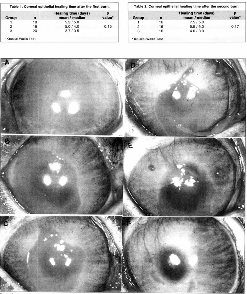
Fig. 1 - Case 2 of Group 1 (removal of corneal-conjunctival limbal flap). Corneal healing o n day 1 (A), day 3 (B), day 7 (C), day 9 (0), day 1 1 (E) and day 20 (F) after the second burn.
Corneal epithelial healing in rabbit eyes with partia[ corneal-conjunctival and conjunctival limbal deficiency
The healing time after the second burn was on average 6 . 5 ± 1 .0 days for Groups 1 and 2 together. This result showed to be significant (p = 0 . 02) when compared with the
controls.
Table 2 shows the mean healing time for the 3 groups . A great variation, shown by the high standard error, occurred in Group 1 , due probably to the fact that healing o f the epithelial defect of two eyes from this group took 3 1 and 2 1 days (cases 2 and 1 1 ) . The healing sequence o f case 2 (Group 1 ) is shown in Fig. 1 . Healing in one case (#3) of Group 2 took 1 3 days. In spite of the greater delay in healing o f Group 1 , there was no statistically significant difference between the groups.
After the first and second burns in both Group 1 and Group 2, the epithelial defect of the superior corneal third, adj acent to limbus remova! was the last to heal in 69.5% and 75 . 3 % of the cases, respectively (Fig. 2A and 2B). In the control group, 34.0% (after the first burn) and 3 8 . 3 % (after the second burn) of the epithelial defects healed last in the superior third. In most cases of thi s group , the epithelial defect healed centripetally (Fig. 2C and 2D) .
Evaluation o f the epithelial healing curve
The healing curve (which besides time also registers the epithelial defect area) o f the limbal deficient eyes o f Groups 1 and 2, analyzed together, and of the controls is presented in Fig. 3 . There was a significant difference only on the first day after the first burn.
Comparing separately the healing curves of each group after the first burn (Fig. 4 ), eyes submitted to parti ai corneal and conjunctival limbal deficiency (Groups 1 and 2) healed more slowly than controls (p = 0.04 between groups), but
there was no significant difference on comparing Group 1 with Group 2. There was a significant difference between Group 1 and the control groups at ali times observed, while this difference was only significant between Group 2 and the control group on day 3 of observation. The method of "repeated measurement analysis" and Wilks ' test was used in this analysis to evaluate multiple comparisons between groups. The same analysis was used to evaluate the results shown in Fig. 6.
The healing curve of the operated eyes (Groups 1 and 2), and of the controls after the second burn is shown in Fig. 5. There was significant difference until day 5 of observation.
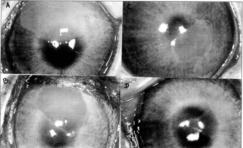
Fig. 2 -Case 5 of Group 2 (removal of conjunctival l i m bal flap) on day 5 (A) and day 7 (B) after the second burn. Case of the control group on day 5 (C) and day 7 (D) atter the second burn.
Corneal epithelial healing in rabbit eyes with partia/ cornea/-conjunctivat and conjunctiva/ limbal deficiency
300
250
- 200 N
E
É. 1 50 "'
� 1 00
50
day 3 day 5 day 7
Fig. 3 - Corneal heal i n g cu rve of eyes with limbal deficiency and controls after the fi rst burn. There was a statistically significant
difference between the two groups (p < 0.05) on day 1 .
250
200
N' E 1 50 E ro ClJ 1 00
.... C1l
50
day 1 day 3 day 5 day 7
Fig. 4 -Corneal healing cu rve of the three studied groups after the first burn. There was a statistically significant difference (p < 0.05) between Group 1 and controls on ali days of observation (*). Between Group 2
and the controls only on day 3 was there a statistically significant difference ( • ).
On analyzing the epithelial healing curve between the three groups, after the second burn (Fig . 6 ) , a more evident delay in corneal healing of Groups 1 and 2 as compared to the controls can be observed (p = 0 . 02 between the 3
groups) . Regarding the different times, it can be observed that Group I healed more slowly than the controls, with significant difference on ali days of follow-up . Comparing Group 2 with the control group, there was statistically significant difference only on day 3. Groups 1 and 2 healed simi larly, mainly after day 5 .
Evaluation o f th e corn eal vascula rization
After the first burn we observed superior peripheral corneal vascularization in one case ( 5 . 5%) o f Group 1 and in 2 cases ( 1 1 . 1 %) o f Group 2. Ali these eyes presented vessels, maximally at 2 mm from the limbus, c1assified as 1 +. There was complete regression of the vessels at the end of the fol low-up. No vascularization occurred among the controls.
532 · A R Q . B R A S . O FTA L . 6 1 (5), O U T U B R 0/ 1 9 9 8
250
200
N' E ..§.. "'
� 1 00 "'
50
o
day 1 day 3 day 5 day 7 day 9 day 1 1
Fig. 5 - Corneal healing cu rve of eyes with l i m bal deficiency and the control group after the second burn. There was a statistically significant
difference between the two groups (p < 0.05) until day 5.
300
250
200 N' E
E 1 50 'iií
�
"' 1 00
50
o
day 1 day 3 day 5 day 7 day 9 day 1 1
Fig. 6 -Corneal healing c u rve of the three groups after the second burn. Up to day 9 of observation there was a statistically significant difference between Group 1 and controls (*). Between Group 2 and the control
group only on day 3 was there a statistically significant difference ( � ).
After the second burn , 3 ( 1 8 . 7%) eyes o f Group 1 presented vessels at the superior cornea with I +, 2+ and 3+ vascularization, respectively. Also in 3 eyes of Group 2 vessels were observed at the superior periphery, ali classified as 1 +. One eye (6.2%) of the control group developed vessels (1+ ). In ali eyes of the three groups which showed vessels at 4 mm from the limbus ( 1 + and 2+) there was regression. In case 2 of Group I , with 3+ vascularization, the vessels remained up to the end o f follow-up (Fig. 1 F).
After the two burns, the comparative statistical ana1ysis showed that there were no differences between the three groups regarding induction of vascularization (p > 0.05). Comparing Groups 1 and 2 together with the control group, there was a1so no significant difference.
Evaluation o f corneal transparen cy
Corneal epithelial healing in rabbit eyes with partia/ corneal-conjunctival and conjunctival limbal deficiency
3 days ) . ln Group 2, three transi ent opacities ( 1 6 . 6%) were observed lasting on average 4 days (ranging from 2 to 6 days). Ali cases, i n both groups, appeared soon after epithelial defect healing and were classified as discrete, according to Hughes 20. No rabbit from the contrai group presented stromal opacity during the observation period. Comparison between the three groups was s ignificant (p < 0 . 0 5 ) . The difference found between Groups I and 2 and between Group 2 and the contrai group was not stati stically significant, however, that between Group I and the contra i group was significant (p = 0 . 04)".
A fter the second burn, there were 7 eyes (43 . 7%) with c orneal opac ity in Group I , lasting on average 2 1 days (ranging from 3 to 60 days ) . Of these cases, 2 8 . 6% were c lassified as severe and 7 1 .4% as discrete. One of the two severe cases persi sted until the end of fo llow-up (60 days). In Group 2, 7 ( 43 . 7%) cases o f transient come a! opacity were observed, persisting on average for 1 2 days (ranging from 3 to
1 7 days ) . Only one ( 1 4 . 3 % ) of these cases was severe and the
rem a i n i n g 8 5 . 7 % were discrete . ln Group 3, the contrai group, there was only one ( 6 . 2 '%) case o f discrete opacity lasting 9
day s . Comparative stati stical analysis between Groups I and 2
and the contrai showed a borderl ine significance (p = 0 . 0 7 ) .
Comparison of frequency of stromal opacities among the operated eyes ( G roups I and 2 ) with contrais showed a significant d i fference (p = 0.04) after the two burns.
Cytologic evaluation o f tlte ocular surface
Corneal cytology performed after healing of the epithelial defect showed the presence of normal squamous epithelial cel l s i n ali cases of the three studied groups . In case 2 (Group
I ). the only case of pers istent corneal "conj unctivalization" in
addition to squamous epithelial cells a small number o f goblet cel l s was evi denced in the superior corneal third.
Appearance of complications
A persistent corneal stromal opacity remained only in one eye o f Group I ( case#2 ) which presented recurrent erosions and 3 + vasc ularizatio n until the end ofthe study (60 days after the second burn) (Fig. I F ) .
DISCUSSION
As shown by Tsai et a i . 22 limbal transplantation is more effi c i ent regarding reconstruction when compared with bulbar con.J u nctival transplantati on . These results were determinant in demonstrating that limbal stem cells are the most qualified to functi onally restare "conj unctivalized" ocular surface in d i seases with limbal d amage .
S i nce then severa) clinicai studies showed good results uti lizing the new limbal transplantation techn i que. Many series uti lized the original technique of Kenyon and Tseng 7 for autologous limbal transplantations g. 1 0· 23 or for allogeneic transplantations 1 1 . The technique utilized in those studies recommends a peripheral keratectomy inc luding 0.5 to 1 mm
of clear cornea to obtain the donor graft. Thi s surgical technique was utilized in Group I animais.
Concomitantly, other authors reported equally satisfactory results of reconstructing the c o rneal s urface u t i l i zi n g conj unctival limbal grafts ( autologous or allogeneic from live donors) without peripheral keratectomy 1 1 · 1 6- 1 8· 24· 25 • In ali these reports the remova! of a conj unctival limbal donor graft, of different sizes, is described, but being as close as possible to its corneal insertion. In the present study this technique was utilized in Group 2 animais.
In most c l inicai series publi shed on l imbal transplantation, be it autologous or allogeneic, uti lizing a live donor, no reports on complications regarding donor eyes are found, since these studies analyze primari ly receptor eyes and do not emphasize follow-up o f donors 6· 23-25
Regarding reports describing complications in eyes with partia! or total limbal defici ency, the fo l lowing can be noted : granuloma-type lesion 1 1 , epitheliopathy, corneal vascul ariza tion and "conj unctivalization" 1 0· 26 .
Earlier studies by Huang and Tseng 6 refer to corneal healing in rabbit eyes with total deficiency of the surgically removed corneal scleral limbus . In this experimental model, the corneas showed a healing delay, vascularization and recurrent erosions after two consecutive central epithelial defects .
Chen and Tseng 1 4 in an experimental study with rabbits, removed two thirds of the corneal-conj unctival l imbus and showed that the ocular surface may significantly decompensate after a great epithelial defect with irreversible signs o f corneal "conj unctivalization" . However, there was no destabilization o f
the ocular surface after two sma l l consecutive epithelial defects. The same authors 27 also proved that partia! l imbal destructi on may cause lighter forms o f limbal deficiency and an abnormal corneal epithelization in the presence o f a la ter great epithelial defect.
In our study a partia! deficiency o f one third o f the l imbus was provoked, in the attempt to simulare cases o f l i mbal flap donors, with minimal limbal deficiency, since the above mentioned studies showed the alterations consequent to remova! of a greater limbal circumference.
Ali the 40 eyes submitted to the two types of surgical procedure of removal of one third ofthe limbus (Groups I and 2) presented rapid epithel izati on. During a 2 1 -day follow-up period there was no sign of destab i 1 ization of the ocular surface in the two groups, probably due to the fact that the remaining SC and T AC were sufficient to maintain cell renewal of the corneal epithelium. However we are not sure how many corneal insults these remaining cells can tolerare .
In arder to study the residual proliferative abi lity of the eyes of our series, with partia] corneal-conj unctival limbal (Group 1 ) and conj unctival limbal deficiency (Group 2), we applied two consecutive alkaline burns. We chose this type of corneal insult because it represents an i mportant cause of ocular trauma to which donor eyes are exposed in daily life. We used two drops of a 0.5 N NaOH solution for 30 seconds,
Corn eal epithelial healing in rabbit eyes with partia! corneal-conjunctival and conjunctival limbal deficiency
a lower concentration and less time than most experimental models of alkaline burn 28· 2". Our purpose was the complete remova! of the corneal epithelium without causing large
I imbal ischemia. The same type o f burn was applied to the normal eyes, which presented no sequelae.
In this study, eyes with partial limbal deficiency (a similar s i tuation to l imbus donor eyes) presented corneal healing time stat i s t i c a lly similar to that of the contro l group . This d i fference probab ly would have been signifi c ant with a greater number of cases, since the remova! of corneal conjunctival flap is more aggressive and probably causes a greater limbal deficiency .
When the area of the epithelial defect is taken into acc ount ( healing curve), we noted that eyes with limbal defi c i ency. after the two burns, have different from normal beh a v i o r, presenting greater defects with greater healing d e l ay (p < 0 . 0 5 ) . Analyzing Groups I and 2 in respect to the controls, we note that Group I (corneal-conj untival graft) healed more slowly than the contra i with a significant d i fferent at ali studi ed time s . Group 2 (conj unctival graft) behaved similarly to the contro l with the exception of day 3 of observation, after the two burn s . The more aggressive surg i c a l technique, uti l ized in G roup I may be responsible for the re sults.
In addition to the eyes with limbal deficiency having s i gni ficantly greater epithelial defects after the burns, these defects presented a delayed healing at the superior corneal third, adj acent to limbus remova! . This fact is due to se dep letion of the excised limbus and the great TA e reduction at the corneal periphery thus leading to epithelial migration from the unharmed limbal periphery as has been shown previously 30. Regarding corneal vascularization, there was no statistical d i fference between the group s . In the cases of vessel development, vascularization was always adj acent to the su perior limbus, demonstrating that, even transiently, the supe rior limbal barrier was interrupted and that the epithelium with conj unctival characteristics advanced over the cornea 1 9•
Corneal opacities are the direct consequence of the burn ( extracellular glycosaminoglycan precipitation and alterations in collagen fibers) associated with stromal inflammation and persistence of the epithelial defects. After epithelial healing there occurs a decrease in inflammation and consequently in the cellular infi ltrate 3 1 • Transient leukomas occurred much more in the eyes with partia! limbal deficiency as compared with the contrais. The difference was significantly greater in Group 1
after the first burn.
There was only one case (case 2 o f Group 1) presenting vesse ls invading beyond 2 mm from the superior limbus, with leukoma and recurrent erosions which persisted until the end o f fo llow-up (Fig. 1 F). Sequelae such as vessels, recurrent e p i t h e l ial defects and corneal opac ity indicate corneal "'conj unctivalization" and are directly proportional to se impairment . This eye was submitted to the most invasive surgical procedure .
53(> -A R Q . B R A S . O FTA L . 6 1 ( 5 ) . O U T U B R 0/ 1 998
Impression cytology, used in cases o f presence o f goblet cells on the corneal surface, has been the method o f choice to diagnose and monitor corneal diseases with limbal deficiency 3 2 . Since no material required for impression cytology was available, we opted for cytology using cytocentrifugation of a corneal epithelium sample removed by means of a nylon brush (eytobrush®) 2 1 • This method was adequa te for the detection of goblet cells in case 2 of Group 1. Absence o f goblet cells, in the remaining cases evaluated, was not surprising, since at the time o f examination only case 2 (Group 1) presented signs o f conj unctival invasion in the superior corneal third (Fig. ! F) .
Studies utilizing specific monoclonal antibodies for differentiated corneal epithelial cells (AE-5), characteristic of the corneal phenotype, and for mucin (AM-3), typical of the conj unctival phenotype, succeeded to prove that eyes with deficiency o f two thirds o f the limbus, submitted to two small epithelial defects developing vessels at the corneal periphery adjacent to limbus remova! had a mixed conj unctival and corneal phenotype, at the corneal periphery, while the center remained normal 14• Some o f our cases in Groups 1 and 2 vascularized at the superior corneal periphery and perhaps analysis using mono clonal antibodies could show a mixed phenotype pattem, proving that there was localized "conj unctivalization" which could be the subject for further studies in this area.
In view of our results, we conclude that, if possible, consi dering that we are dealing with auto logous and allogeneic transplantation utilizing related live donors, we should opt for a graft remova! technique causing the least damage to the donor eye. We believe that conj unctival limbal graft remova!, without keratectomy, is the best option in those cases where tis sue from healthy eyes is removed from patients who are afraid of the procedure in their normal eye. Our results show that eyes with partia! corneal conj unctival limbal deficiency, in the presence of corneal insults (burns), show a greater delay in corneal epithelization (healing curve) and greater chances of presenting complications than eyes with conj unctival limbal deficiency.
The good clinicai results obtained in the series utilizing limbal transplantation 1 1 · 16-18· 24• 25 corroborate our impressions that the peripheral limbal conj unctiva carries a sufficient number o f Se to supply the receptor eye. We believe that this technique of obtention of donor grafts is easier to be technically performed, not presenting ali risks due too peripheral keratectomy, including perforation .
RESUMO
Obj etivo : Comparar a cicatrização epitelial cornean a de olhos de coelhos com deficiência parcial de limbo (DPL) c ó rn e o - c o nj u n ti v a / e c o nju n ti v a / c o m o lhos n o rm a is (controles) .
Método s : A DPL foi induzida pela retirada cirúrgica de 1/3
do limbo superior realizan do uma ceratecto m ia a 2 mm do
Corneal epithelial healing in rabbit eyes with partia! corneal-conjunctival and conjunctival limbal dejiciency
ceratectomia (Grupo 2). Posteriorm en te, todos os o lhos
op erados e o s c o n troles fo ram s u b m etidos a du a s
queimaduras alcalinas consecutivas.
Resultados: Após a 1" e 2" queimaduras, os olhos do Grupo 1
tiveram uma curva de cicatrização epitelial s ign ifica
tivamente mais lenta que os controles, os olhos do Grupo 2 e
os controles tiveram curvas de cicatrização similares. As
córneas dos Grupos 1 e 2 apresentaram mais vascularização
periférica que os controles porém sem diferença significativa.
A incidência de leucomas transitórios foi maior no Grupo 1
quando comparado aos controles (p < 0. 05) . Houve apenas
um caso de leucoma permanente, vascularização e defeito
epiteliais persistentes que pertencia ao Grupo 1 . A avaliação
c itológica dem onstrou células epiteliais n orma is, com
exceção de um caso do Grupo 1 que apresentou células
caliciformes.
Conc lusões : Nossos resultados susten tam o conceito de que
n a s c ó rn eas com DPL o ep itélio a i n da p o de estar
comprom etido após uma queimadura moderada e que a
remoção de limbo córn eo-conjun tiva/ causa uma DPL mais
gra ve do que a remoção de lim bo conjun tiva/. A lém disso,
tra ta-se de um procedimento tecn icamente mais dificil e
in vasivo para o olho doador.
REFERENCES
I . Thoft RA, Friend J . The X, Y, Z hypothesis o f comeal epithelial maintenance. l n vest Ophthalmol Vis Sei 1 98 3 ;24: 1 442-3.
2. Hall PA, Watt FM. Stem cel ls: the generation and maintenance of cellular activity. Development 1 989; I 06:6 1 9- 3 3 .
3 . Schenner A, Galvin S , Sun TT. Differentiation-related expression of a major 64K corneal keratin in vivo and in culture suggests limbal location of comeal epithelial stem cells. J Cell Biol 1 986; 1 03 :49-62.
4 . Ebato B, Friend F, Thoft RA. Comparision of central and peripheral human corneal epithelium in tissue culture. Invest Ophthalmol Vis Sei 1 987;28 : 1 450-6.
5. Ebato B , Friend F, Thoft RA. Comparision of limbal and peripheral human corneal epithelium in tissue culture. lnvest Ophthalmol Vis Sei 1 988;29: 1 533-7.
6 . Huang AJW, Tseng SCG. Corneal epithelial wound heal ing in the absence of l i m b a l e p i the l i a l . A RV O A b stracts . lnvest Ophthalmol Vis S e i 1 9 8 8 ; 2 9 ( S u ppl ) : 1 90 .
7 . Kenyon K R , Tseng SCG. Limbal autograft transplantation for ocular surface d isorders. Ophthalmology 1 989;96:709-23.
8 . Durand L , F ages F, Burillon C. Greffe lamellaire cornéo-conjonctivale "inlay", premier temps préparatoire du tra itement chirurgical des séquelles de brillures ele cornee. J Fr Ophthal mol 1 990; 1 3 : 1 7-23.
538 -ARQ. BRAS. OFTA L. 6 1 (5 } , OUTUBR0/ 1 9 9 8
9. Ronk J F , Ruiz-Esmenjaud S, Osorio M e t a i . Limbal conjunctival autograft in a subacute alkaline corneal bum . Cornea 1 994; 1 3 :465-8.
1 0. Tan DTH, Ficker LA, Buckley RJ. Limbal transplantation. Ophthalmology 1 996; I 03 :29-36.
1 1 . Kwitko S, Marinho DR, Barcaro S, et ai. Allograft conjunctival transplantation for bilateral ocular surface disorders. Ophthalmology 1 995 ; 1 02 : 1 020-5. 1 2 . Tsai RJF, Tseng SCG. Human Allograft Limbal Transplantation for comeal
surface reconstruction. Comea 1 994; 1 3 : 3 89-400.
1 3 . Tsubota K, Toda I, Sai to H et ai. Reconstruction of the corneal epithelium by l imbal a l l o graft tran s p l antation for s evere ocular surface d isorders. Ophthalmology 1 995; I 02: 1 486-96.
1 4. Chen JJY, Tseng SCG. Comeal epithelial wound healing in partia! limbal deficiency. Invest Ophthalmol Vis Sei 1 990;3 1 : 1 3 0 1 - 1 4.
1 5 . Weise RA, Manni s MJ, Vastine DW et ai. Conjunctival transplantation. Autologous and homologous grafts. Arch Ophthalmol 1 985; I 03 : 1 736-40. 1 6. Bri l l JRS. Cirurgia conjuntiva! por indicación comeal. Arch Chil Oftalmol
1 99 1 ;48:73 -80.
1 7 . Clinch TE, Goins KM, Cobo LM. Treatment of contact lens-related ocular surface disorders with autologous conjunctival transplantation. Ophthalmology 1 992;99:634-8.
1 8 . Pfi s ter RR. Corneal stem cell d i sease: Concepts, categorization and treatment by auto and homotransplantation of limbal stem cells. CLAO J 1 994;20: 64-72.
1 9. Thoft RA, Friend J, Murphy HS. Ocular surface epithelium and corneal vascularization in rabbits. The role of wounding. Invest Ophthalmol Vis Sei 1 979; 1 8 : 85 -92.
20. Hughes WF. Alkali bums of the eye. Review of the literature and summary of present knowledge. Arch Ophtha1mol 1 946;35 :423-49.
2 1 . Tsubota K, Kajiwara K, U gajin S et ai. Conjunctival brush cytology. A c ta Cytol 1 990;34:233-5.
22. Tsai RJF, Sun TT, Tseng SCG. Comparison of l imbal and conjunctival autograft transplantation in corneal surface reconstruction in rabbits. Ophtha1mology 1 990;97:446-55 .
2 3 . Shimazaki J, Yang H Y , Tsubota K. Limbal autograft transplantation for recurrent and advanced pterygia. Ophtha1mic Surg Lasers 1 996;27: 9 1 7-23. 24. Cunha M, Allemann N . Transplante autólogo de conjuntiva no tratamento de
pterígio primário e recidivado. Arq Bras Oftal 1 993;56:78-9 1 .
2 5 . Carvalho MJ, Moura RC, Cunha M et ai. Transplante autógeno de conjuntiva no tratamento de queimaduras graves. Arq Bras Oftal 1 994;57: 1 67-9. 26. Jenkins C, Tuft S, Liu C et ai. Limbal transplantation in the management of
chronic contact-lens associated epithel iopathy. Eye 1 993;7:629-33 . 27. Chen JJY, Tseng SCG. Abnorrna1 comeal epithelial wound healing in partial
thickness remova! of l i mbal e p i thel ium. lnvest Ophthalmol V i s S e i 1 99 1 ;32:22 1 9- 3 3 .
28. Chung J H . Healing o f rabbit comeal alkali wounds i n vitro. Comea 1 990;9 :36-40. 29. Saika S, Uenoyama K, H i roi K et ai. Ascorbic acid phosphate ester and wound healing in rabbit corneal alkali burns: epithelial basement membrane and stroma. Graefes Arch Clin Exp Ophthalmol 1 993;23 1 :22 1 -7.
30. Dua HS, Gomes JAP, Singh A. Corneal epithelial wound healing. Br J Ophthalmol 1 994;78 :40 1 -8 .
3 1 . Wagoner MO. Chemical injuries of the eye: current concepts in pathophysiology and therapy. Surv Ophthalmol 1 997;4 1 :275-3 1 3 .