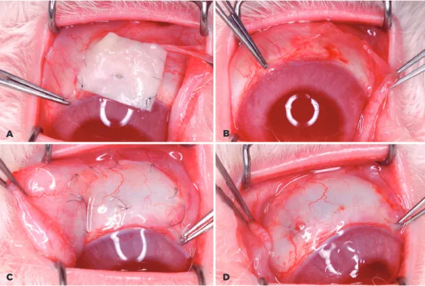
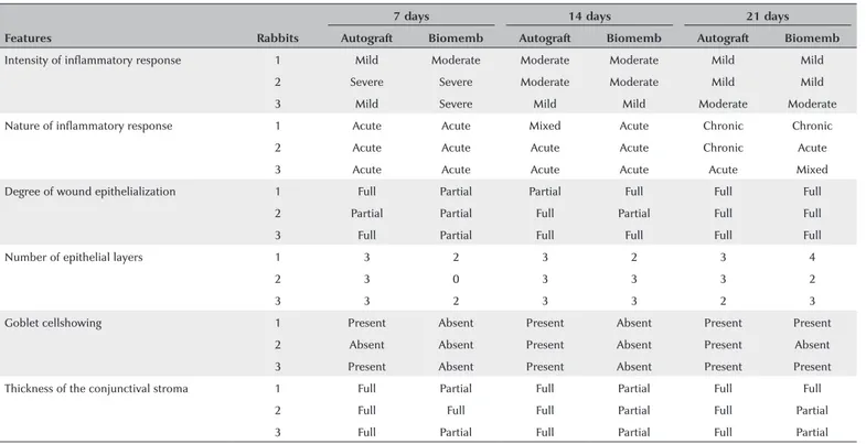
The study of the use of a latex biomembrane and conjunctival autograft in rabbit conjunctiva wound healing
Texto
Imagem
Documentos relacionados
Ainda, em 26 domicílios chefiados por homens há crianças cuja relação não é de filiação para com o chefe e não estão aí acompanhados de pai ou mãe, significando 15,4%
The most significant decrease in force values oc- curred in the first hour, for both latex and non-latex elastics, with the difference percentage higher for non-latex elastic,
The probability of attending school four our group of interest in this region increased by 6.5 percentage points after the expansion of the Bolsa Família program in 2007 and
Objective: To estimate the prevalence of sickle cell disease in adults with delayed diagnosis, receiving treatment at hematology outpatient clinics in the health network of the
The lack of a significant genotypic and phenotypic correlation between growth vigor and the total number of latex vessel rings and between yield and latex vessel size indicated
The results showed that aqueous extract and latex preparation were effective against ten clinical strains of Cryptococcus neoformans in vitro (Latex and extract MIC range of 3.2 -
The objective of this study was assessing effects of clone type, harvest period and climate in the productivity and technological properties of the latex and natural rubber
Na hepatite B, as enzimas hepáticas têm valores menores tanto para quem toma quanto para os que não tomam café comparados ao vírus C, porém os dados foram estatisticamente
