INTRODUCTION
Eye burns are common and may be caused by various chemical and physical agents including acids, alkalis, high temperatures, and fire(1). They are most generally a consequence of chemical handling accidents and may result in permanent damage to the ocular surface and visual function(1).
Corneal alkali burns are considered an ophthalmologic emer-gency. Therefore, timely recognition and implementation of the appropriate treatment represent important steps in controlling the progression of early and late complications(2). The literature describes various forms of treatment for corneal alkali burns. These include arti-ficial tears, collagenase inhibitors, therapeutic contact lenses, topical fibronectin, topical vitamin C, topical citrate(2), conjunctival transplan-tation(1), amniotic membrane patching(3,4),limbal transplantation(5), and autologous serum eye drops(6) as well as treatment of the severe inflammatory processes with topical or systemic corticosteroids. AbsTRACT
Objective: To evaluate the effect of riboflavin-ultraviolet-A-induced cross-linking (CXL) following corneal alkali burns in rabbits.
Methods: The right corneas and limbi of ten rabbits were burned using a 1N so lution of NaOH and the animals were then divided into two groups: a control group submitted to clinical treatment alone and an experimental group that was treated 1 h after injury with CXL, followed by the same clinical treatment as administered to the controls. Clinical parameters were evaluated post-injury at 1, 7, 15, and 30 days by two independent observers. Following this evaluation, the corneas were excised and examined histologically.
Results: There were no statistically significant differences in clinical parameters, such as hyperemia, corneal edema, ciliary injection, limbal ischemia, secretion, cor neal neovascularization, symblepharon, or blepharospasm, at any of the time-points evaluated. However, the size of the epithelial defect was significantly smaller in the CXL group (p<0.05) (day 15: p=0.008 and day 30: p=0.008) and the extent of the corneal injury (opacity lesion) was also smaller (day 30: p=0.021). Histopathology showed the presence of collagen bridges linking the collagen fibers in only the CXL group.
Conclusions: These results suggest that the use of CXL may improve the prognosis of acute corneal alkali burns.
Keywords: Cross-linking reagents; Riboflavin; Ultraviolet therapy/methods; Cornea/ drug effects; Rabbits; Animal
RESUMO
Objetivo: Avaliar o efeito de ligações covalentes de colágeno (cross-linking [CXL]) induzidas pelo tratamento com riboflavina e radiação ultravioleta A após queimaduras por álcali em córneas de coelhos.
Métodos: Dez coelhos foram submetidos a queimadura ocular direita abrangendo estruturas da córnea e limbo usando uma solução de NaOH a 1N. A seguir, os animais foram divididos em dois grupos: um grupo controle submetido a tratamento clínico pós dano corneano e um grupo experimental que foi tratado com CXL uma hora após o dano, seguido pelo mesmo tratamento clínico administrado aos controles. Os parâmetros clínicos foram avaliados 1, 7, 15 e 30 dias após a lesão, por dois obser vadores independentes. Na etapa seguinte, foi realizada a excisão e o exame histológico das córneas.
Resultados: Não houve diferenças estatisticamente significantes nos parâmetros clí nicos de hiperemia, edema da córnea, injeção ciliar, isquemia límbica, secreção, neo-vascularização da córnea, simbléfaro ou blefaroespasmo, em qualquer dos momentos da avaliação. Entretanto, o grupo CXL apresentou um defeito epitelial menor (p<0,05) (dia 15: p=0,008 e dia 30: p=0,008) e menor extensão da lesão na córnea (lesão opaca) (dia 30: p=0,021). O exame histopatológico revelou a presença de pontes de colágeno conectando as fibras de colágeno somente no grupo CXL.
Conclusões: Estes resultados sugerem que o uso de CXL pode melhorar o prognóstico de queimaduras agudas da córnea causadas por alcáli.
Descritores: Reagentes para ligações cruzadas; Riboflavina; Terapia ultravioleta/ mé todos; Córnea/efeito de drogas; Coelhos; Animal
Chemical burns may lead to devastating complications, including corneal perforation due to the rapid degradation of collagen fibers(1). Recently, riboflavin-ultraviolet-A-induced cross-linking (CXL) was de veloped as a technique for enhancing collagen cross-linking in the treatment of corneal wounds(7). CXL has been reported to be a safe and effective method for controlling the progression of corneal ectasia(8,9). The procedure stops the melting process of the cornea(10) and has been shown to increase the resistance of porcine corneas by inhibiting enzymatic degradation(11). CXL was previously used in five cases of corneal necrosis following a bacterial infection refractory to clinical treatment(12), and it was demonstrated that this technique constitutes a useful alternative to emergency keratoplasty by increa-sing corneal resistance against the action of collagenolytic enzymes. Currently, CXL is used to stabilize degenerative corneal disorders such as corneal ectasia (in keratoconus patients and following refractive surgery), where it acts to increase the degree of rigidity of the stromal collagen fibers(13,14).
Induction of corneal collagen cross-linking in experimental corneal alkali burns in rabbits
Indução de ligações covalentes de colágeno em queimaduras corneanas experimentais por álcali em coelhos
Marcello coloMbo-barboza1,2,3,GuilherMe coloMbo-barboza1,2, SerGio FelberG2, Paulo eliaS corrêa DantaS2, elcio hiDeo Sato4
Submitted for publication: February 28, 2014 Accepted for publication: June 16, 2014
Study conducted at Santa Casa de Misericórdia de São Paulo, São Paulo, SP, Brazil. 1 Hospital Oftalmológico Visão Laser, Santos, SP, Brazil.
2 Department of Ophthalmology, Santa Casa de Misericórida de São Paulo, São Paulo, SP, Brazil. 3 Centro Universitário Lusíada, Santos, SP, Brazil.
4 Department of Ophthalmology and Visual Sciences, Paulista School of Medicine (EPM), Federal University of São Paulo (UNIFESP), São Paulo, SP, Brazil.
Funding: No specific financial support was available for this study.
Disclosure of potential conflicts of interest: None of the authors have any potential conflicts of interest to disclose.
Corresponding author: Marcello Colombo Barboza. Av. Conselheiro Nebias, 355 - Santos, SP - 11015-001 - Brazil - E-mail: [email protected]
Considering these aspects, the objective of the present study was to evaluate the effect of CXL in a rabbit model, initiated one hour after a corneal alkali burn.
METHODs
This was a comparative, randomized, blinded experimental study conducted in the animal laboratory and the animal experimentation surgical center of the Santa Casa de São Paulo. The institutional Ani-mal Research Ethics Committee of UNIFESP and the Santa Casa de São Paulo approved the protocol (approval letter 1476/09). The animal study was carried out in compliance with the recommendations of the Association for Research and Vision in Ophthalmology (ARVO).
A
NIMALSELECTIONTen male New Zealand white rabbits weighing 3-4 kg were ran-domly divided into two groups, with five rabbits in each group (the experimental and control groups). The animals were provided with food and water ad libitum and pertinent veterinary supervision.
E
XPERIMENTALMODELOFCORNEALALKALIBURNAn experimental model of corneal burn that has already been described in previous studies was used(15,16). This model was modified only with respect to the diameter of the paper disc in order for it to include the total limbal area. A grade IV ocular burn, according to the Roper Hall classification(17,18), was induced in the right eye of all the rabbits.
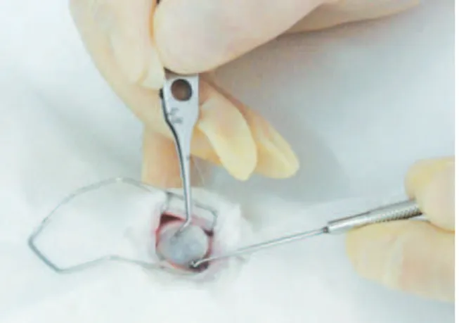
Under the supervision of a veterinarian, general anesthesia was induced by an intramuscular injection of ketamine hydrochloride (25 mg/kg of weight) associated with 2% xylazine hydrochloride (4 mg/kg of weight). Appropriate ventilatory support was provided. Corneas were anesthetized using a drop of 1% proparacaine hydro-chloride ophthalmic drops in the animals’ right eyes. A corneal alkali burn was then induced by applying a 14 mm diameter filter paper disc (Whatman filter paper, # 40) soaked in 1N NaOH (Figure 1). This disc was maintained in contact with the cornea and the limbus for one minute, after which the eyes of the animals were washed with 1.0 cc of 0.9% NaCl.
Sixty minutes after the injury procedure, 0.1% riboflavin drops (Ophthalmos®, São Paulo, Brazil) were applied every five minutes for thirty minutes to the animals in the experimental group. This step was then followed by irradiation with ultraviolet light (UVA 370 nm, with an irradiance of 3 mW/cm2 and a surface dose of 5.4 J/cm2; X-link, Opto, Brazil) for 30 minutes at a distance of 45 mm from the right eye, and including the total cornea and limbus area. The epithelium
was not touched during the procedure. During UVA irradiation, 0.1% riboflavin drops continued to be applied concomitantly every five mi-nutes. Next, clinical treatment was initiated with the application of a 1% prednisolone acetate ophthalmic suspension (Pred Fort, Allergan, Guarulhos, SP, Brazil), preservative-free lubricant eye drops (Optive UD, Allergan), and 0.3% ofloxacin eye drops (Oflox, Allergan). These were all administered three times a day (tid) for the first 15 days (early phase). After this period, the corticosteroid treatment was stopped and only the lubricant eye drops and the antibiotic were maintained for another 15 days (regenerative phase).
The control group (5 eyes in 5 rabbits) was submitted to the same clinical treatment described above after the burn had been induced and the eye washed with 1.0 cc of 0.9% NaCl.
F
OLLOW-
UPANDEXAMINATIONOFTHEANIMALSThe animals underwent a 30 day follow up, with evaluations being conducted by two independent, blinded observers immediately after the burn, and then on days 1, 7, 15, and 30 following the injury. Evaluation of the corneal surface was conducted by external exa-mination and then by biomicroscopy using a hand-held slit lamp (Heine-HSL 100, USA).
Thirty days after the ocular burn, all the rabbits were sacrificed using a 3 ml intramuscular injection of 0.2% acepromazine, followed 15-20 minutes later by 5 ml intravenous thionembutal and 3 ml intravenous potassium chloride. The corneas were then excised and histological sections were prepared and examined following hema-toxylin/eosin (HE) and trichrome Masson staining for assessing the collagen stroma in the two groups.
C
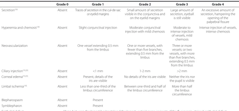
LINICALPARAMETERSUSEDClinical parameters are described on table 1(16,18-20). An example of epithelial defect evaluation can be observed on figure 2.
S
TATISTICALANALYSISKappa coefficients were calculated to estimate observer agree-ment for all the clinical variables investigated. Fisher’s exact test was used to compare differences between the experimental and control groups. For the continuous variables, means, standard deviations, medians, and ranges were calculated. The Mann-Whitney U test was used to compare the continuous variables between groups, with p values <0.05 being considered statistically significant. The complete statistical analysis was conducted using the STATA statistical software package, version 10 (College Station, Texas, USA).
REsULTs
There was high agreement between the two independent obser-vers, as shown by the kappa coefficient range of 0.32-1.0.
There was a statistically significant difference between the two groups with respect to the mean size of the corneal epithelial defect at 15 and 30 days after the injury procedure (Table 2). There was also a statistically significant difference in the mean extent of corneal injury (opacity lesion) at day 30 (Table 3).
No statistically significant differences were found between the experimental and control groups with respect to clinical parameters such as conjunctival hyperemia, ocular secretion, corneal neovascu-larization, ciliary injection, central corneal edema, limbal ischemia, blepharospasm, or symblepharon during the post-injury followup (Table 4).
In the CXL group, histology revealed collagen bridges linking collagen fibers that were arranged in an organized pattern (Figure 3). However, these bridges were absent in the control group (Figure 4). Table 5 shows the presence or absence of bridges in the corneas evaluated.
Table 1. Grading of clinical parameters evaluated
Grade 0 Grade 1 Grade 2 Grade 3 Grade 4
Secretion(18) Absent Traces of secretion in the cul-de-sac
or eyelid margins
Small amount of secretion visible in the conjunctiva and
on the eyelid margins
Large amount of secretion, eyeball
is still visible
An excessive amount of secretion, hampering the
opening of the palpebral fissure
Hyperemia and chemosis(18) Absent Slight conjunctival injection Moderate conjunctival
injection with mild chemosis
Moderate to intense injection
of vessels, mild chemosis
Intense injection of vessels, intense chemosis
Neovascularization Absent One vessel extending 0.5 mm from the limbus
One or more vessels, with fewer than five branches, extending 0.5 mm from the
limbus
Three or more vessels; or two vessels, with more than five branches, extending 0.5 mm from the limbus
Ciliary injection(19,20) Absent <1 mm 1-2 mm >2 mm
Corneal edema(19,20) Absent Present, details of the
iris are visible
No details of the iris are visible Neither the iris nor the pupil is visible
Limbal ischemia(16) Absent Less than one-third of the
limbus circumference
Between one-third and half of the limbus circumference
More than half the limbus circumference
Blepharospasm Absent Present
Symblepharon Absent Present
Epithelial defect Mean between the largest horizontal and the largest vertical measurements of the fluorescein-stained epithelial defect, as measured in millimeters using a surgical caliper. (Figure 2)
Corneal injury Mean between the largest horizontal and the largest vertical measurements of the opacity lesion created by the burn, measured in millimeters using a surgical caliper
DIsCUssION
In the present study, we used 14 mm alkali-immersed filter discs (1 N NaOH) to create severe ocular burns (Roper Hall criteria, grade IV). This methodology was chosen because it has been well described and involves clinically comparable parameters that can be easily measured.
We hypothesized that CXL would allow covalent bonds to be formed between the collagen fibrils, thus promoting thickening of the collagen fibrils through the deposit of structural molecules such as proteoglycans(21), and possibly making the cornea more resistant to the effect of collagenolytic enzymes. In the CXL group, histopatho-logical examinations revealed collagen bridges linking the collagen fibers in the corneal stroma. This may indicate that the collagen fibers in the CXL group were more resistant to collagenolytic enzymes than those in the control group. Furthermore, the arrangement pattern of the stromal collagen fibers was more organized in the CXL group compared to the control group. This may also indicate a greater re-sistance of the collagen fibers to collagenolytic enzymes in the CXL group, resulting in improved wound healing. Nevertheless, further investigation is required with respect to these collagen bridges and wound healing. Some case reports have also shown the antimicrobial
effect of ultraviolet light associated with riboflavin in the treatment of infectious keratitis. The mechanism is presumably either due to the bactericidal effect of ultraviolet light or to the increased resistance of the collagen fibers of the cornea, which prevents the infectious agent from proliferating(12,21,22).
A statistically significant difference was found between the groups regarding the mean extent of the corneal injury on day 30 following the injury (p=0.021). At this time point the lesion caused by the ocular burn is at a late phase of tissue repair; the effect of collagenolytic enzymes has been largely overcome, and the tissue has undergone regeneration of the fibroblasts, with migration of myofibrils to the site and progressive re-epithelization taking place. The associated effects of the increase in resistance to the collagenases in the first few weeks, and the increase in the rigidity of the corneal tissue due to cross-linking, suggests a better recovery in the CXL group compared to the control group. It should be noted that even con-sidering the statistical significance of our results with respect to the mean extent of the corneal injury, when the absolute data is taken into consideration the difference in the mean size of the lesions is close to 1 mm.
The magnitude of this measurement is imperceptible in clinical practice when considering devastating grade IV burns.
Statistically significant results regarding the mean size of the cor-neal epithelial defect were also found on post-injury days 15 (p=0.008) and 30 (p=0.008). Therefore, we can conclude that the cen tripetal movement of the corneal epithelial cells that occurs in the regenera-tion phase of the burned cornea occurred faster in the experimental group compared to the control group. The centripetal movement is associated with an increase in the rigidity of the cornea and the resis-tance of the tissue to enzymatic degradation through the formation of stronger stroma. This serves as a base for the sliding of the epithelial cells during the recovery phase of the corneal epithelial wound.
Some of the difficulties encountered during this study that merit particular mention refer to the objective measurement of the data obtained from the rabbits, and the manipulation of the animals’ eyes
Table 2. Mean size of the corneal epithelial defect in the rabbits follo-wing ocular alkali burn
Time post-injury
size of the corneal epithelial defect (Mean ± sD) (mm)
p-value* Experimental group Control group
01st day 13.20 ± 0.20 13.20 ± 0.20 1.000 07th day 09.85 ± 0.87 10.95 ± 0.71 0.089
15th day 06.40 ± 0.92 09.10 ± 0.87 0.008
30th day 04.05 ± 0.94 6.40 ± 0.65 0.008
Experimental group= cross-linking plus clinical treatment; Control group= only clinical treatment; P*= Mann-Whitney U test, with p-values <0.05 being considered statistically significant.
Table 3. Mean extent of the corneal injury (opacity lesion) in the rabbits after ocular alkali burn
Time post-injury
Extent of the corneal injury (Mean ± sD) (mm)
p-value* Experimental group Control group
01st day 13.10 ± 0.22 13.00 ± 0.11 0.881 07th day 13.05 ± 0.00 12.95 ± 0.11 0.317
15th day 12.20 ± 0.44 12.00 ± 0.58 0.737
30th day 10.35 ± 0.74 11.60 ± 0.54 0.021
Experimental group= cross-linking plus clinical treatment; Control group= only clinical treatment; P*= Mann-Whitney U test, with p-values <0.05 being considered statistically significant.
Table 4. Mann-Whitney U-test p-values for the various clinical parameters evaluated at the diferent time-points following the injury procedure
Clinical parameters (p-values)
Time post-injury
1st day 7th day 15th day 30th day
Ocular secretion 0.206 0.444 0.444 0.444 Conjunctival hyperemia 0.444 n/c 1.000 0.167
Neovascularization n/c 0.167 0.524 0.524
Ciliary injection 0.444 0.167 0.444 1.000
Edema 1.000 1.000 1.000 1.000
Limbal ischemia n/c n/c n/c n/c
Blepharospasm n/c n/c n/c n/c
Symblepharon n/c n/c 0.206 0.206
P*= Mann-Whitney U test, with p-values <0.05 being considered statistically signi-ficant; n/c= not calculated.
Table 5. Presence or absence of stromal interibrillar bridges in histolo-gical sections of alkali-burned rabbit corneas according to the procedu-re performed (CXL or none)
Animal number Corneal procedure Interibrillar bridges
01 CXL Yes
02 CXL No
03 CXL Yes
04 CXL Yes
05 CXL No
06 Control No
07 Control No
08 Control No
09 Control No
10 Control No
when using a hand-held slit lamp. Measuring the greatest horizon-tal and vertical diameters also proved difficult, since although the lesions were circular and symmetrical throughout their extension,
in some cases there were slight discrepancies in re-epithelialization. This occasionally gave rise to asymmetry in the shape of the healing wound. In addition, the sample size used in this study was small (5 rabbits in the CXL group and 5 in the control group). However, it is im-portant to stress that these numbers were agreed upon following an in-depth discussion with the ethics committee for animal research.
The mechanism underlying the statistically significant differences found at later post-injury time-points remains largely unclear, and we therefore recommend that further studies be conducted to clarify the physiopathogenesis of cross-linking in ocular burns.
In conclusion, these results suggest that the use of CXL may im -prove the prognosis of acute corneal alkali burns.
REFERENCEs
1. Wagoner MD. Chemical injuries of the eye: current concepts in pathophysiology and therapy. Surv Ophthalmol. 1997;41(4):275-313.
2. Bechara SJ, Garcia IA, Kobinger E, Rodrigues CJ, Kara José N, Caldeira JA. [Simulta-neous use of acetylcysteine and vitamin C in the therapeutics of cornea burns by alkali]. Arq Bras Oftalmol. 1986;49(4):109-11.
3. Joseph A, Dua HS, King AJ. Failure of amniotic membrane transplantation in the treatment of acute ocular burns. Br J Ophthalmol. 2001;85(9):1065-9.
4. Gomes JA, Romano A, Santos MS, Dua HS. Amniotic membrane use in ophthalmology. Curr Opin Ophthalmol. 2005;16(4):233-40.
5. Nishiwaki-Dantas MC, Dantas PE, Reggi JR. Ipsilateral limbal translocation for treat-ment of partial limbal deficiency secondary to ocular alkali burn. Br J Ophthalmol. 2001;85(9):1031-3.
6. Cypel MC, Goulard DA, Lima FA, Lake JC, Uesugui E, Nishiwaki-Dantas MC, et al. [Sub-conjunctival injection of autogenous blood in the treatment of ocular alcali burn in rabbits]. Arq Bras Oftalmol. 2004;67(5):801-5.
7. McCall AS, Kraft S, Edelhauser HF, Kidder GW, Lundquist RR, Bradshaw HE, et al. Mechanisms of corneal tissue cross-linking in response to treatment with topical riboflavin and long-wavelength ultraviolet radiation (UVA). Invest Ophthalmol Vis Sci. 2010;51(1):129-38.
8. Spoerl E, Mrochen M, Sliney D, Trokel S, Seiler T. Safety of UVA-riboflavin cross-linking of the cornea. Cornea. 2007;26(4):385-9.
Figure 3. Alkali-burned corneal stroma following 30 days of CXL treatment. Note the organized pattern of collagen bridges (arrow) between the stromal ibers. HE staining (A) and trichrome Masson staining (B).
A b
b A
9. Kolli S, Aslanides IM. Safety and efficacy of collagen crosslinking for the treatment of keratoconus. Expert Opin Drug Saf. 2010;9(6):949-57.
10. Schnitzler E, Spörl E, Seiler T. Irradiation of cornea with ultraviolet light and riboflavin administration as a new treatment for erosive corneal processes, preliminary results in four patients. Klin Monbl Augenheilkd. 2000;217(3):190-3.
11. Spoerl E, Wollensak G, Seiler T. Increased resistance of crosslinked cornea against enzymatic digestion. Curr Eye Res. 2004;29(1):35-40.
12. Iseli HP, Thiel MA, Hafezi F, Kampmeier J, Seiler T. Ultraviolet A/riboflavin corneal cross-linking for infectious keratitis associated with corneal melts. Cornea. 2008;27(5):590-4. 13. Hersh PS, Greenstein SA, Fry KL. Corneal collagen crosslinking for keratoconus and
corneal ectasia: one-year results. J Cataract Refract Surg. 2011;37(1):149-60. 14. Jankov MR 2nd, Hafezi F, Beko M, Ignjatovic Z, Djurovic B, Markovic V, et al. [Corneal
cross-linking for the treatment of keratoconus: preliminary results]. Arq Bras Oftalmol. 2008;71(6):813-8.
15. Sangwan VS, Akpek EK, Voo I, Zhao T, Pinar V, Yang J, et al. Krill protease effects on wound healing after corneal alkali burn. Cornea. 1999;18(6):707-11.
16. Siganos CS, Frucht-Pery J, Muallem MS, Berenshtein E, Naoumidi I, Ever-Hadani P, et al. Topical use of zinc desferrioxamine for corneal alkali injury in a rabbit model. Cornea. 1998;17(2):191-5.
17. Roper-Hall MJ. Thermal and chemical burns. Trans Ophthalmol Soc U K. 1965;85:631-53. 18. Santos NC, Sousa LB, Freitas D, Rigueiro MP, Scarpi MJ. [Corneal and conjunctival toxi-city of povidone-iodine eye drops]. Arq Bras Oftalmol. 2003;66(3):279-88. Portuguese. 19. Laria C, Alió JL, Ruiz-Moreno JM. Combined non-steroidal therapy in experimental
corneal injury. Ophthalmic Res. 1997;29(3):145-53.
20. Oztürk F, Kurt E, Cerci M, Emiroglu L, Inan U, Türker M, et al. The effect of propolis ex tract in experimental chemical corneal injury. Ophthalmic Res. 2000;32(1):13-8. 21. Al-Sabai N, Koppen C, Tassignon MJ. UVA/riboflavin crosslinking as treatment for
corneal melting. Bull Soc Belge Ophtalmol. 2010(315):13-7.