CASE REPORT
Endovascular repair of an aorto-iliac aneurysm succeeded by
kidney transplantation
Tratamento endovascular de aneurisma aorto-ilíaco sucedido por transplante renal
Marcelo Bellini Dalio
1, Matheus Bredarioli
2, Edwaldo Edner Joviliano
3, Jesualdo Cherri
3, Haylton Jorge Suaid
3, Carlos Eli Piccinato
4Introduction
Treatment of patients with aorto-iliac aneurysm
asso-ciated with chronic renal failure (CRF) under hemodialysis
is a challenge, due to increased morbimortality
1,2. Several
authors reviewed open aneurysm repair in association
with kidney transplantation (RT) and proposed a staged
approach with aorto-iliac disease being treated irst
3-5.
Endovascular aneurysm repair (EVAR) has been replacing
open repair in patients with a high surgical risk, with the
advantages of less morbimortality and shorter operative
time and hospital stay
6.
here are several reports of successfully EVAR
perfor-med in RT recipients
7-9, but there are just two reported cases
of EVAR followed by successful RT
10,11. hese were
perfor-med on aortic aneurysms. here are no reported cases of
aorto-iliac aneurysm.
his paper reported a case of aorto-iliac aneurysm in a
patient with CRF under hemodialysis successfully
correc-ted by EVAR succeeded by RT.
Abstract
We present the case of aorto-iliac aneurysm in a patient with chronic renal failure requiring dialysis who were treated with an endovascular stent graft and, later on, submitted to kidney transplantation. A 53-year-old male with renal failure requiring dialysis presented with an asymptomatic abdominal aorto-iliac aneurysm measuring 5.0cm of diameter. He was treated with endovascular repair technique, being used an endoprosthesis Excluder®.
After four months, he was successfully submitted to kidney transplantation (dead donor), with anastomosis of the graft renal artery in the external iliac artery distal to the endoprosthesis. he magnetic resonance imaging, carried out 30 days after the procedure, showed a good positioning of the endoprosthesis and adequate perfusion of the renal graft. In the follow-up, the patient presented improvement of nitrogenous waste, good positioning of the endoprosthesis without migration or endoleak. he endovascular repair of aorto-iliac aneurysm in a patient with end-stage renal failure under hemodialysis treatment showed to be feasible, safe and eicient, as it did not prevent the success of the posterior kidney transplantation.
Keywords: Aortic aneurysm; iliac aneurysm; blood vessel prosthesis; kidney transplantation.
Resumo
Apresentamos o caso de aneurisma aortoilíaco em um paciente com insuiciência renal crônica dialítica tratado com uma endoprótese vascular, sendo, após, submetido a transplante renal. Um homem de 53 anos com insuiciência renal dialítica apresentava um aneurisma abdominal aortoilíaco assintomático com 5,0cm de diâmetro. Foi tratado com técnica endovascular com uma endoprótese Excluderâ. Após quatro meses, foi submetido
a transplante renal (doador cadáver) com sucesso, com anastomose da artéria renal do enxerto na artéria ilíaca externa distal à endoprótese. A ressonância magnética 30 dias após o procedimento mostrou a endoprótese bem posicionada e o enxerto renal bem perfundido. No seguimento, o paciente evoluiu com melhora das escórias nitrogenadas, bom posicionamento da endoprótese, sem migração ou endoleak. O reparo endovascular do aneurisma aortoilíaco em paciente com insuiciência renal terminal em hemodiálise mostrou-se exequível, seguro e eicaz, e não comprometeu o sucesso do transplante renal posterior.
Palavras-chave: Aneurisma aórtico; aneurisma ilíaco; prótese vascular; transplante de rim.
Department of Surgery and Anatomy of Ribeirão Preto Medical School of Universidade de São Paulo (USP), Ribeirão Preto (SP), Brazil.
1 PhD; Vascular and Endovascular Surgeon of Department of Surgery and Anatomy of Ribeirão Preto Medical School of Universidade de São Paulo (USP), Ribeirão Preto (SP), Brazil. 2 Vascular and Endovascular Surgeon of Department of Surgery and Anatomy of Ribeirão Preto Medical School of Universidade de São Paulo (USP), Ribeirão Preto (SP), Brazil. 3 PhD; Associate Professor of Department of Surgery and Anatomy of Ribeirão Preto Medical School of Universidade de São Paulo (USP), Ribeirão Preto (SP), Brazil. 4 PhD; Titular Professor of Department of Surgery and Anatomy of Ribeirão Preto Medical School of Universidade de São Paulo (USP), Ribeirão Preto (SP), Brazil. No conlicts of interest declared concerning the publication of this article
EVAR – kidney transplantation - Dalio MB et al. J Vasc Bras 2010, Vol. 9, Nº 3 165
Case report
A 53-year-old man presenting with end-stage CRF
se-condary to hypertensive nephrosclerosis treated with
he-modialysis. He did not present
diabetes mellitus
, but had
a 40-year smoking history. His bilateral limb pulses were
palpable and symmetric. He referred an asymptomatic
ab-dominal mass, and an investigation with computed
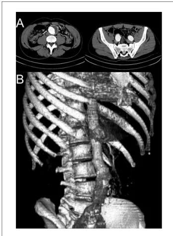
tomo-graphy (CT) scan revealed an infrarenal aortic aneurysm
(5.0cm transverse diameter), extending to both common
iliac arteries (3.0cm transverse diameter each) until its
bi-furcations (Figure1). He underwent EVAR through bilateral
groin incisions, with placement of an Excluder
®endopros-thesis (W. L. Gore & Associates, Flagstaf, Arizona) with the
following measures:
• right main trunk 31mm/14mm/15cm;
• let contralateral leg 16mm/14mm/14cm;
• right extension 16mm/10mm/7cm;
• let extension 16mm/14mm7cm.
In order to completely exclude the aneurysm, both
hypogastric artery oriices were covered by the extensions.
No embolization coils were used. Completion angiogram
showed adequate positioning of the endograt and no en
-doleak. He made a good recovery and, beside some
tran-sient thigh paresthesia, no complications were observed.
Postoperative rectosigmoidoscopy showed no signs of
bo-wel ischemia. he patient went on hemodialysis on the day
ater and was discharged three days ater the procedure.
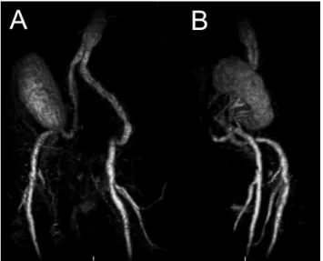
One-month CT scan showed good grat positioning and no
endoleak (Figure 2).
Four months ater EVAR, he underwent RT. hrough
a right Gibson incision; he received a let cadaveric renal
transplant with HLA matching A0B1DR2 and negative
crossmatch. he gratf had two arteries on a Carrel patch,
single vein and single ureter. At dissection, it was possible
to palpate the endograt in the right external iliac artery,
which had a normal aspect and pulse at its distal portion.
No retroperitoneal ibrosis or inlammation was observed.
he transplant arterial patch was sutured in the right
exter-nal iliac artery 0,.5cm proximal to inguiexter-nal ligament by an
end to side anastomosis using continuous polypropilene
6-0. he venous anastomosis was performed end-to-side
in the external iliac vein using continuous polypropilene
Figure 1 - Pre-EVAR axial (A) and 3D reconstruction (B) CT scan.
Figure 2 - Post-EVAR axial (A) and 3D reconstruction (B) CT scan,
EVAR – kidney transplantation - Dalio MB et al. J Vasc Bras 2010, Vol. 9, Nº 3
166
Figure 3 - Post-RT MRI-A, showing good positioning of the aorto-iliac
endograft and adequate perfusion of the renal graft.
5-0. he ureter was implanted using Lich-Gregoir
techni-que. Just ater the procedure, the kidney started to pro
-duce urine. here was no displacement of the endograt
during surgery. No complications were observed and the
patient was placed on an immunosuppression regimen of
mycophenolate, tacrolimus and prednisone. Magnetic
re-sonance imaging angiography (MRI-A) obtained ive days
ater the surgery demonstrated good positioning of the
aorto-iliac endograt and adequate perfusion of the renal
grat (Figure 3). Creatinine levels dropped from a preope
-rative value of 6.2mg/dL to 1.6mg/dL at discharge eight
days ater transplantation. At oice visits, creatinine levels
were stable. he four-year follow-up with yearly MRI-A
showed good positioning of the endograt without migra
-tion or endoleak.
Discussion
Aneurysmal disease involving aorta and both iliac
ar-teries has not a simple treatment. Either open or
endovas-cular approaches require advanced surgical skills. When
associated renal disease is present, the future possibility of a
RT must be considered and the ideal treatment must
provi-de aprovi-dequate conditions for it. Iliac vessels and pelvic space
must be preserved.
Our patient was on hemodialysis and had a
5cm-aorto-iliac aneurysm and was in the waiting line for RT. Although
EVAR and RT simultaneous performance was described
12,
we decided to perform aneurysm repair irst, according to
published series
3-5. he main advantage to treat the
aneu-rysm before RT is to avoid ischemic and nephrotoxic
com-plications in the renal grat, fact that would occur if RT were
irstly performed. he choice of EVAR was based on reports
of increased morbimortality in open surgery performed in
patients like ours
1. Another important reason is that EVAR
does not require extensive dissection of the pelvic space,
thus preserving it for a future RT. In fact, we also purposed
open surgery, but the patient’s choice was EVAR.
In order to completely exclude the aneurysm, covering
of both hypogastric artery oriices were necessary. here are
several description of ancillary procedures to provide low
to hypogastric arteries in EVAR such as surgical bypass
13or endograt associated with crossover femorofemoral
bypass
14. However, these procedures involve respectively
pelvic dissection and use of aorto-mono-iliac endograt,
which would harm posterior RT. hus, we chose covering
hypogastric artery bilaterally. As in our case, Zander et al.
15reported time-limited and insigniicant complications with
bilateral hypogastric artery occlusion in EVAR. Our patient
had only transient thigh paresthesia and full recovery. No
bowel ischemia and no buttock claudication were observed.
Bilateral hypogastric artery occlusion, although risky, may
be acceptable in cases with short therapeutic options, like
ours. Recently available branched endograt for iliac arte
-ries
16would be suitable solution for preserve hypogastric
low in our case.
During RT ater EVAR, possible complications are en
-dograt dislodgment or migration, causing endoleak, and
endograt limb thrombosis due to vascular clamping. hese
complications, which could lead to renal grat loss and
EVAR failure, were not observed in our case. Care should
be taken not to apply vascular clamps over the endograt.
Ater RT, MRI-A was used in the follow-up (six months
ater and then yearly) in order to avoid nephrotoxic efect of
iodinated contrast.
Conclusion
Our experience with aorto-iliac aneurysm corrected by
EVAR succeeded by RT concurs with previous reports of
aortic aneurysm treated in the same way. In patients with
CRF and aorto-iliac aneurysm, EVAR succeeded by RT is
feasible and provides good results.
References
1. Bown MJ, Norwood MG, Sayers RD. he management of abdo-minal aortic aneurysms in patients with concurrent renal impair-ment. Eur J Vasc Endovasc Surg. 2005;30:1-11
EVAR – kidney transplantation - Dalio MB et al. J Vasc Bras 2010, Vol. 9, Nº 3 167
3. Brekke IB, Lien B, Sødal G, et al. Aortoiliac reconstruction in prepa-ration for renal transplantation. Transplant Int. 1993;6:161-3.
4. Piquet P, Berland Y, Coulange C, Olmer M, Mercier C, Rampal M.. Aortoiliac reconstruction and renal transplantation: staged or si-multaneous. Ann Vasc Surg. 1989;3:251-6.
5. Gouny P, Lenot B, Decaix B, et al. Aortoiliac surgery and kidney transplantation. Ann Vasc Surg. 1991;5:26-31.
6. Rutherford RB, Krupski WC. Current status of open versus endo-vascular stent-graft repair of abdominal aortic aneurysm. J Vasc Surg. 2004;39:1129-39.
7. Moon IS, Park SC, Kim SN, et al. Abdominal aortic aneurysm repair in kidney transplant recipients. Transplant Proc. 2006;38:2022-4.
8. Forbes TL, DeRose G, Kribs S, Abraham CZ, Harris KA. Endovascular repair of abdominal aortic aneurysm with coexisting renal allograft: Case report and literature review. Ann Vasc Surg. 2001;15:586-90.
9. Lacombe M. Surgical treatment of aortoiliac aneurysms in renal transplant patients. J Vasc Surg. 2008;48:291-5.
10. Shrestha BM, McKane WS, Raftery AT. Renal transplantation af-ter endovascular repair of abdominal aortic aneurysm. Transplant Proc. 2007;39:1670-2.
11. George P, Tan HP, Beebe H, Ratner LE. Successful renal transplanta-tion after endovascular bifurcated stent graft repair of an abdomi-nal aortic aneurysm. Transplantation. 2001;72:533-4.
12. Adamec M, Matia I, Janousek L, et al. Renal transplantation in pa-tients with abdominal aortic aneurysm-a new surgical approach. Transpl Int. 2004;17:647-50.
13. Unno N, Inuzuka K, Yamamoto N, Sagara D, Suzuki M, Konno H.. Preservation of pelvic circulation with hypogastric artery bypass in endovascular repair of abdominal aortic aneurysm with bilateral iliac artery aneurysms. J Vasc Surg. 2006;44:1170-5.
14. Bergamini TM, Rachel ES, Kinney EV, Jung MT, Kaebnick HW, Mitchell RA. External iliac artery-to-internal iliac artery endograft: A novel approach to preserve pelvic inlow in aortoiliac stent graf-ting. J Vasc Surg. 2002;35:120-4.
15. Zander T, Baldi S, Rabellino M, Rostagno R, Isaza B, Llorens R, et al. Bilateral hypogastric artery occlusion in endovascular repair of ab-dominal aortic aneurysms and its clinical signiicance. J Vasc Interv Radiol. 2007;18:1481-6.
16. Haulon S, Greenberg RK, Pfaf K, Francis C, Koussa M, West K. Branched grafting for aortoiliac aneurysms. Eur J Vasc Endovasc Surg. 2007;33:567-74.
Correspondence:
Department of Surgery and Anatomy of Ribeirão Preto Medical School of USP Avenida Bandeirantes, 3.600 – Campus Universitário CEP: 14048-900 – Ribeirão Preto (SP), Brazil E-mail: [email protected]
Authors’s contributions
Conception and design: MBD Analysis and interpretation: MBD, MB and JC
Data collection: MBD, EEJ and NRAD Writing the article: MBD, EEJ, CEP and HJS Critical revision of the article: CEP, EEJ