Vol.58, n.5: pp. 667-675, September-October 2015 http://dx.doi.org/10.1590/S1516-89132015050185
ISSN 1516-8913 Printed in Brazil
BRAZILIAN ARCHIVES OF BIOLOGY AND TECHNOLOGY
A N I N T E R N A T I O N A L J O U R N A L
Detection of Specific Polypeptide(s) Synthesized during
the Sequential Stages of Differentiation in
Dioscorea
species
Ashwani Kumar
1,2*, Suresh Chand Goyal
1, Pooja Dhansu
1, Parvender Sheoran
2, Arvind
Kumar
2, Ekta Verma
3, Jagdish Parshad
4and Anshuman Singh
21Department of Botany & Plant Physiology; CCS HAU, Haryana – India. 2ICAR-Central Soil Salinity Research
Institute, Haryana – India. 3Department of Biochemistry, CCS HAU, Haryana – India. 4Department of Microbiology, CCS HAU, Haryana - India
ABSTRACT
The present investigation was aimed to detect the specific polypeptide(s) appeared during the sequential stages of differentiation. Among different explants, only nodal explants showed good results for callusing. Depending on the fresh and dry weight, best callus growth was observed on MS medium supplemented with NAA (2.5 mg/L) in Dioscorea alata and 2, 4-D (2.0 mg/L) in D. deltoidea, respectively. This callus was used for the regeneration. Roots differentiation was observed on MS medium + NAA (2.0 mg/L) + IBA (0.5 mg/L) and shoots on MS medium + BAP (2.0 mg/L) + NAA (0.5 mg/L) in D. alata while in D. deltoidea, roots on RT medium + IAA (1.0 mg/L) and shoots on RT medium + BAP (1.0 mg/L) + NAA (0.5 mg/L). Continuous decrease was seen in the total soluble protein during the differentiation in D. alata whereas in D. deltoidea, the protein content decreased upto initiation stage. Four root specific polypeptides (MW 25.56, 24.35, 19.13 and 18.2 kDa) and three shoot specific polypeptides (MW 53.7, 25.12 and 19.13 kDa) were synthesized during the differentiation in D. alata. Similarly, two root specific (MW 33.9 and 31.69 kDa) and one shoot specific (MW 16.98 kDa) polypeptide band were appeared during differentiation in D. deltoidea.
Key words: Dioscorea alata, Dioscorea deltoidea, callus, differentiation, polypeptides
*Author for correspondence: [email protected]
INTRODUCTION
Medicinal plants provide the most exclusive source of life saving drugs for the majority of the world’s population. World Health Organization (WHO) has enlisted over 21,000 plants, which have medicinal value. More than 2000 plant species are used in the traditional medicines as
evident from the Charak Samhita and Sushruta
Samhita and 159 pharmaceutical companies and 3.5 billion people rely on these traditional
medicines (Chandel et al. 1996). Dioscorea, an
important medicinal plant belonging to the family
Dioscoreaceae, comprises 600 species and divided into 23 sections based on the stem twining, leaf morphology, inflorescence, seed wings, bulbil formation, tuber morphology and chemical content (Dahlgren et al. 1985). On the basis of importance
for human being, the genus Dioscorea embraces
Dioscorea species, which are a very important source of secondary metabolites, used in the pharmaceutical industry and medicines. Most of these are source of steroid saponins, whose aglycons (sapogenins) are used as precursors in the synthesis of sex hormones, corticosteroids, fertility
control compounds, anabolic agents and
cardiatonic glucosides (Van-Stadin and Fowlds 1992).
Dioscorea alata L., a cultivated species, commonly known as ratalu, is a rich source of starch, albuminoids, fat, fibre and P2O5. It is a prickly climber with puberulous or pubescent sometimes villous, stem twining to the left with alternate leaves. In India, it is cultivated in the states of Rajasthan, Bihar, Kerala, Assam, Bengal and Malabar. Underground tubers are yams, which are the source of agents used to treat varied conditions as such inflammation, joint pain, diabetes, infection and dysmenorrhea. Yams with a purple tint are sometimes used for colouring and flavouring ice creams. They are starchy, can be dried and ground into meal. They are used as vegetable in the same manner as potato (Furmanowa and Guzewska 1989).
D. deltoidea Wall. (wild species) occurs throughout the north-western Himalayas and extends from Kashmir and Punjab eastward to Nepal and China at the altitudes of 3,000-10,000 ft. It is an extensive climber with unarmed stem twining to the right. Leaves are opposite, rhizome horizontal, borne close to the surface of the soil, with scattered roots, skin light chestnut brown. The tubers though large, are not edible. They are rich in saponin and are used for washing silk, wool and hair, and in dyeing. They are reported to kill the lice. It accumulates 3-5% diosgenin under moderate temperatures in Kashmir and 7-8% diosgenin in tropical Bangalore, the economically feasible concentration being considered to be 3%. The species is second to tose from Mexico in diosgenin content (Furmanowa and Guzewska 1989).
Plant tissue culture techniques have been successfully used for a rapid clonal multiplication of high yielding genotypes or for the production of specific virus-free plants. Although remarkable progress has been made in the area of gene transfer technology, little is known as to how plant cells differentiate in the cultures or about molecular mechanism of in vitro differentiation (Goyal et al. 2009). This unique property also offers an opportunity to investigate the cellular and
molecular basis of differentiation. Little is known
about the intervening biochemical events
occurring in the cultured cells undergoing organogenesis (plant regeneration), therefore elucidation of biochemical changes accompanying the differentiation could decipher the underlying mechanism (Singh et al. 2006). During the process of differentiation, protein profile studies would allow the identification of embryogenic potential or serve as an indication of the loss of regeneration capacity with the culture age. Detection of specific protein(s) synthesized during the sequential stages of differentiation would give an insight into the
biochemical changes occurring during the
differentiation (Mhatre et al. 1991). Thus, the aim of the present study was to evaluate the differential
behavior in callus cultures of D. alata L.
(cultivated species) and D. deltoidea Wall. (Wild species) for polypeptide resolution.
MATERIAL AND METHODS
Plant material and callus induction
D. alata was procured from Central Plantation Crops Research Institute, Kassaragod (Kerala) and
D. deltoidea from the Regional Station, National Bureau of Plant Genetic Resources (NBPGR) at Srinagar (Jammu & Kashmir).Murashige and Skoog’s (1962) (MS) medium and Revised Tobacco (RT) medium (Kaul and Staba 1968) were used as basal medium with 3% sucrose, myo-inositol (100 mg, w/v) and 0.8% agar during the course of present investigation. The node explants obtained from in vitro grown plants of D. alata
and D. deltoidea were cut into small pieces (1.0 cm) and immediately inoculated, aseptically in the flasks containing MS and RT basal medium with different concentration and combination of plant growth regulators for callus initiation. The cultures were kept under photoperiod (2000 lux) of 16 h light at 26 ± 2 C. The high growth value callus thus obtained was sub-cultured on the roots and shoots induction medium for indirect roots and shoots differentiation, respectively. The protein profile studies were done in the high growth value callus raised from nodal explants on MS medium
supplemented with NAA (2.5 mg/L) in case of D.
alata (Table 1 and Fig. 1) and MS medium supplemented with 2, 4-D (2.0 mg/L) in case of
1. Control- undifferentiated callus before kept on differentiating medium.
2. Part of the same callus used for the control was kept on root differentiation medium [MS + NAA
(2.0 mg/L) + IBA (0.5 mg/L)] for D. alata (Table
3; Fig. 2) and [RT + IAA (1.0 mg/L)] for D.
deltoidea (Table 4; Fig. 5). Sampling was done at 4, 8, 12 and 16th day and 2, 4, 6 and 8 day intervals, respectively.
3. Part of the same callus used for the control was kept on shoot differentiation medium [MS + BAP
(2.0 mg/L) + NAA (0.5 mg/L)] for D. alata (Table
3 and Fig. 3) and [RT + BAP (1.0 mg/L) + NAA
(0.5 mg/L)] for D. deltoidea (Table 4 and Fig. 6).
Sampling was done at 4th, 8th, 12th and 16th day and at 3, 6, 9and 12thdays intervals, respectively.
Extraction and determination of soluble proteins
One gram of callus tissue was homogenized with the help of pre-chilled pestle and mortar in 2.5 mL of chilled 0.1M Tris-buffer containing 0.1% PVP, pH 8.0. The homogenate was centrifuged at 10,000 g at 4°C for 15 min. The supernatant containing the soluble proteins was taken in chilled test tube. The amount of protein in the extract was determined following Bradford (1976)
Protein profile resolution (SDS-PAGE)
A 25 μL of crude protein extract, containing 50 μg of protein extract was transferred to an equal volume of Laemmli’s 2X sample buffer (0.5 M Tris-HCl, pH 6.8) containing 20% glycerol, 4%
SDS, 0.5% bromophenol blue (w/v) and 10% β
-mercaptoethanol and heated at 100°C for 3 min and cooled. Electrophoresis was carried out by the method of Laemmli (1970). The cooled samples were then loaded on to a SDS-discontinuous gel system with a 0.1 mm thick stacking gel of 4% polyacrylamide in Tris-HCl buffer (pH 6.8) and a resolving gel of 10% polyacrylamide in Tris-HCl buffer (pH 8.8). The gels were run at 15 mA in the stacking gel and 25 mA in the resolving gel. After electrophoresis, gels were fixed and stained with 0.25% (w/v) Coomassie Brilliant Blue R-250 in 40% (v/v) methanol with 7% glacial acetic acid (v/v) and then destained in 10% methanol (v/v) with 7.5% glacial acetic acid (v/v). After destaining, the gels were stored at 7% glacial acetic acid (v/v).
Statistical analysis
All the experiments were repeated at least two times, using 8-10 replicates (flasks) each containing three explants. The data were analyzed statistically using completely randomized design and the significance was tested at 5% level of critical difference using OPSTAT software (CCS HAU, Hisar).
RESULTS AND DISCUSSION
Callus induction and differentiation
Best callus induction was obtained from the nodal explants on MS medium supplemented with NAA (2.0 and 2.5 mg/L) alone and NAA (2.0 mg/L) along with IBA (0.25 and 0.5 mg/L) in D. alata
(Table 1) whereas in D. deltoidea, best callus was
obtained (Table 2) on MS medium supplemented with 2, 4-D (2.0 and 3.0 mg/L) and RT medium supplemented with 2, 4-D (2.0 and 3.0 mg/L). Depending on fresh and dry weight of the callus, best callus growth was observed on MS medium
supplemented with NAA (2.5 mg/L) in D. alata
(Fig. 1) and 2, 4-D (2.0 mg/L) in D. deltoidea (Fig. 4). Explants required an optimum concentration of growth regulators for their proliferation into unorganized callus. In many other plants, such as
Lycopersicon esculentum (Magdoleen et al. 2010),
Solanum xanthocarpum (Sundar and Jawahar 2011), D. alata (Kumar et al. 2014) NAA alone was used for callus induction. Similarly, 2, 4-D alone has also been used for callus induction and proliferation in other plants, such as Dioscorea deltoidea (Furmanowa and Guzewska 1989), D. alata (Belarmino and Gonzales 2008), and
Dioscorea spp. (Asha and Nair 2009).
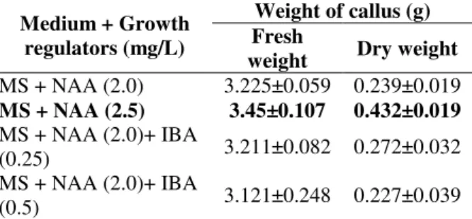
Table 1 - Fresh and dry weight of 4-weeks old calli derived from nodal explant on various concentrations of NAA alone or in combination with IBA supplemented to MS medium in Dioscorea alata.
Medium + Growth regulators (mg/L)
Weight of callus (g) Fresh
weight Dry weight
MS + NAA (2.0) 3.225±0.059 0.239±0.019
MS + NAA (2.5) 3.45±0.107 0.432±0.019
MS + NAA (2.0)+ IBA
(0.25) 3.211±0.082 0.272±0.032 MS + NAA (2.0)+ IBA
(0.5) 3.121±0.248 0.227±0.039
Table 2 - Fresh and dry weight of 4-weeks old calli derived from nodal explant on various concentrations of 2, 4-D supplemented to MS and RT media in Dioscorea deltoidea.
Medium + Growth regulators (mg/L)
Weight of callus (g) Fresh weight Dry weight MS + 2, 4-D (2.0) 2.792±0.159 0.368±0.121
MS + 2, 4-D (3.0) 2.812±0.097 0.298±0.106 RT + 2, 4-D (2.0) 2.567±0.136 0.286±0.217 RT + 2, 4-D (3.0) 2.638±0.167 0.307±0.067
Data represent means of three replicates.
Differentiation process is very complex in plant tissue and controlled by many factors, such as type and concentration of hormones, nutrient medium
and developmental stage of explant during in vitro
culture (Kumar 2011). The callus formed from nodal explants (based on fresh and dry weight) was selected for further studies of differentiation. Roots were regenerated (Fig.
2) on MS medium supplemented with NAA (2.0 mg/L) and IBA (0.5 mg/L) while shoots (Fig. 3) were formed on MS medium containing BAP (2.0
mg/L) and NAA (0.5 mg/L) in Dioscorea alata.
RT medium having IAA (1.0 mg/L) was used for regeneration of roots (Fig. 5) and RT medium supplemented with BAP (1.0 mg/L) and NAA (0.5 mg/L) was used for shoots (Fig. 6) in Dioscorea deltoidea. Differentiation was dependent on the synergistic effect of auxin along with cytokinins in the medium, which evoked good results for indirect shoot proliferation. Such variability in the regeneration from the callus has also been reported in Dioscorea alata (Belarmino et al. 1991),
Dioscorea esculenta (Nair and Chandrababu
1996), Chlorophytum borivilianum (Singh et al.
2006), Cardiospermum halicacabum (Kumar et al.
2008), and Dioscorea spp. (Asha and Nair 2010).
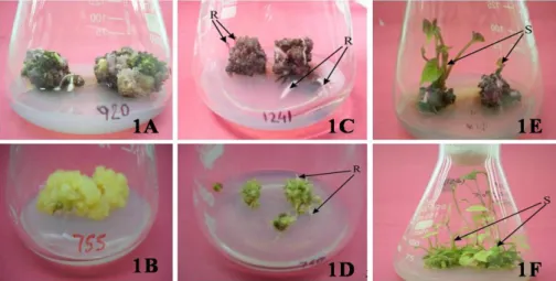
Figure 1 - Callus growth from nodal explants on MS medium supplemented with NAA (2.5 mg/L) in Dioscorea alata (1A) and 2, 4-D (2 mg/L) in D. deltoidea (1B); root differentiation (1C) in D. alata [MS medium + NAA (2.0 mg/L) + IBA (0.5 mg/L)] and (1D) in D. deltoidea [RT medium + IAA (1.0 mg/L)]; shoot differentiation (1E) in D. alata [MS medium + BAP (2.0 mg/L) + NAA (0.5 mg/L)] and in D. deltoidea (1F) [RT medium + BAP (1.0 mg/L) + NAA (0.5 mg/L)].
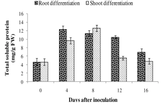
Total soluble protein content
In root differentiating calli of D. alata, protein
content was increased upto 4th day, Thereafter, the
protein content decreased till 16th day, while in case of shoot differentiating calli, the content
increased gradually upto 8th day but a sharp
decrease was observed by 12th day. Thereafter, the
protein content further decreased upto 16th day (Fig. 2). In D. deltoidea, the protein content decreased till the initiation of root and shoot (Fig.
3). After that, the content increased, which showed that the cells were quantitatively changing their
activities to synthesize certain amino
Table 3 - Effect of growth regulators supplemented to MS medium on nodal explants derived callus from D. alata for differentiation (observations recorded upto 6-weeks after inoculation).
Medium + growth regulator (mg/L)
Visual growth of callus after 4-weeks *
Colour and Texture of callus
Differentiation (Days required )
Control ― Brown, compact --
2, 4-D (0.5) + Yellow, fragile --
(1.0) + Yellow, fragile --
NAA (0.5) + Black, compact --
(1.0) + Black, compact --
(2.0) ++ Brown, compact --
(2.5) +++ Pinkish brown, compact --
(3.0) ++ Brown, compact --
NAA (2.0)
+ IBA (0.1) + Brown, compact --
(0.5) +++ Pinkish brown, compact Roots (15)
(1.0) ++ Brown, compact Roots (34)
NAA (2.5)
+ 2, 4-D (1.0) ++ Brown, compact --
(2.0) +++ Brown, compact Roots (28) & Shoots (31)
(3.0) ++ Brown, compact --
BAP (2.0)
+ NAA (0.5) +++ Pinkish brown, compact Shoots (16)
(1.0) +++ Pinkish brown, compact --
(1.5) ++ Green, fragile Abnormal shoots (34)
(2.0) + Black, compact --
BAP (2.0)
+ IBA (0.2) + Green, fragile Shoots (42)
(1.0) ++ Green, fragile Shoots (36)
*– No callus, + Poor callus, ++ Moderate callus, +++ Good callus. Data represent means of three replicates
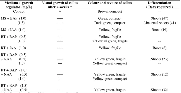
Table 4 - Effect of growth regulators supplemented to MS and RT medium on nodal callus for differentiation (observations recorded upto 6 weeks after inoculation).
Medium + growth regulator (mg/L)
Visual growth of callus after 4-weeks *
Colour and texture of callus Differentiation ( Days required )
Control + Brown, compact --
MS + BAP (1.0) +++ Green, compact Shoots (47)
(1.5) +++ Dark green, compact Abnormal shoots (41)
MS + IAA (1.0) ++ Yellow, fragile Roots (19)
RT + BAP (0.5) ++ Yellow, fragile --
(1.0) +++ Yellowish green, fragile --
RT + IAA (1.0) +++ Yellow, fragile Roots (8)
RT + BAP (0.5)
+ NAA (0.5) +++ Yellow green, fragile Shoots (23)
(1.0) ++ Yellow green, compact --
RT + BAP (1.0)
+ NAA (0.5) +++ Yellow green, fragile Shoots (12)
(1.0) ++ Yellow green, compact --
RT + BAP (1.5)
+ NAA (0.5) +++ Yellow green, fragile Shoots (32)
0 2 4 6 8 10 12 14 16
0 4 8 12 16
To ta l s o lu b le p r o te in (m g /g F W)
Days after inoculation
Root differentiation Shoot differentiation
Figure 2 - Total soluble protein content in callus of Dioscorea alata prior to inoculation (0 day) and 4, 8, 12and 16th days of inoculation on rooting and shooting media (Bar represent SE of the mean of 3 replicates).
0 5 10 15 20 25
0 2D-3D 4D-6D 6D-9D 8D-12D
To ta l so lu b le p r o te in s (m g /g F W)
Days after inoculation
Root differentiation Shoot differentiation
Figure 3 - Total soluble protein content in callus of Dioscorea deltoidea prior to inoculation (0 day) and on 2, 4, 6 and 8th days of inoculation on rooting and on 3, 6, 9 and 12th days of inoculation on shooting media (Bar represent SE of the mean of 3 replicates).
Protein profile resolution (SDS-PAGE)
Resolution of the polypeptides by SDS-PAGE (10%) in the present study exhibited several differences during the differentiation of root and shoot from the calli in D. alata and D. deltoidea.
Dioscorea alata
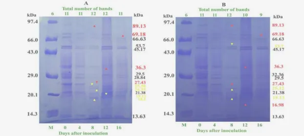
The protein profile pattern using SDS-PAGE (10%) showed differential expression of the proteins during root differentiation (Fig. 4A). Undifferentiated callus used as control on MS medium supplemented with NAA (2.5 mg/L) showed a total number of 11 polypeptides of molecular weight, ranging from 89.13 to 13.63 kDa. The banding pattern from the callus remained unchanged upto 4th day when it was transferred on rooting medium [NAA (2.0 mg/L) + IBA (0.5
mg/L)]. On 8th day, three new bands of molecular
weight 25.56, 24.35 and 18.2 kDa appeared, while two polypeptide bands of MW 89.13 and 27.43 kDa disappeared.
However, on 12th day, the number of bands
remained unchanged though there was appearance of one new band of MW 19.13 kDa and disappearance of one band of 36.3 kDa. Thus, a
total of 12 bands were observed on 8 and 12th day
after inoculation. On 16th day, one polypeptide band of MW 69.18 kDa disappeared (Fig. 4A). Similarly, the numbers of bands remained unchanged, i.e., same as control (11 ranging from 89.13 to 13.63 kDa) up to 4th day of inoculation on shoot differentiation medium [MS + BAP (2.0 mg/L) + NAA (0.5 mg/L)]. However, on 8th day on shoot differentiation medium, there was appearance of three new polypeptide bands of MW 53.7, 25.12 and 19.13 kDa, whereas two polypeptides of 89.13 and 27.43 kDa disappeared.
On 12th day, disappearance of two bands out of 12
polypeptide bands of MW 36.3 and 16.98 kDa was
observed, while on 16th day, one more polypeptide
of MW 69.18 kDa disappeared (Fig. 4B).
The comparison of protein profile of the root and shoot differentiating calli indicated that there was synthesis of four root specific polypeptides (25.56, 24.35, 19.13 and 18.2 kDa) and three shoot specific polypeptides (53.7, 25.12 and 19.13 kDa) during the differentiation. The synthesis of common polypeptide of MW 19.13 kDa observed during the differentiation of root and shoot was of special interest. This suggested that the differentiation process was dependent on the concentration and combination of hormones. The timing of the synthesis of particular polypeptides coincides with the protein content during the differentiation associating their involvement in differentiation process.
Dioscorea deltoidea
The undifferentiated calli used as control on MS medium + 2, 4-D (2.0 mg/L) showed a total number of nine bands with molecular weight ranging from 85.11 to 14.69 kDa. When transferred on the rooting medium [RT + IAA (1.0 mg/L)], three new polypeptide bands of MW 58.7, 47.86 and 24.84
kDa appeared on 2nd day after inoculation. Likewise
on 4th day, two new polypeptide bands of 93.35 and
number and quality of bands remain unchanged (Fig. 5A).
Before transferring the callus on the shooting medium [BAP (1.0 mg/L) + NAA (0.5 mg/L)], the total numbers of polypeptide bands were 12 but on
3rd day, five new polypeptide bands of MW 93.35,
58.7, 47.86, 23.38 and 16.98 kDa appeared.
However, on 6th day, one polypeptide band of 67.66
kDa disappeared. However, on 9th day with the appearance of green patches in the callus, one more
polypeptide band of 42.69 kDa disappeared. On 12th
day with the visual appearance of shoots, four polypeptide bands of MW 36.31, 33.9 and 31.69 kDa disappeared and total number of bands remained 11 (Fig. 5B).
Figure 4 - Protein profile resolution in callus cultures of Dioscorea alata through SDS-PAGE (10%) (A) on rooting medium and (B) on shooting medium. (M, Protein molecular weight marker; yellow arrow indicates appearance of new polypeptide and red arrow indicates disappearance of polypeptide).
The comparison of polypeptide profile of root and shoot differentiating calli showed that four polypeptide bands (MW 93.35, 58.7, 47.86 and 24.84 kDa) appeared during differentiation of root and shoot. However, it was of interest to note that appearance of two root specifc polypeptide bands (MW 33.9 and 31.69 kDa) and one shoot specific polypeptide band (MW 16.98 kDa) was also observed during differentiation. Synthesis of new specific polypeptides of 56 kDa in Arachis hypogaea (Venkatachalam et al. 1997), 61.7 kDa in Chlorophytum borivilianum (Goyal et al. 2009)
and of 85.1 kDa in Cardiospermum halicacabum
(Kumar and Goyal 2010) has been reported during rhizogenesis and polypeptide bands of 97 kDa in
Arachis hypogaea specific to shoot formation (Venkatachalam et al. 1997), 75 kDa in Centella asiatica (Agrawal and Subhan 2003), 117.5 and
112.2 kDa in Chlorophytum borivilianum (Goyal
et al. 2009) and 102.3 and 36.31 kDa in
Cardiospermum halicacabum (Kumar et al. 2013)
have been reported. This suggested that
polypeptide expression varied depending upon the different developmental stages and the differential gene expression of the concerned structural or
regulatory gene(s). Polypeptides could be
potentially used as marker to decipher the
differentiation pathway and selection of
organogenic potential callus or tissue.
CONCLUSIONS
SDS-PAGE studies revealed that there was synthesis of four root specific polypeptides (MW 25.56, 24.35, 19.13 and 18.2 kDa) and three shoot specific polypeptides (MW 53.7, 25.12 and 19.13 KDa) during the differentiation in D. alata. In case of D. deltoidea, four polypeptide bands (MW 93.35, 58.7, 47.86 and 24.84 kDa) appeared during the differentiation of the root and shoot. Appearance of two root specific polypeptide bands (MW 33.9 and 31.69 kDa) and one shoot specific polypeptide band (MW 16.98 kDa) was also observed during the differentiation. The synthesis of common polypeptide of MW 19.13 kDa observed during the differentiation of root and shoot was of special interest. In summary, expression of root/shoot specific polypeptides could be used as markers to characterize the differentiation pathway and to augment the selection of regenerating potential callus for rapid
in vitro propagation.
ACKNOWLEDGEMENT
The authors are thankful to the Head, Department of Botany and Plant Physiology for providing the required research facilities. The National Testing Service Doctoral Fellowship provided by the CIIL, Mysore, Ministry of HRD, GOI to the senior author is duly acknowledged.
REFERENCES
Agrawal V, Subhan S. In vitro plant regeneration and protein profile analysis in Centella asiatica (Linn.) Urban: a medicinal plant. Plant Cell Biotech Mol Biol. 2003; 4: 83-90.
Asha KI, Nair GM. Standardization of callus induction and shoot regeneration in twelve species of Dioscorea. Indian J Plant Genet Resour. 2009; 22(3): 270-280.
Belarmino MM, Gonzales JR. Somatic embryogenesis and plant regeneration in purple food yam (Dioscorea alata L.). Ann Trop Res. 2008; 30(2): 22-33.
Belarmino M, Rosario del AG, Del Rosario AG. Callus induction and organogenesis in Dioscorea species. Jap J Breeding 1991; 41(4): 561-569.
Bradford MM. A rapid and sensitive method for the quantification of microgram quantities of protein utilizing the principle of protein-dye binding. Anal Biochem. 1976; 72: 248-254.
Chandel KPS, Shukla G, Sharma N. Biodiversity in Medicinal Plants in India: Conservation and Utilization. 1996; pp. 1-239, NBPGR, New Delhi. Dahlgren RMT, Clifford HT, Yeo PF. The families of
the monocotyledons, structure, evolution and taxonomy. 1985; Springer, Berlin Heidelberg, New York, Tokyo.
Furmanowa M, Guzewska J. Dioscorea: in vitro culture and the micropropagation of diosgenin containing species. In: Bajaj, Y.P.S. (ed.) Biotechnology in agriculture and forestry, Vol.7, Medicinal and aromatic plants II. Springer, Berlin Heidelberg, New York, Tokyo. 1989; pp. 162-183.
Goyal SC, Singh R, Jain V. Differential expression of specific proteins during in vitro organogenesis in Chlorophytum borivilianum Sant. Et Fernand. J Ind Bot Soc. 2009; 88 (3&4): 111-115.
Jeyaseelan M, Rao MV. Biochemical studies of embryogenic and non-embryogenic callus of Cardiospermum halicacabum L. Ind J Exp Biol. 2005; 43: 555-560.
Kumar A. Morphogenetic and Biochemical Studies in Callus Cultures of Dioscorea Species. Ph.D. thesis, 2011; CCS HAU, Hisar.
Kumar A, Goyal SC. Biochemical changes during organogenesis in callus cultures of Ballon Vine (Cardiospermum halicacabum L.). 2010; Lambert Academic Publishing GmbH & Co. KG, Germany. Kumar A, Dhingra HR, Goyal SC. Protein profile
resolution during in vitro organogenesis in balloon vine (Cardiospermum halicacabum L.). J Ind Bot Soc. 2013; 92 (1 & 2): 103-108.
Kumar A, Goyal SC, Kajla S, Sharma N. Rapid protocol for callus induction and differentiation of roots and shoots in Dioscorea alata-a medicinal plant. Ind J Agric Sci 2014; 84(1): 107-111.
Laemmli UK. Cleavage of structural proteins during the assembly of the head of the bacteriophage Ty. Nature. 1970; 277: 680-685.
Magdoleen G, Osman E, Elhadi A, Khalafalla MM. Callus formation and organogenesis of tomato (Lycopersicon esculentum Mill, C.V. Omdurman) induced by thidiazuron. Af J Biotech. 2010; 9 (28): 4407-4413.
Mhatre M, Rao PS, Bapat VA. Electrophoretic analysis of sandal wood (Santalum album L.) proteins during morphogenesis. Ind J Exp Biol. 1991; 29: 1150-1151. Mohapatra HP, Rath SP. In vitro studies of Bacopa monnieri - An important medicinal plant with reference to its biochemical variations. Ind J Exp Biol. 2005; 43: 373-376.
Murashige T, Skoog F. A revised medium for rapid growth and bioassay with tobacco tissue cultures. Physiol Plant. 1962; 15: 473-497.
Nair NG, Chandrababu S. In vitro production and micropropagation of three species of edible yams. In: Kurup GT, Palaniswamy MS, Potty VP, Padmaja G, Kabeerathumma S and Pillai VS (eds.). Tropical Tuber Crops: Problems, Prospects and Future Strategies. Oxford & IBH, New Delhi, 1996; p 55-60. Singh R, Dhingra HR, Goyal SC. Biochemical changes during shoot differentiation in callus cultures of Chlorophytum borivilianum Sant. Et Fernand. Ind J Plant Physiol. 2006; 11 (2): 130-135.
Sundar AN, Jawahar M. In vitro plant regeneration from leaf and stem explants of Solanum xanthocarpum Schard & Wendl. – an important medicinal herb. J Agric Tech. 2011; 7 (2): 301-306. Van Stadin J, Fowlds DL. Micropropagation of
medicinal Dioscorea species. In: Bajaj YPS (ed.) Biotechnology in agriculture and forestry high tech and micropropagation 111. Springer Berlin Heidelberg, New York 1992; 19: 425-442.
Venkatachalam P, Geetha N, Jayabalan N. In vitro regeneration from immature cotyledon explant and protein profile changes during organogenesis in groundnut (Arachis hypogaea L.). Trop Agri (Trinidad) 1997; 74: 140-145.