Tânia Sofia dos Santos Vieira
Mestre em Ciências Biomédicas
Development of a new nanostructured
scaffold for neural stem/progenitor cell
transplantation
Dissertação para a obtenção do grau de Doutor em
Bioengineering Systems
–
MIT Portugal Program
Orientador: Dr Célia Henriques, Profª auxiliar, FCT-UNL
Co-orientador: Dr João Paulo Borges, Prof auxiliar, FCT-UNL
Co-orientador: Dr Ana Sofia Falcão, Pos-doc, CEDOC
Júri:
Presidente: Prof.Doutor Luís Paulo da Silva Nieto Marques Rebelo
Arguentes: Profª. Doutora Maria Helena Mendes Gil
Doutor Hugo Agostinho Machado Fernandes
Vogais: Prof. Doutor António Alfredo Coelho Jacinto
Profª. Doutora Maria Helena Figueiredo Godinho
Prof. Doutor Frederico Castelo Ferreira
Prof. Doutora Célia Maria Reis Henriques
Doutora Ana Paula Gomes Moreira Pêgo
iii
Development of a new nanostructured scaffold for
neural stem/progenitor cell transplantation
Copyright © Tânia Sofia dos Santos Vieira, Faculdade de Ciências e Tecnologia - Universidade Nova de Lisboa.
v
First of all I want to express my gratitude to my thesis advisors, professor Célia Henriques, professor João Paulo Borges and Ana Sofia Falcão for the presentation of this fascinating project and their effort to ensure all the necessary conditions for their development. I also thank their guidance, encouragement and time during these years. I would like to thank to Professor Jorge Silva for the availability and teaching during the development of this project.
I appreciate the financial support from Fundação para a Ciência e Tecnologia – FCT that funded the PhD grant - SFRH/BD/90682/2012.
I also want to thank the members of the thesis committee, professor Helena Godinho and professor Domingos Henrique for their suggestions throughout the development of the work.
I would like to thank to Professor Elvira Fortunato for the opportunity to use the CENIMAT/i3N research facilities: thanks to Ana Pimentel and Alexandra Gonçalves for DSC/TGA acquisition; Joana Pinto for XRD acquisition and Daniela Gomes for SEM acquisition.
I would like to express my gratitude to Doctor Cecília Bonifácio, from REQUIMTE – FCT/UNL, for NMR acquisition and to Professor Ana Rego, from IST, for the XPS acquisition and for their guidance and help in the interpretation of the results.
I am especially grateful to all the GREAT LAB students that provided moments of fun and laughs and helped to overcome the difficulties. However, I am indebted to Luisa Fialho, Ana Fradinho, Zeliha Güler, Carolina Rufino and Ana Nogueira for their friendship and support beyond the lab.
In the Polymers lab, I’d like to thank to Paula Soares, Susete Fernandes and Coro Echeverria for scientific support and advices, you were always available to help. Augusta thanks for your affection and happy guffaws. Ana Almeida thanks for the help in contact angle measurements. I´d like to thank to all the colleagues and students that passed the Polymers Lab for their contribution to a great lab atmosphere. Special thanks to Mariana Amaro for all the support, encouragement and friendship.
vi
and Marta Costa, for learning, support and happy moments during the first year of the PhD program.
vii
Abstract
Tissue engineering investigates new therapeutic approaches for spinal cord regeneration. Biodegradable scaffolds are employed aiming at creating an appropriate environment to support cell regrowth and transplantation. The transplantation of neural stem/progenitor cells (NSPCs) is a promising strategy under investigation. The main objective of this work was the synthesis of new soft materials for the production of nanostructured scaffolds able to support NSPCs transplantation and enable spinal cord regeneration.
Polyurethanes (PUs) are segmented polymers, with tunable properties. PUs were synthesized using polycaprolactone-diol (PCL-diol) as soft segment, and isophorone diisocyanate and dimethylol propionic acid (DMPA) as hard segment. To introduce biological cues in the polymer backbone, chitosan (CS) and gelatin (Gel) were used to substitute DMPA as chain extender. The PUs were characterized regarding their chemical composition and thermal properties.
Electrospun fibrous mats are convenient structures for cell support. In particular, aligned nanofibers provide a guidance cue to axon regrowth. Electrospinning was used to produce scaffolds of randomly oriented and aligned fibers from the different PU formulations. Scaffolds were characterized regarding their morphology, mechanical behavior, crystallinity, surface properties and hydrolytic degradation. Their impact on cells was evaluated in vitro using human
fibroblasts. Cell adhesion and proliferation was highest for scaffolds produced from PUs containing CS or Gel as the only chain extender.
Stem cell interaction with PU-CS and PU-Gel scaffolds was studied using human umbilical cord mesenchymal stem cells (MSCs) and human fetal spinal cord neural stem cells (NSCs). MSCs proliferated best on PU-Gel randomly oriented fibers whereas NSCs proliferated best on PU-CS with aligned fiber morphology. Neuronal differentiation of NSCs was confirmed using neuronal markers. Neurites aligned along the fibers direction.
The physical, chemical and biological properties of PU-CS and PU-Gel fibrous mats make them promising substrates for NSPC in order to promote neural regeneration.
ix
A engenharia de tecidos investiga novas abordagens para a regeneração da espinal medula. Estruturas biodegradáveis são usadas para criar um ambiente que suporte o crescimento e transplante de células. O transplante de células neurais estaminais/progenitoras (NSPCs) é uma estratégia promissora sob investigação. O principal objetivo deste trabalho foi a síntese de novos materiais macios para produzir matrizes nano-estruturadas capazes de suportar o transplante de NSPCs e permitir a regeneração da espinal medula.
Poliuretanos (PUs) são polímeros segmentados, com propriedades ajustáveis. Foram sintetizados PUs utilizando policaprolactona-diol como segmento macio, e diisocianato de isoforona e ácido dimetilol propiónico (DMPA) como segmento duro. Para introduzir sítios para interação biológica na estrutura do polímero, foram utilizados quitosano (CS) e gelatina (Gel) substituindo o DMPA. Os PUs foram caracterizados química e termicamente.
Matrizes fibrosas eletrofiadas, são estruturas convenientes para o suporte celular. Em particular, nanofibras alinhadas guiam o crescimento dos axônios. Matrizes de fibras orientadas aleatoriamente e alinhadas dos diferentes Pus foram obtidas por eletrofiação e caracterizadas quanto à morfologia, comportamento mecânico, cristalinidade, propriedades de superfície e degradação hidrolítica. O seu impacto nas células foi avaliado in vitro utilizando
fibroblastos humanos. A adesão e proliferação celular foram mais elevadas nas matrizes de PUs contendo CS ou Gel como único extensor de cadeia.
A interação de células estaminais com matrizes de PU-CS e de PU-Gel foi estudada usando células humanas estaminais mesenquimais do cordão umbilical (MSCs) e células estaminais neurais da espinal medula fetal (NSCs). As MSCs proliferaram melhor nas fibras orientadas aleatoriamente de PU-Gel, enquanto as NSCs proliferaram melhor em matrizes de fibras alinhadas de PU-CS. A diferenciação neuronal das NSCs foi confirmada usando marcadores neuronais. As neurites alinharam ao longo das fibras.
As propriedades físicas, químicas e biológicas das matrizes de PU-CS e PU-Gel tornam-nas substratos promissores para NSPCs, na promoção da regeneração neural.
xi
Abstract ... vii
Resumo ... ix
List of Figures ... xv
List of Tables ... xix
List of Acronyms ... xxi
1. Introduction ... 2
2. Literature Review... 8
2.1 Spinal Cord Injury ... 8
2.1.1 Primary injury ... 8
2.1.2 Secondary injury ... 8
2.2 Limited spinal cord regeneration capacity ... 10
2.3 Therapeutic/regenerative strategies ... 11
2.3.1 Drugs ... 12
2.3.2 Stem cells therapy ... 13
2.3.3 Tissue engineering ... 14
2.4 Interaction of scaffolds with NSPCs ... 21
2.4.1 In vitro studies ... 21
2.4.2 In vivo studies ... 25
2.5 Role of scaffold topography in stem cell differentiation ... 26
2.5.1 Nano/micro-scale scaffolds ... 28
2.5.2 Self-assembly nanofibers ... 28
2.5.3 Phase separation fibrillar structures ... 30
2.5.4 Lithographic patterned substrates ... 30
2.5.5 Carbon-based nanomaterials ... 32
2.5.6 Electrospinning ... 32
2.6 References ... 45
3. Electrospun biodegradable chitosan based-poly(urethane urea) scaffolds for soft tissue engineering ... 60
xii
3.2.1 Depolimerization of chitosan and determination of molecular weight ... 62
3.2.2 Synthesis of Polyurethane extended with chitosan ... 63
3.2.3 Characterization of synthesized polyurethanes ... 63
3.2.4 Electrospinning and film casting ... 64
3.2.5 Physico-chemical characterization of fibrous mats and films ... 65
3.3 Results and Discussion ... 69
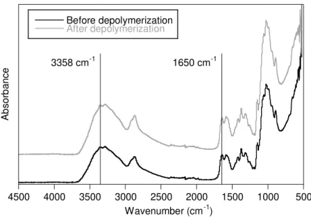
3.3.1 Depolymerization of Chitosan ... 69
3.3.2 Polyurethanes Characterization ... 70
3.3.3 Optimization of the electrospinning process ... 79
3.3.4 Characterization of the electrospun nanofibers ... 82
3.3.5 Aligned vs. random electrospun mats ... 97
3.4 Conclusions ... 104
3.5 References ... 104
4. A new biodegradable gelatin based-poly(ester urethane urea): synthesis, characterization and electrospun scaffolds for soft tissue engineering... 112
4.1 Introduction ... 112
4.2 Materials and methods ... 113
4.2.1 Synthesis of PU-Gel ... 113
4.2.2 Characterization:of PU-Gel ... 114
4.2.3 Electrospinning and film casting ... 114
4.2.4 Characterization of PU-Gel electrospun fibers ... 115
4.3 Results and discussion ... 117
4.3.1 PU-Gel Characterization... 117
4.3.2 Characterization of fiber mats ... 123
4.3.3 Random vs Aligned fibrous mats ... 131
4.4 Conclusion ... 135
4.5 References ... 135
5. Biocompatibility evaluation of electrospun mats from chitosan or gelatin based poly(urethane urea) ... 140
5.1 Introduction ... 140
xiii
5.2.2 Characterization of polyurethanes ... 142
5.2.3 Production of fibrous mats ... 142
5.2.4 Characterization of fibrous mats ... 142
5.2.5 Cell culture experiments ... 143
5.3 Results and discussion ... 146
5.3.1 PU-CS and PU-Gel characterization ... 146
5.3.2 Fibrous mats characterization ... 147
5.3.3 Proliferation of 3T3 fibroblasts ... 151
5.3.4 MSCs adhesion and proliferation on fibrous mats ... 152
5.3.5 NSCs proliferation on the fibrous mats ... 156
5.4 Conclusion ... 161
5.5 References ... 162
6. Conclusions and Future Work ... 166
6.1 Conclusions ... 166
6.2 Future Work ... 169
xv Chapter 2
Figure 2.1 – Pathophysiological events occurring after SCI, including the primary, secondary
and chronic phases. ... 10
Figure 2.2 – Constituents and route of production of PUs. ... 18
Figure 2.3 – Cells mechanosensors are stimulated by external mechanical forces. ... 27
Figure 2.4 – Scheme of the electrospinning setup. ... 34
Chapter 3 Figure 3.1 – IR spectra of CS before and after depolymerization. ... 70
Figure 3.2 – Chemical structure of PU-DMPA/CS showing the urea bond between IPDI and the amine group of CS. ... 71
Figure 3.3 –1H NMR spectra of PCL-diol and IPDI used in the chemical synthesis of PUs. ... 72
Figure 3.4 –1H NMR spectra of the synthetized PUs. ... 72
Figure 3.5– FTIR spectra of the precursors and the intermediate pre-polymer to reach PU-DMPA/CS. ... 74
Figure 3.6– FTIR spectra of the synthetized PUs. ... 74
Figure 3.7 – Carboxyl region (1600 – 1800 cm-1) of PU-CS IR spectrum: Absorbance spectrum (A); second-derivative spectrum (B). ... 75
Figure 3.8– ATR-FTIR spectra in the carbonyl group stretching and the deconvoluted curves of (A) PU-DMPA, (B) PU-DMPA/CS and (C) PU-CS. ... 76
Figure 3.9– ATR-FTIR spectra in the amine group stretching and the deconvoluted curves of(A) PU-DMPA, (B) PU-DMPA/CS and (C) PU-CS. ... 77
Figure 3.10 – Mass losses (A) and the corresponding derivatives (B) vs. temperature of PCL-diol, CS, PU-DMPA, PU-DMPA/CS and PU-CS. ... 78
Figure 3.11 – DSC curves of PCL-diol, CS and PUs with an increasing CS content substituting DMPA as chain extender. ... 79
Figure 3.12 – SEM images of the electrospun fibers produced from 20% PU-DMPA/CS solution with THF:DMF at different ratios. ... 81
Figure 3.13– SEM images of the electrospun fibers produced from PU-DMPA/CS solution at different concentrations with 50:50 THF:DMF solvent system. ... 81
xvi
PU-DMPA, PUU-DMPA/CS and PU-CS. ... 84
Figure 3.16– Hyteresis loops after 10 cycles stretching and recovering of electrospun fibrous mats. ... 85
Figure 3.17 – X-Ray diffractograms of PCL-diol, CS, and films and fiber mats from PU-DMPA, PU-DMPA/CS and PU-CS. ... 86
Figure 3.18– Fitting of the characteristic peaks of the PU-DMPA/CS fibrous mat diffractogram with Voigt functions (red) and a cubic background (green). ... 87
Figure 3.19 – The C1s, N1s and O1s XPS spectra and the respective fitted peaks for PU-CS films (top line) and fibrous mats (botoom line) at 0º take-off angle.. ... 88
Figure 3.20– Water contact angle values for the PUs films and mats and the respective water drop images. ... 91
Figure 3.21 – Hydrolitic degradation of PU films (F_) and fibrous mats (M_) produced from the synthetized PUs immersed in PBS (A) and in lipase solution (B). ... 92
Figure 3.22 – FTIR spectra of PU-DMPA, PU-DMPA/CS and PU-CS films and fibrous mats after degradation in PBS and in lipase solution. ... 93
Figure 3.23– Results of HFFF2 cells’ viability, obtained in a cytotoxicity assessment of PU -DMPA, PU-DMPA/CS and PU-CS (A) films and (B) electrospun mats. ... 94
Figure 3.24– Optical microscope images of the HFFF2 cells seeded in 96 well plate in contact with pure extracts ... 94
Figure 3.25 – HFFF2 cell population. ... 96
Figure 3.26– Fluorescent images of the cells stained with phalloidin (red) and DAPI (blue). .. 97
Figure 3.27 – SEM images of randomly oriented electrospun fibrous mats (column 1) from PU-DMPA (A), PU-PU-DMPA/CS (B) and PU-CS (C), and the respective histograms of the fiber diameter (column 2) and the angular distribution (column 3). ... 99
Figure 3.28– SEM images of aligned electrospun fibrous matrices (column 1) from PU-DMPA (A), PU-DMPA/CS (B) and PU-CS (C), and the respective histograms of the diameter (column 2) and the angular distribution (column 3). ... 100
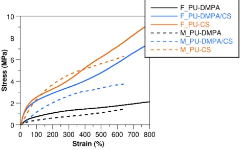
Figure 3.29– Stress-strain curves of the electrospun random and aligned fibrous matrices produced from PU-DMPA, PU-DMPA/CS and PU-CS (A). ... 101
Figure 3.30– Water contact angle values of the PU-DMPA, PU-DMPA/CS and PU-CS electrospun fibrous mats with random and aligned morphology after 1 min recording time. .. 102
Figure 3.31 – Viability assay of HFFF2 cells ... 103
xvii
Figure 4.1– Synthesis route of polyurethane based gelatin. ... 114
Figure 4.2–1H NMR spectra of PU-Gel 1.0 g and its precursors in the range between 0.5 ppm
to 5.0 ppm. ... 118
Figure 4.3– FTIR spectra of PUs synthetized with different amounts of gelatin and their constituents. ... 119
Figure 4.4– C=O stretching band analysis for PU-Gel with different gelatin contents: (A) 5%; (B) 7.5%; (C) 10%. ... 120
Figure 4.5 – N-H stretching band analysis for PU-Gel with different gelatin contents: (A) 5%; (B) 7.5%; (C) 10%. ... 121
Figure 4.6– DSC thermograms of PUs synthesized with different amounts of gelatin. ... 122
Figure 4.7 – Thermal analysis of PU-Gel synthesized with different amounts of gelatin: (A) thermogravimetric analysis spectra; (B) Derivative weight loss curves. ... 123
Figure 4.8 – SEM images of the fibrous mats ... 124
Figure 4.9 – SEM images of the fibrous mats produced from PU-Gel-5... 125
Figure 4.10 – Typical stress-strain curves of PU-Gel films (F_PU-Gel) and fiber mats (M_PU-Gel) (A) and the respective hysteresis loops (B and C) after 10 cycles stretching and recovery. ... 126
Figure 4.11 – X-ray diffractograms of PU-Gel film and fibrous mat (A).. ... 127
Figure 4.12 – Water contact angle values of the PU-Gel films and electrospun fibrous mats and the respective water drop images. ... 128
Figure 4.13 – Degradation profile of the PU-Gel films (A) and fibrous mats (B) in PBS, lipase and trypsin. ... 129
Figure 4.14 – Cytotoxicity assessment of HFFF2 cells cultured with extracts from PU-Gel films and mats at concentrations of 15, 10, and 5 mg/mL. ... 130
Figure 4.15 – Optical microscope images of the HFFF2 cells seeded in 96 well plate in contact with pure extracts. ... 130
Figure 4.16 – (A) Proliferation of HFFF2 cells ... 131
Figure 4.17 – SEM images of random (A) and aligned (D) PU-Gel fibrous mats, and the respective histograms of the fiber diameter distribution (B and E) and the angle distribution (C and F). ... 132
Figure 4.18 – Stress-strain curves of the random (R_) and aligned (A_) PU-Gel fibrous mats. ... 133
xviii
with random and aligned morphology ... 134
Chapter 5
Figure 5.1 – FTIR spectra of PU synthetized with gelatin or chitosan as chain extenders. .... 146
Figure 5.2 – SEM images of the PU-CS (1A, 3A) and PU-Gel (2A, 4A) fibrous mats with random (1A, 2A) and aligned (3A, 4A) morphology. (B) Histogram of the fiber diameter distribution on the mats. (C) Histogram with the angular distribution and the pixel intensity (from FFT analysis) with the acquisition angle for the produced mats. ... 148
Figure 5.3 – Typical stress-strain curves of random and aligned PU-CS and PU-Gel fibrous mats under (A) dry and (B) wet conditions. ... 150
Figure 5.4 – Water contact angle values of the electrospun random and aligned CS and PU-Gel mats and the representative picture of the water drop on the mats’ surface. ... 151
Figure 5.5 – Resazurin proliferation assay of 3T3 fibroblasts seeded on the electrospun PU-CS and PU-Gel random and aligned fibrous mats. ... 152
Figure 5.6 – The average values of MSCs number (A) and growth area (B) seeded on the electrospun fibrous mats during 4 h in the presence and the absence of PL in culture medium ... 153
Figure 5.7 – Fluorescent images of immunofluorescent staining for cytoskeleton (phalloidin, red) and cell nuclei (DAPI, blue) of MSCs seeded on electrospun fibrous mats ... 155
Figure 5.8 - Microscopic fluorescent images of NSCs seeded on electrospun nanofibrous mats from R_PU-CS (a, e), A_PU-CS (b, f), R_PU-Gel (c, g) and A_PU-Gel (d, h) ... 157
Figure 5.9 – Scanning electron microscopy images of NSCs seeded on electrospun nanofibrous mats ... 158
Figure 5.10 – Laser scanning confocal images of NF70 (red) and DAPI (blue) (A – E) and MAP2 (red) and DAPI (blue) (F – J) stained NSCs seeded on electrospun nanofibrous mats. ... 159
xix
List of Tables
Chapter 2
Table 2.1 – Interaction of 3D scaffolds from different polymers with NSPCs from different
sources. ... 23
Table 2.2 – Effects of the electrospun nanofibers on nerve cells. ... 38
Table 2.3 – Effects of the nanofibers alignment and diameter on the NSCs behavior. ... 41
Table 2.4 – Effects of the nanofibers functionalization on the NSCs behavior. ... 43
Table 2.5 - Effects of the nanofibers conductivity on the NSCs behavior. ... 44
Chapter 3 Table 3.1– Electrospinning set-up parameters used in the production of fibrous mats from synthesized PUs. ... 65
Table 3.2 – Wavenumber (v) and relative area (A) of the 5 components of the C=O stretching band and the percentage of carbonyl hydrogen bonded. ... 76
Table 3.3 – Wavenumber (v) and relative area (A) of the 3 components of the N-H stretching band and the percentage of amine hydrogen bonded. ... 77
Table 3.4– Thermal analysis data of the synthetized polyurethanes ... 79
Table 3.5– Results from XRD and tensile tests of films and fibrous mats.. ... 87
Table 3.6 – XPS atomic percentage composition of different PU films and fibers surfaces. .... 89
Table 3.7–Adhesion ratio of HFFF2 cells to films (F_) and fiber mats (M_). ... 96
Chapter 4 Table 4.1– Wavenumber (v) and relative area (A) of the 5 components of the C=O stretching band and the percentage of carbonyl hydrogen bonded. ... 120
Table 4.2– Frequency (v) and relative area (A) of the 4 components of the N-H stretching band and the percentage of amine hydrogen bonded. ... 121
Table 4.3– Thermal analysis data of PU-Gel. From DSC:. ... 122
Table 4.4 – Results from XRD and tensile tests of PU-Gel films and fibrous mats.. ... 127
xxi
3D Three dimensional space
3T3 3-day transfer, inoculum 3×10⁵ cells
AFM Atomic force microscopy ATR Attenuated total reflectance
ATP Adenosine triphosphate
BD Butanediol
BDNF Brain-derived neurotrophic factor BMP4 Bone morphogenic protein bFGF Basic fibroblast growth factor cAMP Cyclic adenosine monophosphate CH3COOH Acetic acid
CH3COONa Sodium acetate
ChABC Chondroitinase ABC
CNS Central nervous system
CNTF Ciliary neurotrophic factor
CNTs Carbon nanotubes
CS Chitosan
CSPGs Chondroitin sulfate proteoglycans
DAPI 4,6-Diamidino-2-Phenylindole dihydrochloride
DD Degree of deacetylation
DHD 2,5-dimethyl-3-hexine-2,5-diol
DMAc Dimethylacetamide
DMEM Dulbecco’s modified Eagle’s medium
DMF N,N-dimethylformamide
DMPA Dimethylol proprionic acid DOPA 3,4-diihydroxy-L-phenylalanine DSC Differential scanning calorimetry DTG Derivative thermo-gravimetric ECM Extracellular matrix
EDTA Ethylenediaminetetraacetic acid EGF Epidermal growth factor
EGFR Epidermal growth factor receptor eFGF Epidermal fibroblast growth factor ESCs Embryonic stem cells
FAK Focal adhesion kinase
FBS Fetal bovine serum
xxii
FTIR Fourier transform infrared spectroscopy FWHM Full width at half maximum
GDNF Glial cell line-derived neurotrophic factor GPC Gel permeation chromatography HFFF2 Caucasian foetal foreskin fibroblasts HFP 1,1,1,3,3,3-hexafluoro-2-propanol
1H NMR Proton nuclear magnetic resonance
HPSG Heparan Sulfate proteoglycan
HS Hard segments
IgG Immunoglobulin G
IPDI Isophorone diisocyanate
IKVAV Ile-Lys-Val-Ala.Val amino acid sequence iPSCs Induced pluripotent stem cells
LDH Lactate dehydrogenase
MAP2 Microtubule-associated protein 2 MAPK Mitogen-activated protein kinase MDI Methyl di-p-phenyl diisocyanate MEM Alpha-minimum essential medium MIDE 2,2’-(methylimino) diethanol MSCs Mesenchymal stem cells NaHSO3 Sodium bisulfite
NaNO2 Sodium nitrite
NaOH Sodium hydroxide
NF70 70 kDa Neurofilament
NGF Nerve Growth factor
NMP N-methylpyrrolidone
Nogo-A Neurite outgrowth inhibitor NSCs Neural stem cells
NSPCs Neural stem/progenitor cells
NT-3 Neurothophin-3
OHT 4-hydroxytamoxifen
PBS Phosphate buffer saline
PCL Polycaprolactone
PCL-diol Polycaprolactone-diol
PDL Poly-D-lysine
PDMS Poly(dimethylsiloxane)
PEDOT Poly(3,4-ethylenedioxythiophene)
PEG Polyethylene glycol
xxiii
PHPMA Poly[N-2-(hydroxypropyl) methacrylamide]
PL Platelet lysate
PLA Poly(lactic acid)
PLCL Poly[(L-lactide)-co-(Ɛ-caprolactone)]
PLGA Poly(lactic-co-glycolic acid)
PLL Poly-L-lysine
PLLA Poly(L-lactic acid)
PU-CS Polyurethane extended with chitosan
PU-CS/DMPA Polyurethane extended with dimethylol proprionic acid and chitosan PU-DMPA Polyurethane extended with dimethylol proprionic acid
PU-Gel Polyurethane extended with gelatin
PUs Polyurethanes
RADA-16 Ac-(Asp-Ala-Asp-Ala)4-CONH2 peptide
REST RE-1 silencing transcriptional factor SCI Spinal cord Injury
SEM Scanning electron microscopy SiRNA Small interference ribonucleic acid
SPC-01 Conditionally immortalized neural stem cell line derived from human fetal spinal cord tissue
SS Soft segments
STEP Spinneret based tunable engineered parameters TCP Tissue culture plate
TEA Triethylamine
TGA Thermogravimetric analysis
THF Tetrahydrophuran
TMS Tetramethylsilane
TrkC Tropomyosin receptor kinase C
UV Ultraviolet
VEGF Vascular endothelial growth factor
WCA Water contact angle
WST-1 [2-(2-methoxy-4-nitrophenyl)-3-(4-nitrophenyl)-5-(2,4-disulfophenyl)-2H-tetrazolium, monosodium salt]
XPS X-ray photoelectron spectroscopy XRD X-ray diffraction
Symbols
A1655 Absorption band at 1655 cm-1
A3450 Absorption band at 3450 cm-1
xxiv
Y Young modulus
ΔHm Enthalpy of fusion
Ɛr Elongation at break
[ƞ] Intrinsic viscosity, θ Diffraction angle
Ia Area of the diffraction peaks resulting from the amorphous reflections Ic Area of the diffraction peaks resulting from the crystalline reflections
K Constant dependent on the solution (solute-solvent system) and temperature
λ Wavelength
Mv Viscosimetric molecular weight
σ600. Tensile stress at 600% strain
ρ Density
τ Crystallite size
Tg Glass transition temperature
Thard Degradation temperatures of soft segments Tm Melting temperature
Tsoft Degradation temperatures of soft segments W1 Specific gravity bottle weight filled with water W2 Specific gravity bottle weight with water and scaffold
W3 Specific gravity bottle weight after removal of water-saturated matrix from W2 Wc,x Crystalline degree
Wi Initial mass
Wk Remaining mass
Chapter 1
2
1. Introduction
Spinal cord injury (SCI), either traumatic or non-traumatic in origin, represent a major health problem affecting not only the patient but also their family and the community. After the injury, loss of nervous tissue and consequently loss of motor and sensory function often produce permanent disabilities such as respiratory failure, pressure sores and autonomic dysreflexia, resulting in complete or partial paralysis (Thuret, Moon et al. 2006; Madigan, McMahon et al. 2009). Worldwide, it is estimated that 2.5 million people live with SCI, with more than 130,000 new SCI reported each year (International Campaign for Cures of Spinal Cord Injury Paralysis, website: http://www.campaignforcure.org/). The main causes of SCI are road traffic accidents, falls, violence and sports activities (Injury 2005), which affects mainly young people with ages between 15 and 29 years (Van den Berg, Castellote et al. 2010). Less than 1% of people who suffered from some type of SCI can recover complete neurological function (Injury 2005).
Unfortunately, there are no actual clinical treatment for this disability. Pain reliefs and surgical decompression are the only procedures realized in clinics, depending on the type of injury, but they are far from ideal to promote the functional regeneration. The transplantation of functional stem cells, mainly neural stem cells (NSCs), to the injury site can lead to minimal improvements at the sensory-motor functions (Tsukamoto, Uchida et al. 2013). However, a few cells survive in the inhospitable injury environment and their differentiation is not controlled. Tissue engineering has been working out in a new therapeutic regenerative approach for the treatment of damaged or missing tissues or organs. In this approach, engineered scaffolds are aimed at creating an appropriate environment to support endogenous cell regrowth and a possible cell transplantation from exogenous sources. Recent studies have point out the implantation of scaffolds as a vehicle for NSCs transplantation as a promising therapeutic strategy to fill in the injury site and promote the spinal cord regeneration (Saglam, Perets et al. 2013; Li, Liu et al. 2016). However, the role of the scaffolds is far beyond that. A scaffold may provide chemical cues (type of polymer and/or functionalization) (Ren, Zhang et al. 2009), mechanical properties (Leipzig and Shoichet 2009) and topographical cues (nano and micro scale topographies) (Kerativitayanan, Carrow et al. 2015) to influence stem cell behavior. Therefore, gather in a scaffold all the characteristics that act in synergy to support the differentiation of NSCs in functional neurons that extent axons over significant distances and form synapses with the host neurons around the injury site is still a challenge.
3
In chapter 2 the SCI problem is described and an overview of the polymers used in tissue engineering scaffolds for spinal cord repair are exposed. The benefits of use scaffolds seeded with NSCs were also detailed. Finally, the effect of the scaffolds topographic and chemical cues were also addressed.
Different techniques were used to create scaffolds with a structure similar to the ECM: phase separation, self-assembly peptide nanofibers and electrospinning. From those, electrospinning has been investigated in the construction of conduits that not only fill in the injury and bridge the lesion site but also contain the topographical signals essential to provide contact guidance to host cells infiltration and axonal outgrowth (Liu, Houle et al. 2012). The easy control over the fiber alignment and diameter as well as their functionalization, make the fibrous substrates suitable to support NSCs (Lim, Liu et al. 2010). The polyurethanes (PUs) are polymers whose their properties can be easily tunable. Therefore, PUs can be designed to have customized chemistry and mechanical properties, resulting in promising biomaterials for a wide range of tissue engineering applications (Guelcher 2008). Electrospun mats from designed PUs are promising substrates for stem cell support in order to promote blood vessels replacement (Wang, Li et al. 2013) and tendon/ligament regeneration (Cardwell, Dahlgren et al. 2012). However, for spinal cord, there are no reports designing and processing through electrospinning a tunable PU to get mats that support and induce the differentiation of NSCs.
To overcome this gap, in chapter 3 is described the synthesis of PUs extended with dimetlylol proprionic acid (DMPA), DMPA and chitosan (CS) and CS, which were characterized with spectroscopic techniques and thermal analysis. CS is widely used in biomedical applications due to its biocompatibility, biodegradability and antimicrobial, antimicrobial, antioxidant and hemostatic properties (Dash, Chiellini et al. 2011). In neural regeneration, CS has been explored as a suitable biomaterial for neural differentiation (Du, Tan et al. 2014). It is also described the optimization of the electrospinning process in order to get mats from the synthetized PUs with random and aligned morphology. Their morphology, mechanical properties, degradation profile, wettability and cytotoxicity were evaluated. The mats were also seeded with caucasian foetal foreskin fibroblasts (HFFF2) cells and the adhesion and proliferation of the cells on the mats was evaluated.
4
morphology were characterized according to mechanical properties, degradation profile, wettability and cytotoxicity. The adhesion and proliferation of HFFF2 fibroblasts in the mats was also studied.
In the chapter 5, the ability of the mats from PUs extended with either chitosan or gelatin to support human mesenchymal stem cells (MSCs) and NSCs is evaluated. Mats were seeded with human MSCs and adhesion and proliferation assay as well as fluorescent staining was performed to evaluate the viability of those cells on the mats. Human NSCs were also seeded on the mats and their proliferation was evaluated. In addition, the ability of the cells to differentiate in neurons on the mats, without additional biomolecules, was evaluated by immnufluorescent analysis.
Finally, the conclusions of this study are described in chapter 6. The results demonstrate the feasibility of the electrospun mats to support human mesenchymal and neural stem cells. Further research on the field is also described.
References
Amadori, S., P. Torricelli, et al. (2015). "Effect of sterilization and crosslinking on gelatin films." Journal of Materials Science: Materials in Medicine 26(2): 1-9.
Cardwell, R. D., L. A. Dahlgren, et al. (2012). "Electrospun fibre diameter, not alignment, affects mesenchymal stem cell differentiation into the tendon/ligament lineage." Journal of tissue engineering and regenerative medicine 8(12): 937–945.
Dash, M., F. Chiellini, et al. (2011). "Chitosan—A versatile semi-synthetic polymer in biomedical applications." Progress in polymer science 36(8): 981-1014.
Du, J., E. Tan, et al. (2014). "Comparative evaluation of chitosan, cellulose acetate, and polyethersulfone nanofiber scaffolds for neural differentiation." Carbohydrate polymers 99: 483-490.
Guelcher, S. A. (2008). "Biodegradable polyurethanes: synthesis and applications in regenerative medicine." Tissue Engineering Part B: Reviews 14(1): 3-17.
National Spinal Cord Injury Statistical Center. (2005). "Spinal Cord Ijury. Facts and Figures at a Glance." The Journal of Spinal Cord Medicine 28(4): 379:380.
Kang, H.-W., Y. Tabata, et al. (1999). "Fabrication of porous gelatin scaffolds for tissue engineering." Biomaterials 20(14): 1339-1344.
Kerativitayanan, P., J. K. Carrow, et al. (2015). "Nanomaterials for engineering stem cell responses." Advanced healthcare materials 4(11): 1600-1627.
Leipzig, N. D. and M. S. Shoichet (2009). "The effect of substrate stiffness on adult neural stem cell behavior." Biomaterials 30(36): 6867-6878.
Li, X., S. Liu, et al. (2016). "Training Neural Stem Cells on Functional Collagen Scaffolds for Severe Spinal Cord Injury Repair." Advanced Functional Materials 26(32): 5835-5847.
Lim, S. H., X. Y. Liu, et al. (2010). "The effect of nanofiber-guided cell alignment on the preferential differentiation of neural stem cells." Biomaterials 31(34): 9031-9039.
5
Madigan, N. N., S. McMahon, et al. (2009). "Current tissue engineering and novel therapeutic approaches to axonal regeneration following spinal cord injury using polymer scaffolds." Respiratory physiology & neurobiology 169(2): 183-199.
Ren, Y.-J., H. Zhang, et al. (2009). "In vitro behavior of neural stem cells in response to different chemical functional groups." Biomaterials 30(6): 1036-1044.
Saglam, A., A. Perets, et al. (2013). "Angioneural crosstalk in scaffolds with oriented microchannels for regenerative spinal cord injury repair." Journal of Molecular Neuroscience 49(2): 334-346.
Thuret, S., L. D. Moon, et al. (2006). "Therapeutic interventions after spinal cord injury." Nature Reviews Neuroscience 7(8): 628-643.
Tsukamoto, A., N. Uchida, et al. (2013). "Clinical translation of human neural stem cells." Stem Cell Res Ther 4(4): 102.
Van den Berg, M., J. Castellote, et al. (2010). "Incidence of spinal cord injury worldwide: a systematic review." Neuroepidemiology 34(3): 184-192.
Chapter 2
8
2. Literature Review
2.1 Spinal Cord Injury
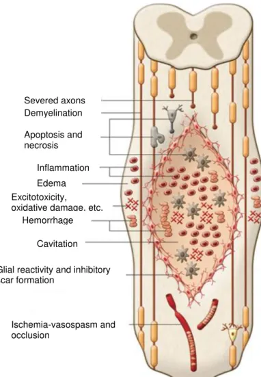
Spinal cord has well-characterized descending and ascending tracts. The ascending tracts are the ones that receive the sensorial inputs and the descending tracts are responsible for a rich variety of quantifiable motor outputs, ranging from simple reflexes to more complex motor patterns, such as scratching, fast paw shake and locomotion (Rossignol and Frigon 2011). In a devastating condition (physical or mechanical trauma) the ascending and/or descending pathways, which connects the brain to the rest of the body, are disrupted. This phenomenon results in a large damage to the spinal cord, leading to paralysis and loss of sensation below the level of injury (Ghosh, Haiss et al. 2009). The initial trauma – primary injury is followed by the secondary injury, consisting of several events including the loss of neuronal and glial cells, which culminates with the formation of cystic cavities and glial scars (Figure 2.1).
2.1.1 Primary injury
The primary injury emerges from the initial physical and/or mechanical trauma to the spinal cord and surrounding vertebral column, caused by blunt impact, compression and penetrating trauma. Blunt impact comes mainly from falls or collisions; compression from hyperflexion, hyperextension, axial loading and severe rotation; and penetrating trauma usually arise from gunshots and stab wounds (Viano, King et al. 1989; Dubendorf 1999; Hulsebosch 2002). After immediate mechanical damage, a cascade of events such as blood vessel damage, dislocation of bones, rupture of intervertebral discs, injury to ligaments and cease of blood flow that deprive the spinal cord of oxygen and nutrients takes place, leading to immediate cell necrosis at the point of impact (Hulsebosch 2002). Without any treatment, the cells and axons in the spinal cord that were not affected by the primary injury can be damaged by secondary injury events spreading to the surrounding tissue (Wang, Zhai et al. 2011).
2.1.2 Secondary injury
The secondary injury is characterized by the events that take place within the spinal cord in response to the primary injury. Those events propagate from the site of injury to unaffected areas of the spinal cord and include:
1- Ischemia and micro-vascular damage, comprising vasospasm, thrombosis, hemorrhage and increased permeability that combined with edema lead to hypoperfusion and necrosis (Tator and Fehlings 1991; Winkler, Sharma et al. 2002; Samadikuchaksaraei 2007).
9
conducting to the depolarization of the neuronal membrane potential (McDonald and Sadowsky 2002; Park, Velumian et al. 2004).
3- Oxidative stress, resulting from free radical formation and lipid peroxidation that can attack membranes and other cell components, disturbing unaffected neurons and oligodendrocytes (Braughler and Hall 1989; McDonald and Sadowsky 2002).
4- Inflammation, recruitment and activation of inflammatory cells associated with secretion of cytokines, which contribute to further tissue damage (Dusart and Schwab 1994; Takami, Oudega et al. 2002).
5- Loss of ionic intracellular balance, increase of the opioids at the injury site, depletion of energy metabolites, conducting to an anaerobic metabolism, an increase of lactate dehydrogenase (LDH) activity and an activation of calpains and caspases, culminating in cellular apoptosis (Samadikuchaksaraei 2007).
After days to weeks from the injury, a fluid filled cystic cavity is formed due to the removal of injured neurons, their axons and necrotic debris. The cyst is expanded to adjacent spinal cord areas, increasing the cell dead and loss of neuronal function, mainly the dead of oligodendrocytes that lead to malfunction and degeneration of the intact axons.
10
Figure 2.1 – Pathophysiological events occurring after SCI, including the primary, secondary and chronic phases. (reproduced with permission from (Mothe and Tator 2013))
2.2 Limited spinal cord regeneration capacity
The inflammatory events in the acute phase are necessary to prevent infections, clear the debris tissue and close the blood-brain barrier, restraining the lesion site. However, in the chronic stage, inflammation, myelin debris and glial scar formation limit the axonal regeneration and consequently, the capacity of the spinal cord to restore their functions after an injury. The scar formed after the injury is a hostile environment with inhibitory molecules and proteoglycans without the ability to support the neuronal cells; therefore, acting as a chemical and physical barrier to the axonal regeneration (Yiu and He 2006).
The inhibitory molecules released after SCI that limit the spinal cord regeneration are: myelin-associated proteins that inhibit axonal growth such as, oligodendrocyte myelin protein – neurite outgrowth inhibitor (Nogo-A), (GrandPré, Nakamura et al. 2000); netrin-1 (Löw, Culbertson et al. 2008); transmembrane semaphoring Sema4D/CD100 (Moreau-Fauvarque, Kumanogoh et al. 2003) and ephrin-B3 (Benson, Romero et al. 2005). In addition, new
Severed axons Demyelination
Apoptosis and necrosis
Inflammation Edema Excitotoxicity,
oxidative damage, etc. Hemorrhage
Cavitation
Glial reactivity and inhibitory scar formation
11
compounds are formed to help the propagation of the inflammation and the remodeling of the ECM. The deposition of inhibitory ECM molecules around the injury sites such as, inhibitory chondroitin sulfate proteoglycans (CSPGs) secreted by reactive astrocytes on the glial scar will impair the spinal cord regeneration and induces the formation of dystrophic cones on injured neurons (Smith-Thomas, Stevens et al. 1995; Niederöst, Zimmermann et al. 1999).
Nevertheless, there are also some endogenous regeneration events that take place after SCI, such as the upregulation of some proteins related to axonal growth at the lesion site contributing to axonal sprouting in short distances and the migration of Schwann cells from spinal roots to the damaged tissue to promote the myelination of spinal cord axons. However, the recovery of the spinal cord is very limited (Duncan and Hoffman 1997; Zawadzka, Rivers et al. 2010). Although it is not a regenerative event, the glial scar formed by the astrocytes derived from ependymal cells is needed to restrict secondary damage on the lesion site (Sabelström, Stenudd et al. 2013). In addition, the ependymal cells also provide neurotrophic effects essential to the survival of the intact neurons after a spinal cord injury. Also, the presence of oligodendrocyte progenitor cells on the glial scar can generate myelinating oligodendrocytes after SCI (Sabelström, Stenudd et al. 2014).
Plasticity within intrinsic spinal cord circuits has been reported as a phenomenon that help in the spinal cord repair. The spinal cord inputs, from descending and peripheral sources, experience functional and anatomical changes that contribute to recovery, mainly by accessing and modulating the modified spinal network in a meaningful way (Raineteau and Schwab 2001; Blesch and Tuszynski 2009). However, these events are almost negligible compared to all the obstacles that prevent the complete spinal cord regeneration.
2.3 Therapeutic/regenerative strategies
Nowadays, there is no treatment for SCI. The standard therapies consist in surgical sta-bilization and decompression of the spinal cord associated with the use of some drugs such as methylprednisolone, to reduce inflammation and consequently, to minimize secondary damage (Hyun and Kim 2010) and Pregabalin to neuropathic pain relief (Sadosky, Parsons et al. 2016). However, the disabilities arising from the SCI such as paraplegia or tetraplegia, respiratory failure and absence of sphincter control, are still a concern as they drastically reduce the patient’s life quality.
12
The complex mechanism involving SCI requires strategies that can overcome the inhibitory environment at the injury, and at the same time can also prevent neuronal loss, promote axonal myelinationn regeneration and the reconnection of the interrupted spinal cord signal, leading to the complete spinal cord regeneration.
2.3.1 Drugs
Several drugs have been tested to limit the spinal cord secondary injury, facilitating regeneration. Riluzole, which have been used in the treatment of amyotrophic lateral sclerosis, is in phase IIB/III clinical trials for the treatment of acute SCI, demonstrating neuroprotective benefits (Fehlings, Nakashima et al. 2016). Rolipram, a phosphodiesterase 4 inhibitor that elevated the cyclic adenosine monophosphate (cAMP) levels, demonstrated anti-inflammatory effects, enhanced axonal growth and functional recovery when administered in rats with SCI (Nikulina, Tidwell et al. 2004; Costa, Pereira et al. 2013). Epothilone B, which is a microtubule-stabilizing drug, encourage the polymerization of microtubules, inducing the axonal growth and functional recovery (Ruschel, Hellal et al. 2015). The bacterial enzyme chondroitinase ABC (chABC), which digests CSPG (contribute to the inhibitory environment), was investigated in a rat SCI model and was effective in the restoration of electrophysiological activities and in the promotion of functional recovery (Bradbury, Moon et al. 2002). When the delivery of the ChABC was carried out via gene therapy into a cervical contusion injury rat model, it was observed improvements in the upper limb and hands function (James, Shea et al. 2015). However, the ChABC therapy combined with treadmill rehabilitation was more effective in promoting tissue regeneration of rats with chronic severe spinal cord contusion by changing neural plasticity (Shinozaki, Iwanami et al. 2016). Purified anti-Nogo-A monoclonal immunoglobulin G (IgG) antibodies, which block the myelin protein Nogo-A, enhanced the neurite outgrowth and axonal regeneration in rat SCI model (Liebscher, Schnell et al. 2005). A Rho GTPase, central regulators of actin reorganization, antagonist – VX-210 – is in phase I/IIA trial and led to motor improvements without safety concerns in SCI (Fehlings, Theodore et al. 2011). Other compounds such as antiserum to dynorphin A (Faden 1990), omega-3 polyunsaturated fatty acids alphalinolenic acid and docosahexaenoic acid (King, Huang et al. 2006) and 4-aminopyridine (Hayes, Blight et al. 1993) were also investigated to prevent the inhibitory environment inside the SCI to facilitate the regeneration process.
Neurotrophic factors such as, neurotrophin-3 (NT-3), nerve growth factor (NGF), brain-derived neurotrophic factor (BDNF), ciliary neurotrophic factor (CNTF) and glial cell line-brain-derived neurotrophic factor (GDNF), enhance neuronal survival, proliferation, migration and differentiation, axonal growth and synaptic plasticity, promoting the repair and recovery at some extent of the central nervous system after injury (Nomura, Tator et al. 2006; Hyun and Kim 2010).
13
the focus should be in the regenerative therapies such as stem cells transplantation and tissue engineering to achieve functional recovery.
2.3.2 Stem cells therapy
Stem cell transplantation has been widely studied in the treatment of several disabilities, including the treatment of spinal cord. Stem cells (embryonic or adult in origin) are cells with the ability to self-renew and differentiate into multiple lineages. Embryonic stem cells (ESCs) and recently, induced pluripotent stem cells (iPSCs) are able to differentiate into the three germ layers (endoderm, ectoderm and mesoderm). Although their high self-renewal and differentiation ability, the in vivo transplantation of ESCs can induce teratomas, indicating the need to strictly control
the proliferation/differentiation processes of those cells. Even more, the use of these cells is related with some ethical issues. On the other hand, the adult stem cells such as mesenchymal stem cells (MSCs) and neural stem cells (NSCs) are more restricted and can only differentiate into specific lineages.
Examples of stem cells that have been investigated for the SCI regeneration include: embryonic or fetal stem cells, NSPCs, oligodendrocyte progenitor cells, MSCs from umbilical cord blood and bone marrow, olfactory ensheathing glia and recently, iPSCs (Romanyuk, Amemori et al. 2015). Several reviews summarized the benefits/effects of employing different stem cells types both in vitro and in vivo in the central nervous system regeneration (Kabu, Gao et al. 2015; Iyer,
Wilems et al. 2017; Zhu, Uezono et al. 2017). Briefly, the stem cells implanted in rats with SCI were able to differentiate into neurons and helped to bridge and restore the signaling in the spinal cord, resulting in sensory- and motor-level improvements (Lu, Woodruff et al. 2014; Iyer, Wilems et al. 2017). In addition, the stem cells secreted factors, which have neuroprotective effects and promote regeneration of the damage axons (Raspa, Pugliese et al. 2016).
From the different stem cells, the NSPCs, found in mammalian brain and spinal cord, were the most promising cell source since they are committed to the neural lineage. The transplanted NSCPs to SCI have the potential to repopulate the damaged area with new neurons, to remielinate the axons and to modulate the environment to neural repair (permitting neural plasticity, trophic factor support and controlling the inflammatory response) (Bonner and Steward 2015). NSPCs, mainly from fetal sources, are in phase I/II clinical trials to the treatment of SCI, improving the sensorial responses (Tsukamoto, Uchida et al. 2013).
14
2.3.3 Tissue engineering
Tissue engineering has been working out in a new therapeutic approach of regenerative medicine for the treatment of damaged or missing tissues or organs. It combines several strategies such as cell transplantation, scaffolds, and biomolecules/drug delivery systems. Therefore, tissue engineering has been investigated in spinal cord regeneration.
After SCI, a structure suitable to connect the two injury ends and to create a suitable environment for cell transplantation is needed. Tissue engineered scaffolds can act as structures that bridges the lesion site and fill in the necrotic areas, creating the suitable cues to provide axonal guidance through the lesion site and connection with the host tissue as well as support the transplanted and endogenous cells and drugs/biomolecules (Cheng, Huang et al. 2007; Potter, Kalil et al. 2008; Olson, Rooney et al. 2009).
The scaffolds seeded with stem cells and transplanted in rat SCI models, improved behavioral recovery and graft survival, reduced the cavitation and increased oligodendrocytic differentiation, compared to cells transplanted without any substrate (Mothe, Tam et al. 2013). The presence of the physical support, facilitate the exchange of oxygen, nutrients, growth factors, and cytokines from the cells with the surrounding environment, improving the cell survival inside the lesion (Bozkurt, Mothe et al. 2010).
Scaffolds can control the delivery of biomolecules such as BDNF (Patist, Mulder et al. 2004; Stokols and Tuszynski 2006) and NT3 (Piantino, Burdick et al. 2006; Fan, Zhang et al. 2011) in the SCI site, which have beneficial effects in spinal cord regeneration, disrupting the inhibitory environment inside the lesion and leading to better axonal growth and improved locomotion. Several reviews are available in the literature explaining the effects of the controlled release of drugs and bioactive agents from scaffolds for spinal cord regeneration (Kwon, Okon et al. 2011; Tator, Hashimoto et al. 2012; Kabu, Gao et al. 2015).
Scaffold properties
15
elastic moduli in order to minimize mechanical parenchymal damage at points in contact between the scaffolds and the host; and 7) be produced on large scale and in a reproducible way (Straley, Foo et al. 2010; Wang, Zhai et al. 2011; He, Wang et al. 2012).
Materials used in scaffold’s preparation
The physico-chemical properties of a scaffold and the interaction of the biological environment with it, depend on the physico-chemical properties of the raw material. Both synthetic and natural polymers have been investigated as materials for the production of scaffolds for spinal cord regeneration.
Natural polymers
Natural polymers are extracted from the natural ECM of humans and animals. They are biocompatible, biodegradable and have motifs to promote cell adhesion, proliferation, and even differentiation. However, the use of natural polymers is associated with some drawbacks such as variability in fabrication and risk of immunogenicity due to the incomplete polymer purification (Straley, Foo et al. 2010; Kubinová and Syková 2012). Moreover, natural polymers usually own weak mechanical properties and rapid degradation rate, which can be advantageous or not depending on the application (Kai, Jin et al. 2013).
Scaffolds from natural polymers, such as collagen, agarose, fibrin and/or fibronectin, silk fibroin and chitosan, were evaluated in the regeneration of the spinal cord. Scaffolds from collagen, which is the main structural protein of connective tissue, improved the forelimb-hindlimb locomotion when implanted into cat spinal cord transection (Goldsmith, Fonseca et al. 2005). Yoshii et al. produced collagen filaments which were implanted in a rabbit spinal cord with a 3 mm transected defect, promoting not only the axonal regeneration but also the function restoration of the transected spinal cord (Yoshii, Ito et al. 2009). Fibrous collagen nerve conduits were repopulated with host cells and prevented astrocyte accumulation in SCI rat models (Liu, Houle et al. 2012). Porous honeycomb collagen sponges filled with PuraMatrix hydrogel conducted to regeneration, migration and differentiation of neural cells in rats with complete SCI transection, resulting in locomotors recovery (Kaneko, Matsushita et al. 2015). Linearly ordered collagen scaffolds surface modified with a collagen biding epidermal growth factor receptor (EGFR) antibody, reduced the glial scar formation and promoted the neuronal differentiation as well as myelination of endogenous NSCs in a transverse thoracic rat SCI, which result in functional neurons exhibiting synaptic activity and conducting functional recovery (Fan, Li et al. 2017).
16
Agarose is extracted from seaweed and has a particular characteristic: the gelling temperature around 37 ºC – the human body temperature. Agarose scaffolds that gels in situ filled
in a hemisection spinal cord defect in adult rats. The gels supported the three dimensional space (3D) neurite extension in vivo, controlled the delivery of trophic factors and anti-scar agents,
enhancing the regeneration (Jain, Kim et al. 2006).
Silk fibroin, a protein found in the silk produced by spiders and other insects, was processed into multichannel scaffolds with hierarchical pore structure and coated with laminin. The construct when implanted in hemisection SCI rat model enhanced the ECM and blood vessels formation guiding the extension of the axons through the injury, with benefits in the locomotor function (Zhang, Yan et al. 2016).
Chitosan is a polysaccharide derived from chitin, which is extracted from the exoskeleton of crustaceans. It was been applied in the medical field due to the hemostatic and antibacterial properties. Chitosan nerve conduits, fabricated by lyophilization, with an internal structure of open channels and coated with laminin were implanted in a rat SCI model to observe the axonal re-growth. The axons growth around the channels, requiring further scaffold optimization (Cheng, Huang et al. 2007).
Synthetic polymers
Synthetic polymers can be processed in large quantities and their chemical and physical properties can be easily controlled. They own good mechanical properties and stability in the body, overcoming the drawbacks of natural polymers. However, synthetic polymers usually are hydrophobic and lack the biological motifs for cell adhesion and proliferation found in the natural polymers (Kubinová and Syková 2012; Kai, Jin et al. 2013).
17
al. 2010) and PLGA/poly-L-lysine (PLL) (Slotkin, Pritchard et al. 2017) improved the remodeling of the tissue into complete lateral thoracic hemisection spinal cord injury of African green monkeys. In the scaffolds with PLL, the recovery of the monkey’s locomotion was observed because the benefic effects of the positive charges of PLL, promoting a positive environment for survival and growth of the axons (Slotkin, Pritchard et al. 2017). Recently, a PCL scaffold was designed to maximize open pore volume using a modified salt-leaching technique. The scaffold is composed of several microtubules inserted into a larger tube. The space into the microtubules as well as the interstitial space between the tubes were filled with neurons and the axonal growth was linear to the scaffolds into transected rat spinal cord model (Shahriari, Koffler et al. 2017).
Polyurethanes
Polyurethanes (PUs), the major class of synthetic elastomers, have been recently investigated as suitable biomaterials to produce scaffolds for tissue engineering. They have three main constituents, a macrodiol or polyol, a diisocyanate, and a chain extender (Figure 2.2).
Usually the synthesis of PUs is realized in two steps. In the first step, the pre-polymer is formed by reacting the hydroxyl groups of a polyol with the isocyanate groups of a polyisocyanate (usually a diisocyanate). The polyisocyanates used in the synthesis can be aromatic, aliphatic or lysine-derived. The polyols are macrodiols with a polyether, polyester or polycarbonate backbone, and hydroxyl functional groups (at least two groups). The second step consists in the extension of the pre-polymer with a short polyol or polyamide extender, resulting in polyurethane or polyurethane urea, respectively (Guelcher 2008; Bagdi, Molná et al. 2011).
18 Figure 2.2 – Constituents and route of production of PUs.
By changing the type and ratio of the PUs constituents, PUs with different physico-chemical properties (mechanical, thermal, and physical), degradation rates and biocompatibility can be obtained and used as biomaterials in different fields (Tatai, Moore et al. 2007; Li, Li et al. 2013).
Mechanical Properties
Soft tissues like heart, blood vessels, skeletal muscle, tendon, and so forth, are very elastic and strong, with non-linear stress-strain behavior (Ma, Hong et al. 2011). Unlike soft tissues, the available polymers are too stiff with low elongation or very soft with low strength (Zhang, Zhang et al. 2006). Modulus mismatch between biomaterials and the surrounding tissue can exacerbate inflammatory reactions that prevent and/or impair the tissue regeneration process (Ma, Hong et al. 2011). PUs constituents can be wisely chosen to synthetize PUs with mechanical properties similar to the tissues intended to repair/replace.
The polyol, either polyester, polyether or polycarbonate, has an important effect on the mechanical properties of the PUs. The use of a polycarbonate as soft segment rendered PUs with superior elongation at break and resilience compared to the PUs based polyester (Ma, Hong et al. 2011). The variations in the molecular weight of the soft segment also affected the mechanical moduli of the PUs (Ma, Hong et al. 2011). In PUs with polyester in their structure, the interaction between hard and soft segments was stronger, resulting in PUs with higher strain hardening tendency as well as larger tensile strength and smaller deformations comparing to PUs based on polyethers (Bagdi, Molná et al. 2011).
The choice of the chain extender also influence the final mechanical properties of the PUs. Chan-Chan et al. (2010) produced biodegradable segmented PUs using either butanediol or dithioerythritol as chain extenders. The use of butanediol, which hinder the phase separation, conducted to a PU with lower strain at break and superior Young modulus comparing to PUs using the dithioerythritol as chain extender, which impart phase separation (Chan-Chan, Solis-Correa et al. 2010). The PUs synthetized with different chain extenders such as, 2,5-dimethyl-3-hexine-2,5-diol (DHD), hexaethylene glycol, glycerin, or castor oil, had distinct mechanical properties due to their distinct chemical structure and the presence of hard-segment crosslinking
OH HO
Polyol: Polyester Polyether Polycarbonate
+
Isocyanate: Aromatic Aliphatic Cycloaliphatic Lysine
NCO OCN +
Chain extender: Diol Diamine
OH
H2N
Pre-polymer
Soft segment Hard segment
19
(Oprea 2010). PUs with longer chain lengths between crosslinks had lower young modulus and high elongation at break (Oprea 2010).
Biodegradation
An appropriate degradation rate is also essential and should be in tune with the rate of new tissue formation: a too fast degradation will compromise the needed support for the forming tissue and a too slow degradation may compromise the healing, contributing to a persistent host inflammatory response (Zhang, Zhang et al. 2006).
The in vivo degradation of PUs occur mainly by hydrolytic, enzymatic and oxidative attack
(Santerre, Woodhouse et al. 2005). The polyesters are the preferred choice as soft segments for PUs. The ester links are hydrolysable and their degradation can be accelerated by the presence of enzymes existent in the organism, such as the esterases (Tokiwa, Ando et al. 1990; Wang, Labow et al. 1997). On the other hand, the polyethers are less prone to hydrolytic and enzymatic attack. However, they are susceptible to oxidative degradation (Schubert, Wiggins et al. 1995).
The urea and urethane linkages are reasonably stable to either hydrolysis, oxidative stress or enzymatic attack, but still they are degradable in vivo. To control the degradation rate of
PUs and to get degradation products without toxicity, the naturally-derived chain extenders have been incorporated in PUs. Those include phenylalanine diester chain extender (Skarja and Woodhouse 2001) and lysine or ornithine chain extenders (Marcos-Fernández, Abraham et al. 2006), rendering biodegradable PU with non-toxic and easily metabolized in vivo degradation
products. The design of enzyme-sensitive chain extenders is another way to control the PUs degradation. The chain extender based on DL-lactic acid and ethylene glycol on the PU backbone accelerated the hydrolytic degradation (Tatai, Moore et al. 2007). A collagenase-sensitive peptide was designed and used as chain extender to control the degradation of PUs through the enzyme collagenase (Fu, Hong et al. 2014).
The choice of the diisocyanate has influence not only on the degradability of the PUs but also, on the toxicity of the degradation products. In order to obtain biodegradable PUs, the aliphatic and lysine-derived diisocyanates have been better choices in detriment of the aromatic ones. The aromatic isocyanates did not meet agreement according to the toxicity of their degradation products. The toxic effects are dependent on the degradation rate of the PUs as well as the clearance rate by the tissue (Guelcher 2008). On the other hand, the biodegradable PUs synthetized with the aliphatic diisocyanates (Park, Gong et al. 2013) and lysine-derived diisocyanates (Wang, Yu et al. 2011) can be easily degraded in phosphate buffer saline (PBS) and enzymatic solution and their degradation products were not toxic in in vitro experiments.
Biocompatibility
20
differentiation. As synthetic polymers, PUs are destitute of biological recognition sites for cell adhesion and proliferation and have poor hydrophilicity and hemocompatibility (Wang, Feng et al. 2012).
The biological response of PUs can be improved by using hydrophilic, polarized and with biological recognition sites chain extenders. Zhang et al. synthetized PUs with methylene di-p-phenyl-diisocyanate (MDI), PCL-diol and with either butanediol (BD) or 2,2’-(methylimino) diethanol (MIDE) chain extenders. The PU with MIDE had superior hydrophilicity and swelling rate, resulting in superior fibroblast’s adhesion and proliferation (Zhang, Zhang et al. 2006). PUs extended with N,N-bis (2-hydorxyethyl)-2-aminoethane-sulfonic acid also supported the adhesion and proliferation of fibroblasts, with additional anticoagulation properties (Zhang, Wen et al. 2008). The use of aminoacids as chain extenders in the PU structures is a way to introduce specific biological functionality, such as enhanced cellular adhesion. Perales-Alcacio and co-workers (Perales-Alcacio, Santa-Olalla Tapia et al. 2013) extended PUs with either glutamic acid, cysteine or glycine aminoacids. The viability of endothelial cells was superior in PUs with either glycine or cysteine in their structure. Endothelial cells were also used to evaluate the compatibility of PUs with glycine, arginine and aspartic acid (Chan-Chan, Tkaczyk et al. 2013) and the PUs extended with arginine support endothelial cells adhesion and viability.
PUs can be processed into scaffolds with tunable mechanical properties and degradation rates as well as with the ability to support the host and/or transplanted cells for tissue engineering. The PUs scaffolds have been widely studied for the cardiovascular and cartilage regeneration, encouraging the formation of new tissue when they are associated with stem cells. For central nervous system regeneration, PUs were used as substrates to guide neuronal differentiation in vitro (Carlberg, Axell et al. 2009; Zandén, Erkenstam et al. 2014). In vivo, the use of the reverse
thermal gel poly(ethylene glycol)-poly(serinol hexamethylene urethane) loaded with bone marrow stromal cells, increased the cells survival inside the lesion, improving the hindlimb motor and sensorimotor recovery in rats with spinal cord contusions (Ritfeld, Rauck et al. 2014). Hsieh et al. 2015 synthetized aqueous PU dispersions with both PCL and PLLA on the soft segment that formed hydrogels at 37 ºC by thermally-induced self-assembly, the gels had an elastic modulus similar to that of the brain tissue. The PU water dispersion was used to embed NSCs before gelation, constituting an ink for 3D printing. The cells proliferate and differentiate properly on the hydrogel, rendering functional recovery of zebrafish neuronal injury (Hsieh, Lin et al. 2015).
Electrical conductive polymers
21
somatosensory evoked potentials, particularly in the presence of iodine-doped pyrrole (Cruz, Mondragón-Lozano et al. 2012). Therefore, iodine-doped pyrrole in a mesoparticles formulation implanted in rats with traumatic SCI were combined with a treadmill training which induced neuroplasticity, promoting the spinal cord functional recovery and preserving the tissues (Alvarez-Mejia, Morales et al. 2015). Recently, poly(3,4-ethylenedioxythiophene) (PEDOT)-coated carbon microfibers functionalized with a multimolecular complex of polylysine, heparin, basic fibroblast growth factor and fibronectin were well integrated into transected rat spinal cord. The construct interacted with the host cells, which provided guidance cues for axonal growth and regeneration and promoted angiogenesis (Alves-Sampaio, García-Rama et al. 2016).
2.4 Interaction of scaffolds with NSPCs
As described before, either the transplantation of NSPCs or the implantation of scaffolds are strategies that lead to improved spinal cord regeneration at some extent. However, using scaffolds seeded with NSCs, improved the axonal regeneration in rat SCI model (Olson, Rooney et al. 2009). Therefore, combining a 3D scaffold with NSCs as a therapeutic approach for SCI, can conduct to better results for spinal cord regeneration, as these structures should support the stem cells, guiding their migration, while supporting phenotype maintenance.
2.4.1
In vitro
studies
The cell behavior is regulated by cell-matrix and cell-cell interactions. The interactions between the scaffolds and the NSPCs have been evaluated to get a cell-scaffold construct that maximizes spinal cord regeneration (Table 2.1). The scaffolds, mainly hydrogels and guiding tubular structures, had a 3D porous macrostructures to support the NSPCs, which ameliorated the communication between cells compared to flat 2D structures (Wang, Ao et al. 2010).
In general, the 3D porous scaffolds made of synthetic polymers such as polyethylene glycol (PEG) and PLGA and natural polymers such as collagen, gelatin, hyaluronic acid and chitosan owns the ability to support the adhesion, proliferation and differentiation of NPSCs into the three neural lineages by adjusting the culture medium composition with specific growth factors and/or others biomolecules.