Contents lists available atScienceDirect
Respiratory Physiology & Neurobiology
j o u r n a l h o m e p a g e :w w w . e l s e v i e r . c o m / l o c a t e / r e s p h y s i o l
Long-term exposure to cigarette smoke impairs lung function and increases
HMGB-1 expression in mice
Frank Silva Bezerra
a,b, Samuel Santos Valenc¸a
c, Karla Maria Pereira Pires
a, Manuella Lanzetti
a,
Wagner Alves Pimenta
a, Aline Cunha Schmidt
b, Luís Cristóvão Porto
a, Walter Araujo Zin
b,∗ aLaboratory of Tissue Repair, Histology and Embryology Department, Roberto Alcantara Gomes Institute of Biology, Rio de Janeiro State University, Rio de Janeiro, Brazil bLaboratory of Respiration Physiology, Carlos Chagas Filho Institute of Biophysics, Federal University of Rio de Janeiro, Rio de Janeiro, BrazilcLaboratory of Inflammation, Oxidative Stress and Cancer, Biomedical Sciences Institute, Federal University of Rio de Janeiro, Rio de Janeiro, Brazil
a r t i c l e
i n f o
Article history: Accepted 23 March 2011
Keywords: Cigarette smoke Lung inflammation Oxidative stress HMGB-1 expression Mice
a b s t r a c t
Cigarette smoke (CS)-induced emphysema is caused by a continuous inflammatory response in the lower respiratory tract. The development of the condition is believed to be mediated by oxidant–antioxidant imbalance. This paper describes the effects of long-term CS exposure on alveolar cell recruitment, antiox-idant defense systems, activity of extracellular matrix metalloelastases, expression of metalloelastase MMP-12, and high mobility group box-1 protein (HMGB-1). Ten C57Bl/6 mice were exposed to 12 cigarettes-a-day for 60 consecutive days, while 10 control animals were exposed to ambient air. After sacrifice, bronchoalveolar lavage fluid (BALF) was removed, and lung tissue underwent biochemical and histological analyses. In CS-exposed animals influx of alveolar macrophages and neutrophils into BALF, lung static elastance, and expression of MMP-12 and HMGB-1 were significantly increased while the activity of antioxidant enzyme was significantly reduced in comparison with control group. Thus, we demonstrated for the first time that long-term CS exposure decreased antioxidant defenses concomitantly with impaired lung function, which was associated with HMGB-1 expression.
© 2011 Elsevier B.V. All rights reserved.
1. Background
Chronic obstructive pulmonary disease (COPD) is caused pri-marily by the inhalation of cigarette smoke (CS), an irritant comprising some 5000 constituents including high concentrations of free radicals and other oxidants (Pryor and Stone, 1993). CS stimulates inflammatory cell recruitment and proteinase produc-tion, both involved in the development of emphysema and chronic bronchitis (Abboud and Vimalanathan, 2008; Churg et al., 2008). Moreover, the continuous inhalation of CS is known to trigger impairment in pulmonary elasticity as well as airway-parenchymal remodeling. These findings result mainly from thickened airway walls and the presence of higher amounts of collagen fibers and reduced content of elastic fibers in the small airways walls (Morris and Sheppard, 2006).
∗Corresponding author at: Universidade Federal do Rio de Janeiro, Centro de Ciên-cias da Saúde, Instituto de Biofísica Carlos Chagas Filho, Laboratório de Fisiologia da Respirac¸ão, Ilha do Fundão, CEP 21941-902, Rio de Janeiro, RJ, Brazil.
Tel.: +55 21 25626557; fax: +55 21 22808193.
E-mail addresses:[email protected](F.S. Bezerra), [email protected](S.S. Valenc¸a),[email protected](K.M.P. Pires), [email protected](M. Lanzetti),[email protected] (W.A. Pimenta),[email protected](A.C. Schmidt),[email protected] (L.C. Porto),[email protected],walter [email protected](W.A. Zin).
CS-induced emphysema is frequently associated with either an imbalance in proteinase and antiproteinase production or an increased oxidative status (Stehbens, 2000). However, the pre-cise role of antioxidant enzymes in CS exposure-induced oxidative stress remains uncertain, and only a few studies address the associ-ation between the activities of these enzymes and oxidative status (Baskaran et al., 1999; Valenca et al., 2008).
Correlations have been reported relating the number of macrophages in histological sections and the levels of morphologic markers of tissue destruction (Eidelman et al., 1990; Finkelstein et al., 1997), but no such correlations have been established regard-ing neutrophil content. A variety of macrophage metalloproteases, including gelatinases A and B (MMP-2 and MMP-9), matrilysin (MMP-7), and MMP-12 are known to degrade elastin and colla-gen (Senior et al., 1989; Senior et al., 1991; Shapiro, 1994). In this context, human emphysematous lungs show higher levels of MMP-1 (interstitial collagenase), MMP-2, MMP-9, and matrix type-MMP-1 (MT1)-MMP compared with their healthy counterparts (Imai et al., 2001; Ohnishi et al., 1998), while the lungs of guinea pigs that were exposed to smoke present increased amounts of MMP-1 (Selman et al., 1996).
The chromatin binding protein high mobility group box chromosomal protein 1 (HMGB-1) has been shown to be a pro-inflammatory cytokine and a mediator of acute inflamma-tory injury to the lungs (Mantell et al., 2006). Thus, HMGB-1
1569-9048/$ – see front matter© 2011 Elsevier B.V. All rights reserved.
intratracheally delivered to mice elicited acute inflammatory lung injury accompanied by neutrophil infiltration, edema formation and increased production of cytokines (Abraham et al., 2000). Fur-thermore, increased levels of HMGB1 have been detected in the plasma as well as in the lung epithelial lining fluid in patients with acute lung injury (ALI) and in mice with lipopolysaccharide-induced ALI (Abraham et al., 2000; Bitto et al., 2010).
To our knowledge, the putative participation of HMGB-1 in CS-induced emphysema has never been described. Therefore, we decided to investigate the expression of HMGB-1 and MMP-12 in CS-induced emphysema, and assess the resulting lung dam-age based on histological, biochemical and pulmonary function analyses.
2. Methods
The study was approved by the Animal Care and Use Committee of the Rio de Janeiro State University.
2.1. Reagents
Potassium dihydrogen phosphate, dipotassium hydrogen phos-phate, sodium chloride, ethylenedinitrilotetraacetic acid (EDTA), hydrogen peroxide, ethanol, acetic acid, and formalin were pur-chased from Vetec (Duque de Caxias, RJ, Brazil). Calcium chloride, sodium dodecyl sulfate (SDS), zinc chloride, acrylamide, adrenaline, bovine serum albumin (BSA), nicotinamide adenine dinucleotide phosphate (NADPH), gelatin, glycerol, mercaptoethanol, Tris–HCl, bromophenol blue, Coomassie blue, Triton X-100, Tween-20, avidin–biotin peroxidase (ABP), 3,3′-diaminobenzidine (DAB) and
rabbit anti-goat IgG biotinylated secondary antibody were bought from Sigma (St. Louis, MO, USA). Goat anti-mouse matrix met-alloproteinase 12 (MMP-12) and goat anti-mouse HMGB-1 were obtained from Santa Cruz Biotechnology (Santa Cruz, CA, USA). Nitrocellulose membranes and Rainbow molecular weight markers were purchased from Amersham Pharmacia Biotech (Pittsburgh, PA, USA), Bradford reagent was acquired from Bio-Rad (Hercules, CA, USA), and Diff-Quik Romanowski stain was bought from Baxter Dade (Dudingen, Switzerland).
2.2. Experimental animals
Twenty C57BL/6 male mice (8 weeks old; weight range: 20–24 g) were purchased from the Veterinary Institute of the Universidade Federal Fluminense (Niterói, RJ, Brazil). Animals were maintained in an environmentally controlled room (25±2◦C;∼80% relative
humidity) under a 12-h light/dark cycle (starting at 6.00 pm daily), and were provided water and foodad libitum.
2.3. Exposure to CS
Two identical chambers were used to expose the animals to either CS or air (Pires et al., 2011). Mice (n= 10) were exposed to the smoke generated by 12 commercial, full flavored, filtered Vir-ginia cigarettes (10 mg of tar, 0.9 mg of nicotine and 10 mg of carbon monoxide per cigarette) on a daily basis during 60 consecutive days. Briefly, CS mice were placed in the inhalation chamber (40-cm long, 30-cm wide and 25-cm high), inside an exhaustion chapel. A cigarette was coupled to a plastic 60 mL syringe so that puffs could be drawn into it and subsequently expelled into the expo-sure chamber. We aspirated 1 L of smoke from one cigarette with this syringe (20 puffs of 50 mL each) and immediately injected each puff into the chamber. The animals were maintained in this smoke-air condition (∼3%) for 6 min, and then the cover of the inhalation chamber was removed, allowing a 1-min smoke evacuation by the chapel exhaustion system. This cigarette exposure procedure
was repeated four times (4×6 min) with 1-min intervals (exhaus-tion). We repeated this procedure three times daily (morning, noon and afternoon) resulting in an overall 72 min of CS exposure to 12 cigarettes. Each cigarette smoked produced 300 mg/m3of total
par-ticulate matter in the chamber (measured by weighing material collected on Pallflex filters). Mice (n= 10) exposed to ambient air over the same time span were used as a control group. Morphom-etry was performed in the right lungs, while BALF collection and enzymatic activity testing were done in the left lungs (n= 5 in each group). Pulmonary mechanics was measured in another group of mice (n= 5 in each sub-group). Please see below.
Carboxyhemoglobin (COHb) concentration was measured after exposure to CS and was not toxic (Beutler and West, 1984).
2.4. Measurement of pulmonary mechanics and lung volume
Twenty-four hours after the last exposure to CS or ambient air, lung mechanics was determined in five animals of each group as previously described (Soares et al., 2007). Lung static elastance (Est,L) was evaluated 10–15 times in each animal over an
exper-imental period of approximately 30 min. Thereafter the animals were euthanized by cervical displacement and exsanguinated by transection of the abdominal aorta. In 10 randomly chosen animals (5 from CS-exposed group and 5 air-exposed mice), the trachea was occluded and the lungs removed. Functional residual capacity (FRC) was determined by volume displacement of saline solution (Scherle, 1970).
2.5. Preparation of samples for histological analysis
Twenty-four hours after the last CS or air exposure, mice (n= 5 in each group) were sacrificed and the right ventricle was per-fused with saline solution (NaCl 0.9%) to remove as much blood as possible from the pulmonary circulation. A surgical thread was carefully passed around the right lung hili structures that were then tightly ensemble ligated; the left lungs were inflated by instill-ing buffered 4% formaldehyde under a pressure of 25 cmH2O for
2 min in order to check for leaks, their hili were then ligated, and the lungs removed and weighed. Inflated lungs were fixed for 48 h before embedding in paraffin. Five-m thick tissue sections were
stained with either hematoxylin-eosin, Sirius red or orcein. Goat anti-mouse MMP-12 and goat anti-mouse HMGB-1 were used as primary antibodies in immunohistochemical analyses. The biotiny-lated secondary antibody, together with ABP and DAB, were used according to the instructions supplied by the manufacturer. After staining for MMP-12 and HMGB-1, lung sections were counter-stained with hematoxylin. Some lung sections underwent the same procedures without the primary antibodies (negative controls); in this case HMGB-1 and MMP-12 were not detected. Analysis of lung slides was done by a skilled blinded pathologist.
2.6. Morphometric analysis
Lung sections were quantitatively assessed with a microscope (Axioplan, Zeiss, Oberkochen, Germany) coupled to a color video camera (TK-C380, JVC, Yokohama, Japan) and monitor (PVM-14N2U Trinitron, Sony, Basingstoke, UK). Pulmonary emphysema was quantified by the mean linear intercept (Lm) (Saetta et al.,
1985). For this purpose, 16 randomly selected fields were observed at 200×magnification in each slide. Stereological analysis used a test-system attached to the video monitor, comprising 21 points and a strictly delineated test-area in order to avoid overestimation of the number of structures (Weibel, 1979). The points (PP) that fell on airspaces (PPair) and elastic fibers (PPef) were counted and
yielding airspaces (Vvair) and elastic fibers (Vvef) volume densities,
respectively.
2.7. BALF collection and preparation of lung homogenates
Following exsanguination and prior to lung removal, the left lung airspaces of both experimental and control animals (n= 5 from each group) were washed with saline solution (final volume col-lected 1.2–1.5 mL) and the BALF was colcol-lected and stored on ice. The left lungs were then immediately removed, homogenized in an ice-cold Ultra-Turrax®model T 8 homogenizer (Toronto, Canada) with 10% (w/v) of 0.1 M potassium phosphate buffer (pH 7.5) con-taining 5 mM disodium EDTA, and centrifuged at 3000×gfor 5 min. Supernatants were stored at−20◦C until required for the analysis
of antioxidant enzyme activities, gelatin zymography and western blotting.
2.8. Analysis of BALF
The total numbers of mono- and polymorphonuclear cells in BALF samples were evaluated using a Zi Coulter counter (Beckman Coulter, Carlsbad, CA, USA). For differential cell counts slides were prepared using BALF samples with the aid of a Shandon (Waltham, MA, USA) Cytospin cytocentrifuge and subsequently treated with Diff-Quik Romanowski stain. At least 200 cells per BALF slide were counted using standard morphological criteria.
2.9. Activity of antioxidant enzymes in lung homogenates
Superoxide dismutase (SOD) was spectrophotometrically assayed at 480 nm by monitoring the inhibition of adrenaline autoxidation (Bannister and Calabrese, 1987). Catalase (CAT) activ-ity was evaluated based on the rate of decrease in hydrogen peroxide absorbance measured at 240 nm (Aebi, 1984). Glutathione peroxidase (GPx) activity was assessed by monitoring the oxida-tion of NADPH (detected at 340 nm) in the presence of hydrogen peroxide (Flohe and Gunzler, 1984). The total protein content in homogenized lung tissue samples was determined using the method ofBradford (1976).
2.10. Gelatin zymography
Aliquots of lung homogenates and placental tissue (positive control), each containing 30g of protein, were used for MMP-2
and MMP-9 determination. They were subjected to non-reducing electrophoresis on an 8% acrylamide stacking gel/7% acrylamide separating gel slab containing 1 mg/mL gelatin in the presence of SDS under non-reducing conditions. Following electrophore-sis, gels were washed twice with 2.5% Triton X-100, rinsed with water, and incubated overnight at 37◦C in 50 mM Tris–HCl (pH
8) containing 5 mM calcium chloride and 2 nM zinc chloride. Gels were stained with Coomassie blue and destained with 25% ethanol and 10% acetic acid solution. Areas associated with gelatinolytic activity appeared as clear bands on a blue background. The molec-ular weights of lung tissue proteins present in the clear bands were estimated by comparison with those of the placental sam-ple. Gelatinolytic activity was densitometrically quantified as the intensity of the negative bands in relation to those determined in the positive control (Niu et al., 2000). For such purpose Scion Image 4.03 software (Scion Corporation, Frederick, MD, USA) was used.
2.11. Western blotting
Aliquots of lung homogenates, each containing 30g of
pro-tein, were denatured in 50 mM Tris–HCl (pH 6.8) containing 1%
SDS, 5% 2-mercaptoethanol, 10% glycerol and 0.001% bromophe-nol blue, and heated in boiling water for 3 min. Samples, together with Rainbow molecular weight markers (GE Healthcare Bio-Sciences Corp., Piscataway, NJ, USA), were submitted to 12% SDS polyacrylamide gel electrophoresis and the separated lung tis-sue proteins transferred to nitrocellulose membranes. Membranes were blocked with Tween-TBS [20 mM Tris–HCl (pH 7.5) containing 500 mM sodium chloride and 0.5% Tween-20] supplemented with 2% BSA, and probed (1:1000) with the specific primary antibod-ies goat anti-mouse MMP-12 and goat anti-mouse HMGB-1. After extensive washing in Tween-TBS, the membranes were incubated with biotinylated secondary antibody and ABP for 1 h and then visualized by DAB staining. The intensities of the bands were den-sitometrically quantified using Scion Image 4.03 software (Scion Corporation, Frederick, MD, USA) after ponceau staining of the membrane.
2.12. Statistical analyses
All data were expressed as mean±S.E.M. or as median and percentiles (10 and 90%), and analyzed using GraphPad Prism 5 data analysis software (GraphPad Software, CA, USA). Normally distributed continuous data (i.e. BALF counts, antioxidant enzyme activities and pulmonary mechanics) were analyzed using Stu-dent t-test with Welch’s correction, while discrete data (Vvair,
Vvef and densitometric measurements) were treated using the
Mann–Whitney test. In all cases, the level of significance was set at 5%.
3. Results
3.1. Carboxyhemoglobin level
The mean (±S.E.M.) COHb level in air-exposed mice was 1.1±0.2%, while that in CS-exposed mice was 13.4±1.3%.
3.2. Lung histology
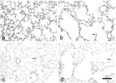
Photomicrographs of lung sections in control animals pre-sented normal alveoli with thin alveolar septa and few alveolar macrophages (Fig. 1a) and elastic fibers displaying fine branching in the alveolar septa (Fig. 1c). On the other hand, mice exposed to CS exhibited enlarged airspaces and thickened alveolar septa (Fig. 1b), a large amount of alveolar macrophages and rupture of elastic fibers in the alveolar septa (Fig. 1d).
3.3. Pulmonary mechanics
Lung static elastance and functional residual capacity were sig-nificantly higher (p< 0.05 and <0.01, respectively) in CS-exposed mice than in control animals (Table 1).
3.4. Lung morphometry
CS group exhibited mean linear intercept and airspace volume density significantly higher (p< 0.05 and <0.01, respectively) than the control group, while the mean elastic fiber volume density in CS-exposed animals was significantly lower (p< 0.05) than in con-trol group (Table 1).
3.5. BALF cellularity
Fig. 1.Photomicrographs of lung sections from control mice (panels a and c) and animals exposed to cigarette smoke (CS) over a 60-day period (panels b and d), showing details of the alveolar septa (a and b) and elastic fibers (c and d). Bar = 10m. Arrows indicate thin and thick septa in control and CS groups, respectively.
Table 1
Pulmonary mechanics, morphometry, bronchoalveolar lavage fluid (BALF) cellularity and antioxidant enzyme activities in control and cigarette smoke exposed (CS) mice.
Parameters Control CS
Pulmonary mechanics
Lung static elastance (Est,L; cmH2O/mL) 34.47±2.23 45.61±2.98a
Functional residual capacity (FRC; mL) 0.17±0.01 0.26±0.01b
Morphometry
Mean linear intercept (Lm;m) 28.5±1.2 36.8±2.2a
Airspace volume density (Vvair; %) 61.0 (55.0–65.0) 74.0 (67.0–84.0)b
Elastic fiber volume density (Vvef; %) 17.0 (15.0–18.0) 10.0 (8.0–12.0)b
BALF cellularity
Macrophages (cells×103/mL) 158.4±15.0 413.8±30.6c
Neutrophils (cells×103/mL) 4.6
±2.0 63.2±6.0c
Enzymatic activities
Superoxide dismutase (U/mg protein) 41.7±1.7 31.1±1.7a
Catalase (U/mg protein) 0.38±0.03 0.23±0.01a
Glutathione peroxidase (mM/min mg protein) 0.74±0.02 0.51±0.05a
Data are expressed as mean±S.E.M. or as median and percentiles (10–90%). Right lungs were used for morphometry, while BALF and enzymatic activities were performed in the left lungs (n= 5 in each group). Pulmonary mechanics was measured in another 10 mice (n= 5 in each group).
ap< 0.05. bp< 0.01. c p< 0.001.
3.6. Activity of antioxidant enzymes
The activities of SOD, CAT and GPx were significantly (p< 0.05) lower in lung homogenates of CS animals than in control group (Table 1).
3.7. Activity of MMP-2 and MMP-9
Fig. 2 displays a representative gelatin zymography in lung homogenates. MMP-2 activity tended to be less intense in CS group animals in control mice, but the difference was not statistically significant (Fig. 3). MMP-9 activity could not be detected in lung homogenates in all instances.
3.8. Expression of MMP-12 and HMGB-1
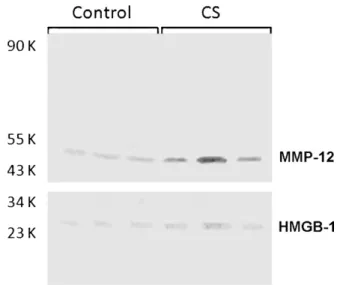
MMP-12 and HMGB-1 stainings were lightly expressed in con-trol group (Figs. 4a and c, respectively); they were easily detected in alveolar macrophages from CS-exposed animals (Figs. 4b and d,
respectively). MMP-12 and HMGB-1 bands were significantly enhanced (p< 0.05) in CS group in comparison with control mice (Figs. 3 and 5).
4. Discussion
Our results confirmed that long-term CS-exposure of mice leads to the development of emphysema, in line with our previous
Fig. 3.Densitometric analyses of the matrix metalloproteinase 2 (MMP-2) bands from gelatin zymography (Fig. 2) and matrix metalloproteinase (MMP-12) and high-mobility group box (HMGB-1) bands from Western blotting (Fig. 5) obtained from control (open columns) and cigarette smoke-exposed (solid columns) animals. ‘*’ indicates statistically significant difference between groups (p< 0.05).N= 10 animals per group.
findings (Pires et al., 2011; Valenca et al., 2006; Valenca et al., 2004). Exposure to CS compromised lung mechanics probably because of the disruption of the elastic fiber network and thickening of alve-olar septa (Figs. 1b and d). Thus static elastance and functional residual capacity were increased in CS animals (Table 1) as pre-viously reported in emphysema (Ross et al., 1962). However, the commonest protocol for emphysema development in mice found in the literature takes 6 months to complete (Churg et al., 2004; Guerassimov et al., 2004; Sato et al., 2008), while in this study we used our previously reported 60-day protocol (Pires et al., 2011). The length of time required to produce emphysema varies from ani-mal to aniani-mal but it generally depends on the method of exposure and on the cigarette dose (Mahadeva and Shapiro, 2005; Wright and Churg, 2002).
Macrophage recruitment into BALF is triggered by various com-ponents of CS, including free radicals (Pryor and Stone, 1993).
Fig. 4.Immunostained lung sections from control mice (panels a and c) and animals exposed to cigarette smoke over a 60-day period (panels b and d), showing matrix metalloproteinase 12 (MMP-12) expression (panels a and b) and high-mobility group box (HMGB-1) expression (panels c and d). Bar = 10m. Arrows indicate
macrophages expressing the proteins.
Fig. 5. Western blots of replicate lung homogenates from control mice and animals exposed to cigarette-smoke (CS) over a 60-day period after probing with goat anti-mouse matrix metalloproteinase 12 (MMP-12) and goat anti-anti-mouse high-mobility group box-1 (HMGB-1) primary antibodies.
Continuous exposure to CS generates a constant chemotactic stimulation of macrophages, which were, indeed, found in large amounts in the BALF of our CS-exposed animals (Table 1). Although a significant influx of macrophages into BALF was observed in an earlier investigation by our group (Valenca et al., 2004), there was no evidence of the substantial recruitment of neu-trophils detected in the present study (Table 1). In previous experiments animals were exposed to fewer cigarettes (9 per day), and probably the different CS doses modulate the influx of inflammatory cells, as we demonstrated during short-term (Silva Bezerra et al., 2006) and long-term (Valenca et al., 2006) CS exposure.
Oxidant–antioxidant balance in BALF is also known to play an important role in the pathogenesis of COPD owing to the oxidant-mediated activation of nuclear factor kappa-B (Rahman, 2006). In this context, exposure to CS decreases SOD, CAT, and GPx activi-ties (Valenca et al., 2008) and contributes additional oxidants by stimulating inflammation, thus augmenting the production of free radicals, especially superoxide anion (O2•−). This radical anion
plays a critical role in oxidative metabolism in the lung, and is a key mediator of the pathophysiological responses that lead to the development of emphysema (Pryor and Stone, 1993). There-fore, we suggest that the increase in O2•− production mediated
by exposure to CS directly affected SOD activity (Table 1) thereby impairing the dismutation of the radical to hydrogen peroxide. CAT activity in the lung is found mainly in alveolar macrophages and epithelium (Fridovich and Freeman, 1986). Exposure to CS led to a significant reduction in CAT activity (Table 1), possi-bly indicating that the epithelial cells surviving lung parenchyma destruction underwent intracellular oxidative damage. Addition-ally, the expression of glutathione peroxidase (GPx), a primary antioxidant enzyme that scavenges hydrogen peroxide and organic hydroperoxides (Flohe and Gunzler, 1984), may also be down regulated by CS since in the present study GPx activity was sig-nificantly reduced in mice that had been exposed to CS for 60 days (Table 1).
in CS mice (Table 1). Although MMP-2 and MMP-9 are believed to be important in the pathogenesis of CS-induced emphysema in humans (Segura-Valdez et al., 2000), they could not be detected in homogenates of lung tissue derived from CS-exposed mice (Fig. 2).
Our results indicate that in mice there is an association between CS-induced emphysema and increased pulmonary HMGB-1 expression (Fig. 3), primarily related to alveolar macrophages. Although the study does not provide evidence that HMGB-1 drives the inflammation, is a consequence of it or, indeed, is directly involved at all, the protein must certainly be considered as a component of emphysema in mice. HMGB-1 was initially iden-tified as a DNA binding protein, but more recent data indicate that it presents potent pro-inflammatory properties (Klune et al., 2008). It is believed that HMGB-1 signals through toll-like recep-tors, although it can activate various other cells involved in the immune response or in inflammatory reactions, and thus acts as a cytokine (Yang et al., 2001). Although HMGB-1 has been shown to be involved in the pathogenesis of acute lung injury (Bitto et al., 2010; Mantell et al., 2006), the demonstration of an associa-tion between expression of the cytokine and mouse emphysema represents an important step towards a deeper understanding of its physiological role and in identifying potential therapeutic targets.
In conclusion, the present study provides, for the first time, evidence that long-term CS exposure leads to emphysema asso-ciated with HMGB-1 expression in mice. The involvement of HMGB-1 in pulmonary emphysema discloses another possible pathway to explain oxidative stress and proteinase action in the mouse lung, and suggests a potential therapeutic target for future studies.
Acknowledgments
This work was supported by Fundac¸ão de Amparo à Pesquisa do Estado do Rio de Janeiro (FAPERJ), Conselho Nacional de Desen-volvimento Científico e Tecnológico (CNPq) and Ministério da Ciência e Tecnologia (MCT).
Appendix A. Supplementary data
Supplementary data associated with this article can be found, in the online version, atdoi:10.1016/j.resp.2011.03.023.
References
Abboud, R.T., Vimalanathan, S., 2008. Pathogenesis of COPD. Part I. The role of protease–antiprotease imbalance in emphysema. Int. J. Tuberc. Lung Dis. 12, 361–367.
Abraham, E., Arcaroli, J., Carmody, A., Wang, H., Tracey, K.J., 2000. HMG-1 as a medi-ator of acute lung inflammation. J. Immunol. 165, 2950–2954.
Aebi, H., 1984. Catalase in vitro. Methods Enzymol. 105, 121–126.
Bannister, J.V., Calabrese, L., 1987. Assays for superoxide dismutase. Methods Biochem. Anal. 32, 279–312.
Baskaran, S., Lakshmi, S., Prasad, P.R., 1999. Effect of cigarette smoke on lipid peroxi-dation and antioxidant enzymes in albino rat. Indian J. Exp. Biol. 37, 1196–1200. Beutler, E., West, C., 1984. Simplified determination of carboxyhemoglobin. Clin.
Chem. 30, 871–874.
Bitto, A., Barone, M., David, A., Polito, F., Familiari, D., Monaco, F., Giardina, M., David, T., Messina, R., Noto, A., Di Stefano, V., Altavilla, D., Bonaiuto, A., Minutoli, L., Guarini, S., Ottani, A., Squadrito, F., Venuti, F.S., 2010. High mobility group box-1 expression correlates with poor outcome in lung injury patients. Pharmacol. Res. 61, 116–120.
Bradford, M.M., 1976. A rapid and sensitive method for the quantitation of micro-gram quantities of protein utilizing the principle of protein-dye binding. Anal. Biochem. 72, 248–254.
Churg, A., Cosio, M., Wright, J.L., 2008. Mechanisms of cigarette smoke-induced COPD: insights from animal models. Am. J. Physiol. Lung Cell Mol. Physiol. 294, L612–631.
Churg, A., Wang, R.D., Tai, H., Wang, X., Xie, C., Wright, J.L., 2004. Tumor necrosis factor-alpha drives 70% of cigarette smoke-induced emphysema in the mouse. Am. J. Respir. Crit. Care Med. 170, 492–498.
Eidelman, D., Saetta, M.P., Ghezzo, H., Wang, N.S., Hoidal, J.R., King, M., Cosio, M.G., 1990. Cellularity of the alveolar walls in smokers and its relation to alveolar destruction. Functional implications. Am. Rev. Respir. Dis. 141, 1547–1552.
Finkelstein, J.N., Johnston, C., Barrett, T., Oberdorster, G., 1997. Particulate–cell interactions and pulmonary cytokine expression. Environ. Health Perspect. 105 (Suppl 5), 1179–1182.
Flohe, L., Gunzler, W.A., 1984. Assays of glutathione peroxidase. Methods Enzymol. 105, 114–121.
Fridovich, I., Freeman, B., 1986. Antioxidant defenses in the lung. Annu. Rev. Physiol. 48, 693–702.
Guerassimov, A., Hoshino, Y., Takubo, Y., Turcotte, A., Yamamoto, M., Ghezzo, H., Triantafillopoulos, A., Whittaker, K., Hoidal, J.R., Cosio, M.G., 2004. The develop-ment of emphysema in cigarette smoke-exposed mice is strain dependent. Am. J. Respir. Crit. Care Med. 170, 974–980.
Hautamaki, R.D., Kobayashi, D.K., Senior, R.M., Shapiro, S.D., 1997. Requirement for macrophage elastase for cigarette smoke-induced emphysema in mice. Science 277, 2002–2004.
Imai, K., Dalal, S.S., Chen, E.S., Downey, R., Schulman, L.L., Ginsburg, M., D’Armiento, J., 2001. Human collagenase (matrix metalloproteinase-1) expression in the lungs of patients with emphysema. Am. J. Respir. Crit. Care Med. 163, 786–791.
Klune, J.R., Dhupar, R., Cardinal, J., Billiar, T.R., Tsung, A., 2008. HMGB1: endogenous danger signaling. Mol. Med. (Cambridge, MA) 14, 476–484.
Mahadeva, R., Shapiro, S.D., 2005. Animal models of pulmonary emphysema. Curr. Drug Targets Inflamm. Allergy 4, 665–673.
Mantell, L.L., Parrish, W.R., Ulloa, L., 2006. Hmgb-1 as a therapeutic target for infec-tious and inflammatory disorders. Shock 25, 4–11.
Morris, D.G., Sheppard, D., 2006. Pulmonary emphysema: when more is less. Physi-ology (Bethesda) 21, 396–403.
Niu, R., Okamoto, T., Iwase, K., Nomura, S., Mizutani, S., 2000. Quantitative analysis of matrix metalloproteinases-2 and -9, and their tissue inhibitors-1 and -2 in human placenta throughout gestation. Life Sci. 66, 1127–1137.
Ohnishi, K., Takagi, M., Kurokawa, Y., Satomi, S., Konttinen, Y.T., 1998. Matrix metalloproteinase-mediated extracellular matrix protein degradation in human pulmonary emphysema. Lab. Invest. 78, 1077–1087.
Pires, K.M., Bezerra, F.S., Machado, M.N., Zin, W.A., Porto, L.C., Valenca, S.S., 2011. N-(2-mercaptopropionyl)-glycine but not Allopurinol prevented cigarette smoke-induced alveolar enlargement in mouse. Respir. Physiol. Neurobiol. 175, 322–330.
Pryor, W.A., Stone, K., 1993. Oxidants in cigarette smoke. Radicals, hydrogen perox-ide, peroxynitrate, and peroxynitrite. Ann. N.Y. Acad. Sci. 686, 12–27 (discussion 27-18).
Rahman, I., 2006. Antioxidant therapies in COPD. Int. J. Chron. Obstruct. Pulmon. Dis. 1, 15–29.
Ross, J.C., Copher, D.E., Teays, J.D., Lord, T.J., 1962. Functional residual capacity in patients with pulmonary emphysema. A comparative study using gas dilu-tion and plethysmographic techniques for measurement. Ann. Intern. Med. 57, 18–28.
Saetta, M., Shiner, R.J., Angus, G.E., Kim, W.D., Wang, N.S., King, M., Ghezzo, H., Cosio, M.G., 1985. Destructive index: a measurement of lung parenchymal destruction in smokers. Am. Rev. Respir. Dis. 131, 764–769.
Sato, A., Hoshino, Y., Hara, T., Muro, S., Nakamura, H., Mishima, M., Yodoi, J., 2008. Thioredoxin-1 ameliorates cigarette smoke-induced lung inflammation and emphysema in mice. J. Pharmacol. Exp. Ther. 325, 380–388.
Scherle, W., 1970. A simple method for volumetry of organs in quantitative stereol-ogy. Mikroskopie 26, 57–60.
Segura-Valdez, L., Pardo, A., Gaxiola, M., Uhal, B.D., Becerril, C., Selman, M., 2000. Upregulation of gelatinases A and B, collagenases 1 and 2, and increased parenchymal cell death in COPD. Chest 117, 684–694.
Selman, M., Montano, M., Ramos, C., Vanda, B., Becerril, C., Delgado, J., Sansores, R., Barrios, R., Pardo, A., 1996. Tobacco smoke-induced lung emphysema in guinea pigs is associated with increased interstitial collagenase. Am. J. Physiol. 271, L734–743.
Senior, R.M., Connolly, N.L., Cury, J.D., Welgus, H.G., Campbell, E.J., 1989. Elastin degradation by human alveolar macrophages. A prominent role of metallopro-teinase activity. Am. Rev. Respir. Dis. 139, 1251–1256.
Senior, R.M., Griffin, G.L., Fliszar, C.J., Shapiro, S.D., Goldberg, G.I., Welgus, H.G., 1991. Human 92- and 72-kilodalton type IV collagenases are elastases. J. Biol. Chem. 266, 7870–7875.
Shapiro, S.D., 1994. Elastolytic metalloproteinases produced by human mononuclear phagocytes. Potential roles in destructive lung disease. Am. J. Respir. Crit. Care Med. 150, S160–164.
Silva Bezerra, F., Valenca, S.S., Lanzetti, M., Pimenta, W.A., Castro, P., Goncalves Koatz, V.L., Porto, L.C., 2006. Alpha-tocopherol and ascorbic acid supplemen-tation reduced acute lung inflammatory response by cigarette smoke in mouse. Nutrition (Burbank, Los Angeles County, CA) 22, 1192–1201.
Soares, R.M., Cagido, V.R., Ferraro, R.B., Meyer-Fernandes, J.R., Rocco, P.R., Zin, W.A., Azevedo, S.M., 2007. Effects of microcystin-LR on mouse lungs. Toxicon 50, 330–338.
Stehbens, W.E., 2000. Proteinase imbalance versus biomechanical stress in pul-monary emphysema. Exp. Mol. Pathol. 69, 46–62.
Valenca, S.S., da Hora, K., Castro, P., Moraes, V.G., Carvalho, L., Porto, L.C., 2004. Emphysema and metalloelastase expression in mouse lung induced by cigarette smoke. Toxicol. Pathol. 32, 351–356.
Valenca, S.S., Silva Bezerra, F., Lopes, A.A., Romana-Souza, B., Marinho Cavalcante, M.C., Lima, A.B., Goncalves Koatz, V.L., Porto, L.C., 2008. Oxidative stress in mouse plasma and lungs induced by cigarette smoke and lipopolysaccharide. Environ. Res. 108, 199–204.
Weibel, E.R., 1979. Stereologic Methods. I. Practical Methods for Biological Mor-phometry. Academic Press, London.
Wright, J.L., Churg, A., 2002. Animal models of cigarette smoke-induced COPD. Chest 122, 301S–306S.