The Effects of NF-
κ
B and c-Jun/AP-1 on the
Expression of Prothrombotic and
Proinflammatory Molecules Induced by
Anti-β
2
GPI in Mouse
Longfei Xia1☯, Hongxiang Xie1☯, Yinjing Yu1, Hong Zhou1,2
*, Ting Wang1, Jinchuan Yan2*
1Jiangsu Key Laboratory of Medicine Science and Laboratory Medicine, School of Medicine, Jiangsu University, Zhenjiang, Jiangsu Province, China,2Department of Cardiology, Affiliated Hospital of Jiangsu University, Zhenjiang, Jiangsu Province, China
☯These authors contributed equally to this work.
*[email protected](HZ);[email protected](JY)
Abstract
Our previous data demonstrated that nuclear factor-κB (NF-κB) and activator protein-1 (AP-1) are involved in the process of anti-β2GPI/β2GPI-induced tissue factor (TF) expression in
monocytes. However, the role of NF-κB and AP-1 in pathogenic mechanisms of antipho-spholipid syndrome (APS)in vivohas been rarely studied. This study aimed to investigate whether NF-κB and c-Jun/AP-1 are involved in anti-β2GPI-induced expression of
prothrom-botic and proinflammatory molecules in mouse. IgG-APS or anti-β2GPI antibodies were
injected into BALB/c mice in the presence or absence of PDTC (a specific inhibitor of
NF-κB) and Curcumin (a potent inhibitor of AP-1) treatment. Our data showed that both IgG-APS and anti-β2GPI could induce the activation of NF-κB and c-Jun/AP-1 in mouse
perito-neal macrophages. The anti-β2GPI-induced TF activity in homogenates of carotid arteries
and peritoneal macrophages from mice could significantly decrease after PDTC and/or Cur-cumin treatment, in which PDTC showed the strongest inhibitory effect, but combination of two inhibitors had no synergistic effect. Furthermore, anti-β2GPI-induced expression of TF,
VCAM-1, ICAM-1 and E-selectin in the aorta and expression of TF, IL-1β, IL-6 and TNF-αin peritoneal macrophages of mice were also significantly attenuated by PDTC and/or Curcu-min treatment. These results indicate that both NF-κB and c-Jun/AP-1 are involved in regu-lating anti-β2GPI-induced expression of prothrombotic and proinflammatory moleculesin vivo. Inhibition of NF-κB and c-Jun/AP-1 pathways may be beneficial for the prevention and treatment of thrombosis and inflammation in patients with APS.
Introduction
The antiphospholipid syndrome (APS) is an acquired autoimmune disease characterized by the occurrence of thrombosis and/or recurrent miscarriages. APS is also associated with the OPEN ACCESS
Citation:Xia L, Xie H, Yu Y, Zhou H, Wang T, Yan J (2016) The Effects of NF-κB and c-Jun/AP-1 on the Expression of Prothrombotic and Proinflammatory Molecules Induced by Anti-β2GPI in Mouse. PLoS
ONE 11(2): e0147958. doi:10.1371/journal. pone.0147958
Editor:Cristoforo Scavone, Universidade de São Paulo, BRAZIL
Received:September 14, 2015
Accepted:January 10, 2016
Published:February 1, 2016
Copyright:© 2016 Xia et al. This is an open access article distributed under the terms of theCreative Commons Attribution License, which permits unrestricted use, distribution, and reproduction in any medium, provided the original author and source are credited.
Data Availability Statement:All relevant data are within the paper.
presence of antiphospholipid antibodies (aPL), including lupus anticoagulant (LA),
anticardio-lipin antibodies (aCL) and anti-β2-glycoprotein I antibodies (anti-β2GPI) [1–2]. There is a
close relationship between aPL and vascular thrombotic events and an array of obstetric
com-plications [3]. In APS, most aPLs are autoantibodies directly against phospholipid binding
pro-teins such asβ2GPI and prothrombin [4]. The majority of studies have demonstrated that
β2GPI is the major antigenic target, and anti-β2GPI autoantibodies are predominantly
respon-sible for the clinical manifestations of APS [5]. High titers of anti-β2GPI antibodies are
fre-quently found in the plasma of the patients, suggesting their important roles in APS [6].
The pathogenic mechanisms of APS have not been fully elucidated, and a number of
mecha-nisms have been proposed. Increasing evidence has indicated that aPL/anti-β2GPI may bind to
cells via its specific binding molecules/receptors, such as annexin A2 (ANX2) and Toll-like receptor 4 (TLR4), causing the activation of endothelial cells (ECs), monocytes and platelets
[7–9]. Some Studies have also suggested that aPL/anti-β2GPI can recognizeβ2GPI binding to
monocytes and ECs, leading to a series of alterations in intracellular signaling pathwaysin
vitro. These alterations can result in prothrombotic and proinflammatory phenotypes of
monocytes and ECs, e.g., secretion of inflammatory cytokines (IL-6, IL-1β, TNF-α, and IL-8),
induction of adhesion molecules (VCAM-1, ICAM-1 and E-selectin) and TF [10–11].
More-over, aPL/anti-β2GPI may cause thrombosis, ECs activation and pregnancy loss in animal
models of APS [12–14]. Furthermore, a multiple of studies illuminated that TLR4/NF-κB
path-way is involved in mediating the pathogenic effects of aPL/anti-β2GPI on ECs and monocytes
[15–16]. Our previous results implicated that both nuclear factor-κB (NF-κB) and activator
protein-1 (AP-1) are involved in anti-β2GPI/β2GPI-induced TF expression in monocytes [17].
However, whether NF-κB and AP-1 are activatedin vivoor whether inhibitors of NF-κB and
AP-1 are effective in reversing prothrombotic and proinflammatory effects of aPL/anti-β2GPI
in vivo, have not been well studied.
NF-κB is one of the key cytoplasmic transcription factors and expressed in almost all cell
types to mediate the expression of more than 100 different genes. Numerous NF-κB target
genes are relevant to immune responses and inflammation [18]. NF-κB is a complex of
hetero-dimeric and homohetero-dimeric transcription factors, and NF-κB family is composed of RelA, c-Rel,
RelB, NF-κB1 (p50 and its precursor p105), and NF-κB2 (p52 and its precursor p100) [18].
NF-κB is normally kept inactive in the cytoplasm by interaction with IκBs. Upon stimuli (such
as inflammatory cytokines, infection with viruses or LPS, stress signals), IκBs are
phosphory-lated and degraded by ubiquitin-proteasome pathway, leading to the activation of NF-κB.
Activated NF-κB can enter into nucleus to regulate the transcription of target genes [19].
Tran-scription factor AP-1 consists of heterologous dimeric complex that contains members of the JUN, FOS, ATF and JDP subunits. The different AP-1 factors may regulate different target genes through interaction with specific protein kinases and a variety of transcriptional coacti-vators, and thus execute distinct biological functions such as cell proliferation and survival
[20]. Generally, the main ingredients of AP-1 are c-Jun and c-Fos in mammals. Similarly, the
activity of NF-κB and AP-1 can be regulated by several upstream kinases, such as
p38-mito-gen-activated protein kinase (p38 MAPK), extracellular signal-regulated kinase (ERK) and
c-Jun N-terminal kinases (JNKs) [21].
However, whether both NF-κB and c-Jun/AP-1 are involved in aPL/anti-β2GPI-induced
expression of TF, adhesion molecules and inflammatory cytokinesin vivohas not yet been
clar-ified. Taking into account these circumstances, we investigated the effects of NF-κB and c-Jun/
AP-1 on aPL/anti-β2GPI-induced expression of prothrombotic and proinflammation
mole-cules by using a specific NF-κB antagonist pyrrolidinedithiocarbamate acid (PDTC) and an AP-1 inhibitor Curcumin in BALB/c mice. The alterations in these molecules were assessed by TF activity/expression and the expression of adhesion molecules (VCAM-1, ICAM-1, and data collection and analysis, decision to publish, or
preparation of this manuscript.
Competing Interests:The authors have declared
E-selectin) and proinflammatory cytokines (IL-1β, IL-6, and TNF-α) in the carotid artery,
aorta and peritoneal macrophagesin vivo.
Materials and Methods
Mice and Chemicals
BALB/c mice weighing approximately 22 g (8–10 weeks of age) were purchased from
Compar-ative Medicine Centre of Yangzhou University (Yangzhou, China). All animals were housed in the Laboratory Animal Research Center of Jiangsu University. All animal experiments were approved by the Laboratory Animal Administration Committee of Jiangsu University and consistent with the Guide for the Care and Use of Laboratory Animals published by the US National Institutes of Health.
Pyrrolidinedithiocarbamic acid (PDTC) (Sigma, Saint Louis, MO, USA) was dissolved in sterilized PBS and Curcumin (Sigma, USA) was dissolved in olive oil, aliquoted and stored at
-20°C for the experimentsin vivo.
Preparation of IgGs
Polyclonal anti-β2GPI antibodies were purified from sera of New Zealand rabbits immunized
with humanβ2GPI. Control antibodies (NR-IgG) from normal rabbits were purified by Protein
G Sepharose columns. Total IgG containing the aPL antibodies (IgG-APS) from patients with primary APS was purified using Protein G Sepharose columns. In addition, the sera from APS
patients displayed high titers of anti-β2GPI antibodies (≧66.9 SGU/mL). Ethical approval was
granted by the Institutional Review Board of the Affiliated Hospital of Jiangsu University. All IgG samples and reagents were subjected to Detoxi-GelTM (Pierce, Rockford, IL, USA) to
remove endotoxin contamination (<0.03 EU/ml) by the Limulus amebocyte lysate assay
(ACC, Falmouth, MA, USA).
Isolation of mouse carotid artery, aorta and peritoneal macrophages
The male BALB/c mice (9–10 animals/group) were injected by intraperitoneal injection (i.p.)
with 500μg of NR-IgG or anti-β2GPI or APS-IgG twice (at 0 and 48 h). In some experiments,
mice were treated with PDTC (100 mg/kg, once a day) in phosphate-buffered saline (PBS) by i. p. or Curcumin (50 mg/kg, once a day) in olive oil by oral gavage daily for 3 consecutive days at 2 h before the IgG injections. BALB/c mice were anesthetized with pentobarbital (50 mg/kg, i.p.) and exposed by surgical procedures. Pieces of approximately 5 mm of carotid arteries were dissected from both sides in each mouse and were collected in a Tris (hydroxymethyl) amino-methane-buffered saline (TBS)/0.1% Triton X-100 buffer containing heparin and homoge-nized. Homogenates of pooled carotid artery from four mice in each group were washed once with TBS-0.1% Triton X-100 containing heparin and twice with TBS-0.1% Triton X-100, and used for experiments. Then aortas from six mice were collected in a TBS/0.1% Triton X-100 buffer containing heparin. Hereafter, vascular membranes were completely peeled under stereo microscope and the remaining blood vessels were used for experiments. Finally, isolation of peritoneal macrophages was done in the animals immediately after the surgical procedures and after they were sacrificed. Peritoneal macrophages of the mice were obtained by flushing the peritoneal cavity of the mice with 10 mL of PBS solution for 5 min. The macrophages were suspended in RPMI 1640 (Gibco BRL, Grand Island, NY, USA) without fetal bovine serum (FBS) (Hyclone, Logan, UT, USA) solution after centrifuged at 1000 rpm, 4°C, for 10 min.
humidified cell incubator. After 30 min, nonadherent cells were removed and the remaining cells were washed twice with PBS, were then used for experiments.
Western blotting analysis
The aortas and macrophages were collected and lysed in 200μL RIPA buffer containing 20
mM Tris-HCl (pH7.5), 150 mM NaCl, 1% Triton X-100, 1 mM PMSF, 2.5 mM EDTA, and homogenized by sonication. The lysates were centrifuged at 10,000 rpm for 20 min (Kubota 6930, Tokyo, Japan). Protein samples underwent 12% SDS-PAGE and were then transferred onto a polyvinylidene difluoride (PVDF) membrane (Bio-Rad, Hercules, CA, USA), which was blocked with 5% fat-free milk in TBST buffer (20 mmol/L Tris-HCl, 137 mmol/L NaCl and 0.1% Tween 20) for 2 h at room temperature (RT), then incubated with primary antibodies
against NF-κB p65, p-NF-κB p65, c-Jun, p-c-Jun, IL-6 and TNF-α(1:1000, Cell Signaling
Technology, Beverly, MA, USA), ICAM-1, VCAM (both at 1:1000, Bioworld, St. Louis, MN,
USA), E-selectin and TF (1:1000, Abcam, Cambridge, UK), IL-1β(1:200, Santa Cruz, CA,
USA) andβ-actin (1:1000, Proteintech Group, Chicago, IL, USA) in TBST buffer overnight at
4°C, then washed and incubated with secondary antibodies for 1 h at 37°C. Finally, the immu-noblots were visualized using ECL Western blotting detection reagents (GE Healthcare, Buck-inghamshire, UK).
Real-time quantitative polymerase chain reaction (RT-qPCR)
amplification
The aortas and macrophages were collected and homogenized by sonication in 200μL TRIzol
(Invitrogen, Carlsbad, CA, USA). Total RNA was isolated by using TRIzol according to the
manufacturer’s protocol. Oligo dT-primers were used for reverse transcription with 1μg of
total RNA in a 10μL reaction volume (TOYOBO Bio-Technology, Osaka, Japan; 2720 Thermal
Cycler). The levels of target mRNA were performed by qPCR using SYBR Green I dye (Takara
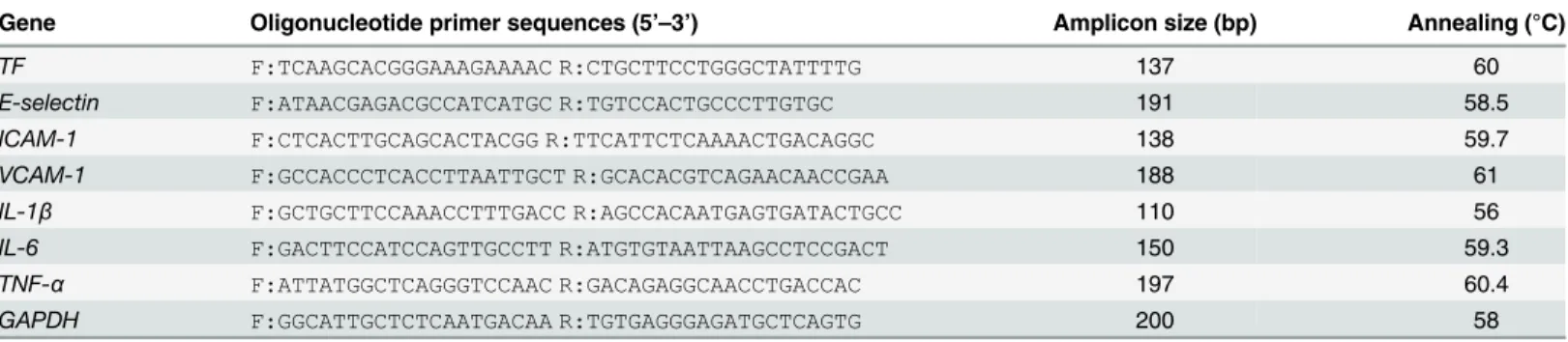
Biotec, Kyoto, Japan). The primers pairs used for PCR were shown inTable 1. Results were
expressed as fold differences relative to the level ofGAPDHusing the 2-ΔΔCTmethod.
Immunofluorescence
Mouse peritoneal macrophages were harvested and cultured in RPMI 1640 supplemented with
1% penicillin/streptomycin in 24 well flat-bottomed plates at 37°C and 5% CO2in a humidified
cell incubator. After 30 min, non-adherent cells were removed by washing. The adherent mac-rophages were fixed in 4% paraformaldehyde for 20 min at 37°C, and permeabilized with ice-cold 0.3% Triton X-100 for 10 min at room temperature (RT), and blocked in PBS containing 5% bovine serum albumin (BSA) for 1 h at RT. The cells were then incubated overnight at 4°C
with TF (1:200; Abcam), IL-1β(1:50; Santa Cruz), IL-6 (1:200; Cell Signaling Technology), and
TNF-αantibodies (1:200; Cell Signaling Technology), respectively. Subsequently, cells were
incubated with Cy3-conjugated goat anti-rabbit IgG (1:200, Santa Cruz) for 1 h at room tem-perature. Nuclear counterstaining was performed with DAPI for 10 min at RT. Immunofluo-rescence images were acquired using A Zeiss fluoImmunofluo-rescence microscope with 20×. All image acquisition parameters were kept constant throughout all experiments.
TF activity measurement
TF activity in mouse carotid artery homogenates and peritoneal macrophages was investigated
in our study. The carotid artery and peritoneal macrophages resuspended in 50μL of a TBS/
factor X activation by TF/VIIa complex by utilizing a commercial chromogenic assay (Assaypro,
Greenwich, CT, USA). The concentration of generated factor Xa was calculated in pM/106cells
in peritoneal macrophages and pM/mg protein in carotid artery homogenates, respectively.
Statistical analysis
All experimental points were performed in triplicate or quadruplicate, and all assays were repeated a minimum of 3 times. Normally distributed variables were expressed as means ± standard deviation (SD). Differences between control and experimental conditions were
assessed using the Student’s two-tailed t test for paired samples. For multiple group
compari-sons, we used ANOVA with Dunnett’s post test. All statistical analyses were performed using
SPSS statistical software package version 20.0 (SPSS, Chicago, IL, USA). Statistical significance
was defined asp<0.05.
Result
IgG-APS and anti-
β
2GPI induce phosphorylation of NF-κ
B and AP-1 in
mouse peritoneal macrophages
Our previous studies demonstrated that NF-κB and c-Jun/AP-1 are involved in anti-β2GPI/
β2GPI-induced tissue factor expression in monocytes [17], but these results have not been
vali-datedin vivo. Therefore, we firstly measured the phosphorylation of NF-κB and c-Jun/AP-1 in
the peritoneal macrophages from the male BALB/c mice injected with NR-IgG, or anti-β2GPI,
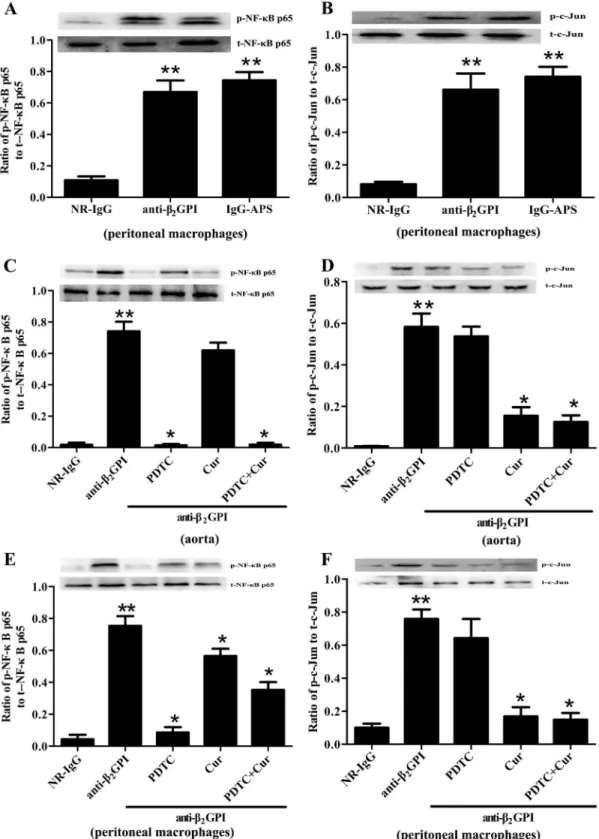
or IgG-APS. As shown inFig 1A and 1B, the phosphorylation of NF-κB p65 and c-Jun/AP-1
was markely elevated in the peritoneal macrophages from BALB/c mice after anti-β2GPI or
IgG-APS treatment, compared to control mice injected with NR-IgG. The phosphorylation
lev-els of NF-κB p65 and c-Jun in mice injected with anti-β2GPI were almost similar to those in
mice injected with anti-β2GPI or IgG-APS.
PDTC and Curcumin abrogate the phosphorylation of NF-
κ
B and AP-1 in
mice treated with anti-
β
2GPI
The anti-β2GPI-induced NF-κB and c-Jun/AP-1 phosphorylations were further validated by
using specific NF-κB and c-Jun/AP-1 inhibitorsin vivo. BALB/c mice were pretreated with
NF-κB inhibitor PDTC (100 mg/kg, once a day) by i.p. or AP-1 inhibitor Curcumin (50 mg/kg,
once a day) by oral gavage for 3 consecutive days at 2 h before anti-β2GPI injections in
subse-quent experiments. Then we harvested aorta and peritoneal macrophages from experimental
animals. Compared with the mice treated with anti-β2GPI alone, PDTC treatment dramatically
inhibited the anti-β2GPI-induced phosphorylation of NF-κB p65 in the aorta (Fig 1C) and
Table 1. Primers used for real-time qPCR analysis.
Gene Oligonucleotide primer sequences (5’–3’) Amplicon size (bp) Annealing (°C)
TF F:TCAAGCACGGGAAAGAAAAC R:CTGCTTCCTGGGCTATTTTG 137 60
E-selectin F:ATAACGAGACGCCATCATGC R:TGTCCACTGCCCTTGTGC 191 58.5
ICAM-1 F:CTCACTTGCAGCACTACGG R:TTCATTCTCAAAACTGACAGGC 138 59.7
VCAM-1 F:GCCACCCTCACCTTAATTGCT R:GCACACGTCAGAACAACCGAA 188 61
IL-1β F:GCTGCTTCCAAACCTTTGACC R:AGCCACAATGAGTGATACTGCC 110 56
IL-6 F:GACTTCCATCCAGTTGCCTT R:ATGTGTAATTAAGCCTCCGACT 150 59.3
TNF-α F:ATTATGGCTCAGGGTCCAAC R:GACAGAGGCAACCTGACCAC 197 60.4
GAPDH F:GGCATTGCTCTCAATGACAA R:TGTGAGGGAGATGCTCAGTG 200 58
Fig 1. The effects of PDTC and Curcumin on anti-β2GPI-mediated NF-κB and AP-1 phosphorylation.BALB/c mice (4 per group) were injected with NR-IgG (500μg) or IgG-APS (500μg) or anti-β2GPI (500μg) in the presence or absence of PDTC (100 mg/kg, once a day) or/and Curcumin (50 mg/kg, once
a day), as described in Materials and Methods. Aorta homogenates (C-D) and peritoneal macrophages lysates (A-B, E-F) were collected for analyzing the phosphorylation levels of NF-κB p65 and c-Jun by western blotting using specific NF-κB p65, p-NF-κB p65, c-Jun, p-c-Jun, and controlβ-actin antibodies, respectively. Shown are the pooled data of three separate experiments with similar results.**Statistically significant difference from NR-IgG group (p<0.05)
*statistically significant difference from the anti-β2GPI group (p<0.05).
peritoneal macrophages (Fig 1E). Curcumin treatment also obviously decreased the
anti-β2GPI-induced phosphorylation of NF-κB p65 in peritoneal macrophages (Fig 1E), and slightly
decreased but not statistically significant in the aorta (p>0.05). Futhermore, the anti-β2
GPI-induced phosphorylation of c-Jun in the aorta and peritoneal macrophages was inhibited
dramatically by Curcumin (Fig 1D and 1E). However, the levels of total NF-κB p65 and c-Jun
in the aorta and peritoneal macrophages from anti-β2GPI-treated mice were not affected
by PDTC and/or Curcumin. In addition, combination of PDTC and Curcumin didn’t show
amplified effects on inhibiting the phosphorylation of NF-κB p65 and c-Jun/AP-1.
PDTC and Curcumin inhibit TF expression and activity in mice treated
with anti-
β
2GPI
It has been widely reported that aPL can stimulate the upregulation of TF expression and
activ-ity in vascular endothelial cells and monocytes, thereby leading to increased thrombosis [10,
22–24]. To demonstrate the important roles of NF-κB and c-Jun/AP-1 in anti-β2GPI-induced
TF expression and its activityin vivo, we sought to examine the effect of PDTC and Curcumin
on TF expression in anti-β2GPI-treated mice. Aorta and peritoneal macrophages were obtained
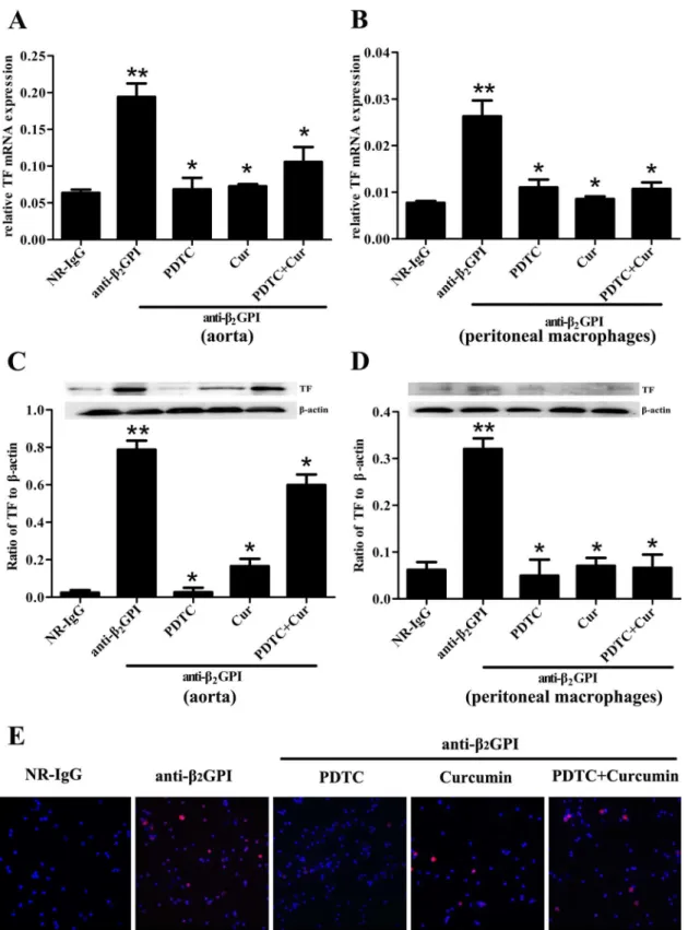
from BALB/c mice after different treatments, and were used to detect expression of TF mRNA and protein by RT-PCR, Western blotting and immunofluorescence. We found that
anti-β2GPI could significantly upregulate the expression ofTFmRNA (Fig 2A and 2B) and protein
(Fig 2C and 2D) in aorta and peritoneal macrophages compared with NR-IgG treated mice
(p<0.05). It seemed that anti-β2GPI-caused expressions of TF protein and mRNA in peritoneal
macrophage are lower than that in the aorta (Fig 2A–2D). Importantly, the pretreatment
with PDTC and/or Curcumin could significantly attenuate anti-β2GPI-induced TF expression
in aorta and peritoneal macrophages (Fig 2A–2D) (p<0.05). Among all treatment groups,
PDTC pretreatment showed the strongest inhibitory effect on the expression of anti-β2
GPI-inducedTFmRNA and protein in aorta and peritoneal macrophages. Furthermore, similar
results of TF protein expression in peritoneal macrophages were observed by
immunofluores-cence (Fig 2E).
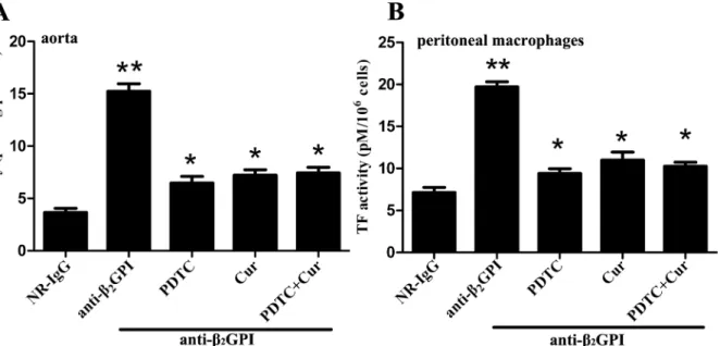
Next, we examined the influence of PDTC and Curcumin on anti-β2GPI-induced TF
activ-ityin vivo. TF activities in carotid artery homogenates (Fig 3A) and peritoneal macrophage
lysates (Fig 3B) from anti-β2GPI-treated mice were significantly higher than those in
NR-IgG-treated mice (p<0.05 vs NR-IgG). The TF activity in macrophage and aorta was almost similar
(Fig 3). However, PDTC and Curcumin pretreatments significantly blocked TF activity in
carotid artery homogenates (Fig 3A) and peritoneal macrophage lysates (Fig 3B) from
anti-β2GPI-treated mice (p<0.05,vs. anti-β2GPI group). However, the combined treatments of
PDTC and Curcumin had no synergistic effects on TF activity, consistent with the trend of NF-κB and c-Jun/AP-1 phosphorylation.
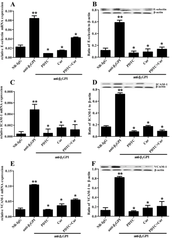
PDTC and Curcumin attenuate anti-
β
2GPI-induced expression ofE-selectin, ICAM-1 and VCAM-1
We further examined whether PDTC and Curcumin can attenuate anti-β2GPI—induced
endo-thelial cell (EC) activation by measuring the expression of E-selectin, ICAM-1 and VCAM-1 in
aortic homogenates. Relative mRNA expression ofE-selectin,ICAM-1andVCAM-1in aortas
is shown inFig 4A, 4C and 4E. Anti-β2GPI injection induced a significant increase in relative
mRNA expression ofE-selectin,ICAM-1andVCAM-1in aortic homogenates (p<0.05,vs.
NR-IgG). The elevated expression ofE-selectin,ICAM-1andVCAM-1mRNA in aortic
homog-enates from anti-β2GPI-treated mice was significantly blocked by PDTC and/or Curcumin
Fig 2. Anti-β2GPI-induced TF expression in mouse is diminished by PDTC or/and Curcumin.BALB/c mice (6 per group) were injected with NR-IgG (500μg) or anti-β2GPI (500μg) in the presence of PDTC (100 mg/kg, once a day) or/and Curcumin (50 mg/kg, once a day), as described in Materials and
group). Interestingly, the combination of PDTC and Curcumin treatment didn’t show enhanced inhibitory effects. Consistent with mRNA expression, the similar changes in protein expression of E-selectin, ICAM-1 and VCAM-1 were observed in aortic homogenates from
anti-β2GPI-treated mice after PDTC and/or Curcumin pretreatments (Fig 4B, 4D and 4F).
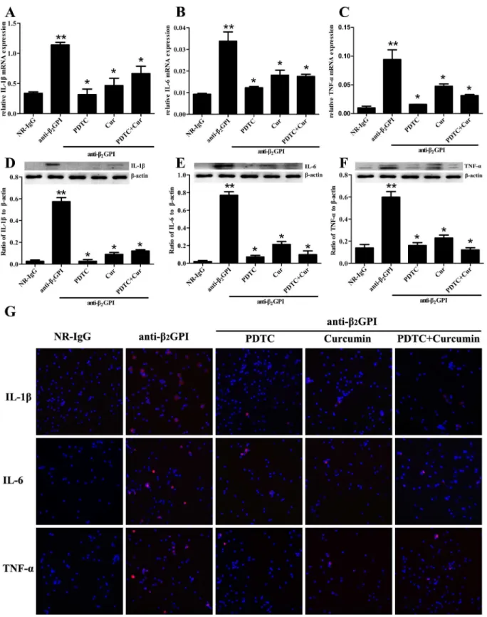
PDTC and Curcumin attenuate anti-
β
2GPI-induced expression of IL-1β
,
IL-6 and TNF-
α
We also determined whether PDTC and Curcumin abrogated anti-β2GPI—induced expression
of IL-1β, IL-6 and TNF-αin peritoneal macrophages by RT-PCR, Western blotting and
immu-nofluorescence staining. We found that anti-β2GPI could induce a significant upregulation
of relative mRNA levels ofIL-1β,IL-6andTNF-αin peritoneal macrophages (Fig 5A–5C,
p<0.05,vs. NR-IgG). However, the upregulated mRNA levels ofIL-1β,IL-6andTNF-αin the
peritoneal macrophages from anti-β2GPI-treated mice were significantly attenuated by
pre-treating the mice with PDTC and/or Curcumin, in which PDTC showed the strongest
inhibi-tory effects (Fig 5A–5C,p<0.05,vs. anti-β2GPI group). Curcumin could significantly restrain
IL-1βexpression, but not completely inhibit upregulation of IL-6 and TNF-αin the peritoneal
macrophages from anti-β2GPI-treated mice. Moreover, the combination of PDTC and
Curcu-min pretreatments didn’t show enhanced inhibitory effects. Western blotting results showed
that the elevated protein levels of IL-1β, IL-6 and TNF-αin peritoneal macrophage lysates
from anti-β2GPI-treated mice were obviously abrogated in the presence of PDTC and/or
Cur-cumin (Fig 5D–5F). Interestingly, the inhibitory effect of Curcumin on anti-β2GPI induced
expression of these inflammatory molecules seemed a little weaker than that of PDTC, but the
red fluorescence represents surface TF immunoreactivity, whereas the blue color confirms the presence of cells stained with DAPI reagent (original magnification ×200).**Statistically significant difference from NR-IgG group (p<0.05)*statistically significant difference from the anti-β2GPI group (p<0.05).
doi:10.1371/journal.pone.0147958.g002
Fig 3. Anti-β2GPI-induced TF activity in mouse is abrogated with PDTC or/and Curcumin treatment.BALB/c mice (4 per group) were injected with NR-IgG (500μg) or anti-β2GPI (500μg) in the presence of PDTC (100 mg/kg, once a day) or/and Curcumin (50 mg/kg, once a day), as described in Materials
and Methods. The carotid arteries (A) and peritoneal macrophages (B) were harvested from mice at 72 h after the frist injection, and TF activity in homogenates was determined by a commercial kit as described in Materials and Methods. Shown are representative of three independent experiments. **Statistically significant difference from NR-IgG group (p<0.05)*statistically significant difference from the anti-β2GPI group (p<0.05).
Fig 4. Inhibition of anti-β2GPI-induced E-selectin, ICAM-1 and VCAM-1 expression by PDTC or/and Curcumin.BALB/c mice (6 per group) were injected with NR-IgG (500μg) or anti-β2GPI (500μg) in the presence of PDTC (100 mg/kg, once a day) or/and Curcumin (50 mg/kg, once a day), as
described in Materials and Methods. Total RNAs were obtained from frozen aortic wall. The relative mRNA levels ofE-selectin(A),ICAM-1(C) andVCAM-1
(E) in aorta were respectively detected by RT-qPCR. Aorta homogenates were collected for analyzing the protein expression of E-selectin (B), ICAM-1 (D) and VCAM-1 (F) by Western blotting using specific E-selectin, ICAM-1, VCAM-1 and controlβ-actin antibodies, respectively. Shown are the pooled data of three separate experiments with similar results.**Statistically significant difference from NR-IgG group (p<0.05)*statistically significant difference from the
anti-β2GPI group (p<0.05).
Fig 5. The effects of anti-β2GPI antibodies on IL-1β, IL-6 and TNF-αexpression by PDTCor/ and Curcumin in mice.BALB/c mice (6 per group) were injected with NR-IgG (500μg) or anti-β2GPI (500μg) in the presence of PDTC (100 mg/kg, once a day) or/and Curcumin (50 mg/kg, once a day), as
described in Materials and Methods. Total RNAs was prepared from mouse peritoneal macrophage, and the relative mRNA levels ofIL-1β(A),IL-6(B) and
TNF-α(C) were respectively measured by RT-qPCR. Peritoneal macrophage lysates from mice of different treatment were collected for detection of IL-1β
combined treatments of PDTC and Curcumin didn’t show enhanced inhibitory effects (Fig
5D–5F). Immunofluorescence staining results (Fig 5G) showed similar expression pattern to
those of IL-1β, IL-6 and TNF-αproteins detected by Western Blot (Fig 5D–5F).
Discussion
It is widely accepted that anti-β2GPI antibodies play a pathogenic role in APS and have been
shown to induce activation of ECs,in vitroandin vivo, which may contribute to
hypercoagula-bility in APS patients [12,25–26]. Thrombin is correlated with the overall functional
coagula-tion status of plasma in APS patients, and induced expression of connective tissue growth
factors in rat vascular smooth muscle cells via the AP-1 pathway [27–28]. NF-κB may be
involved in aPL/anti-β2GPI-induced expression of TF and adhesion molecules in ECs and
monocytesin vitro[16,29–30]. Our previous results demonstrated that both NF-κB and
c-Jun/AP-1 are involved in anti-β2GPI/β2GPI-induced TF expression in monocytes [17]. In the
present study, we show for the first time that NF-κB and c-Jun/AP-1 are also involved in
anti-β2GPI-induced expression of prothrombotic and proinflammatory moleculesin vivo, and that
these effects can be attenuated by PDTC (a specific inhibitor of NF-κB) [31] and Curcumin (a
potent inhibitor of AP-1) [32].
Although the important role of NF-κB in aPL/anti-β2GPI-induced pathogenic mechanisms
has been widely recognized, this study specifically addressed the functions of anti-β2
GPI-induced NF-κB and c-Jun/AP-1 activationin vivo. Interestingly, both IgG-APS from APS
patients and polyclonal rabbit anti-human anti-β2GPI antibodies could activate a similar
degree of NF-κB p65 and c-Jun/AP-1 phosphorylation in the peritoneal macrophages of
BALB/c mice. These findings are consistent with our previousin vitroexperimental data in
monocytic cell line and THP-1 cells [17]. Hence, the polyclonal anti-β2GPI antibodies were
used in subsequent experiments. Furthermore, a specific NF-κB inhibitor PDTC and an AP-1 inhibitor Curcumin were administrated to the mice for a relatively short period of time (72 h).
PDTC and Curcumin could markedly attenuate anti-β2GPI-induced activation of NF-κB and
c-Jun/AP-1 in the aorta and peritoneal macrophages respectively. Meanwhile, Curcumin alone
also showed a significantly inhibitory effect on anti-β2GPI-induced NF-κB phosphorylation
although its inhibitory effect was obviously weaker than that of PDTC. Possible reason is that Curcumin functions are cell specific and also functions on NF-κB. For example, Curcumin can
inhibit LPS-induced inflammation by suppressing nuclear translocation of NF-κB in rat
vascu-lar smooth muscle cells [33]. It was also reported that Curcumin could inhibit the activation of
NF-κB and AP-1 in aortic endothelial cells [34].
Previous studies demonstrated that anti-β2GPI-dependent induction of TF activity and
expression in circulating blood monocytes and vascular endothelium are associated with the
hypercoagulability in APS [24,35]. In this study, we observed that anti-β2GPI-induced TF
pro-tein and mRNA expression in mouse peritoneal macrophage are lower than that in the aorta, but TF activity in macrophage and aorta was almost similar. The possible reason is because TF is mainly expressed by vascular cells, such as monocytes and endothelial cells. In addition, not all of expressed TF is active, and TF is normally expressed in cells or present in the blood in an encrypted form (inactive TF). This difference between TF protein and mRNA expression and
TF activity limits inappropriate activation of the blood coagulation cascade [36]. Moreover, it
is well established that NF-κB activation is required for aPL-induced TF upregulationin vitro
immunoreactivity, whereas the blue color represents cell nuclei stained with DAPI reagent (original magnification ×200). Shown are the pooled data of three separate experiments with similar results.**Statistically significant difference from NR-IgG group (p<0.05)*statistically significant difference from the anti-β2GPI group (p<0.05).
[16,37]. However, whether NF-κB and c-Jun/AP-1 are involved in prothrombotic effects of
anti-β2GPIin vivohas been rarely investigated. Therefore, we hypothesize that PDTC or
Cur-cumin could abrogate the procoagulant effects induced by anti-β2GPI antibodiesin vivo. As
expected, PDTC or Curcumin could significantly inhibit the upregulation of anti-β2
GPI-induced TF activity in carotid artery homogenates and peritoneal macrophages, in which PDTC exerted the strongest inhibitory role. Consistent with these results, treatment with
PDTC or Curcumin could also markedly inhibit anti-β2GPI-mediated expression ofTFmRNA
and protein in aortic homogenates and peritoneal macrophages. Likewise, PDTC showed the strongest inhibitory effect compared to other treatment groups. Interestingly, the combined
treatments of PDTC and Curcumin didn’t show enhanced inhibitory effect on anti-β2
GPI-induced TF expression and its activityin vivo. We speculate that both NF-κB and c-Jun/AP-1
play an indispensable role through their own target molecules in this process, since TF gene
promoter contains two AP-1 sites and a NF-κB site [38]. More importantly, this is the first
time to report that c-Jun/AP-1 can participate in anti-β2GPI-induced TF activity/expressionin
vivo. Therefore, we have sufficient reasons to believe that PDTC or Curcumin can decrease the
thrombogenic effects of anti-β2GPI antibodiesin vivothrough inhibiting the activation of
NF-κB and/or c-Jun/AP-1 signaling pathways.
Thrombosis is a devastating consequence and the most prominent clinical manifestation in
APS, which may affect any organ in the body [39]. Nonetheless, the induction of an endothelial
proinflammation and procoagulation by aPL has been widely accepted as a major pathogenic
mechanism underlying the prothrombotic properties [40]. The aPL-induced ECs activation
leads to loss of its anticoagulant properties and transformation to a pro-adhesive and procoa-gulant phenotype characterized by increased expression of adhesion molecules (E-selectin,
ICAM-1, and VCAM-1) [5,41–42]. Some recent studies reported that anti-β2GPI antibodies
can induce signaling transduction through a multiprotein complex including annexin A2 and TLR4, in which TLR4 can induce the activation of TLR4/myeloid differentiation factor 88
(MyD88)-dependent pathway and NF-κB signaling in ECs and moncytes [41–44]. Indeed, our
data demonstrated that anti-β2GPI antibodies upregulated the expression of ICAM-1,
VCAM-1 and E-selectin in the aortic homogenates of mice. Moreover, these effects were significantly blocked in the presence of PDTC or Curcumin, in which PDTC showed the strongest inhibi-tory effect on the expression of ICAM-1, VCAM-1 and E-selectin in the aorta from
anti-β2GPI-treated mice. We speculate that PDTC or Curcumin may inhibit anti-β2GPI-mediated
expression of ICAM-1, VCAM-1 and E-selectin in the aorta by inhibiting the activation of
NF-κB or/and c-Jun/AP-1, thus blocking the interaction of leukocytes and platelets with
endothe-liumin vivo, and finally suppressing the inflammatory response and the procoagulant state.
It has reported that high levels of IL-1β, IL-6 and TNF-αare present in serum of APS
patients, indicating the presence of a proinflammatory phenotype in the body [11,45]. Our
previous studies found that anti-β2GPI/β2GPI complexes promoted the expression of
proin-flammatory cytokines via TLR4/NF-κB signaling pathwayin vitro[43,46]. Here we further
validate that NF-κB and c-Jun/AP-1 can modulate anti-β2GPI-induced expression of IL-1β,
IL-6 and TNF-αin peritoneal macrophages from mice. The high expression of these
inflamma-tory cytokines can be significantly abolished by pretreatment of PDTC or Curcumin before the
anti-β2GPI injection. Moreover, PDTC can completely block their high expression, comparable
to their in the NR-IgG-treated mice. These data indicate that NF-κB and c-Jun/AP-1 not only
contribute to anti-β2GPI-induced procoagulant activity and expression of adhesion molecules,
but also mediate proinflammatory responsesin vivo. In the pathogenic mechanisms of APS,
NF-κB may play a vital role while AP-1 may play a supporting role, thus there may be an
ordinal relation between NF-κB and AP-1. Fujiokaet al. reported that NF-κB regulates the
Interestingly, the combined treatments of PDTC and Curcumin sometimes showed even less
inhibitory effects on anti-β2GPI-induced expression of prothrombotic and proinflammatory
molecules in mouse compared with their individual treatment alone. Possible explanation is that simultaneous administration of PDTC and Curcumin may arise some kinds of side effects in mouse body, or some interactions between two inhibitors may impair their original role. So
it’s not surprising that the combined pretreatments of PDTC and Curcumin did not show the
enhanced inhibitory effects compared with their individual treatment alone, suggesting that separate administration of PDTC or Curcumin may achieve optimal inhibitory effects on their target molecules.
Recent accumulated evidence has demonstrated that a“two hit hypothesis”has been widely
accepted to explain the clinical observation that thrombotic events occur occasionally in spite of the persistent presence of aPL. The hypothesis is that aPL (first hit) only increases the thrombophilic risk while the thrombosis will occur in the presence of another thrombophilic
conditions (second hit), such as inflammation and infection [22,48]. Our results provide solid
evidence to support this“two hit hypothesis”in APS. Moreover, the activation of NF-κB and
c-Jun/AP-1 is a crucial step in triggering the expression of prothrombotic and proinflammatory molecules during the pathogenesis of aPL-induced clinical manifestation. Additionally, the direct treatments for preventing thromboembolic events using antithrombotic medications and for modulating the immune response using immunotherapy have considerable side effects. However, there are also a lot of APS patients in whom aPL Abs are persistently present in the serum for a long period of time but thrombotic events only occur occasionally. Hence, anticoa-gulation is not always effective for all patients, otherwise it can increase a risk of hemorrhage.
Furthermore, agents like PDTC or Curcumin may act directly on the putative“first hit”to
ameliorate those effects, reduce the risk for developing clinical events when a“second hit”
occurs. However, whether PDTC can be used for clinical patient still needs further investiga-tion. It has been found that PDTC has some neurotoxicity and hepatotoxicity by generating
some toxic compounds, including CS2and pyrrolidine [49].
Conclusion
Our results demonstrated that both NF-κB and c-Jun/AP-1 are involved in regulating
anti-β2GPI-induced expression of prothrombotic and proinflammatory molecules in mouse model.
NF-κB plays an indispensable role in aPL-mediated pathogenic effects in APS, while c-Jun/AP-1 is also involved in this process. PDTC and Curcumin treatment may be considered as a new approach to treat and/or prevent thrombosis in APS patients.
Acknowledgments
The authors would like to acknowledge all members of the Hong Zhou team for collaboration to the project.
Author Contributions
Conceived and designed the experiments: LX HZ. Performed the experiments: LX HX YY. Analyzed the data: LX HX HZ TW. Contributed reagents/materials/analysis tools: LX HZ TW JY. Wrote the paper: LX HZ JY.
References
2. Miyakis S, Lockshin MD, Atsumi T, Branch DW, Brey RL, Cervera R, et al, International consensus statement on an update of the classification criteria for definite antiphospholipid syndrome (APS). J Thromb Haemost. 2006; 4(2): 295–306. PMID:16420554
3. Mehdi AA, Uthman I, Khamashta M. Antiphospholipid syndrome: pathogenesis and a window of treat-ment opportunities in the future. Eur J Clin Invest. 2010; 40(5): 451–464. doi:10.1111/j.1365-2362.
2010.02281.xPMID:20345380
4. Willis R, Pierangeli SS. Anti-β2-glycoprotein I antibodies. Ann N Y Acad Sci. 2013; 1285: 44–58. doi:
10.1111/nyas.12080PMID:23692565
5. Allen KL, Fonseca FV, Betapudi V, Willard B, Zhang J, McCrae KR. A novel pathway for human endo-thelial cell activation by antiphospholipid/anti-β2 glycoprotein I antibodies. Blood. 2012; 119(3): 884–
893. doi:10.1182/blood-2011-03-344671PMID:22106343
6. Arad A, Proulle V, Furie RA, Furie BC, Furie B.β2 glycoprotein-1 autoantibodies from patients with anti-phospholipid syndrome are sufficient to potentiate arterial thrombus formation in a mouse model. Blood. 2011; 117(12): 3453–3459. doi:10.1182/blood-2010-08-300715PMID:21245481
7. Ma K, Simantov R, Zhang JC, Silverstein R, Hajjar KA, McCrae KR. High affinity binding of beta2-glyco-protein I to human endothelial cells is mediated by annexin II. J Biol Chem. 2000; 275(20):15541–
15548. PMID:10809787
8. Raschi E, Borghi MO, Grossi C, Broggini V, Pierangeli S, Meroni PL. Toll-like receptors: another player in the pathogenesis of the antiphospholipid syndrome. Lupus. 2008; 17(10): 937–942. doi:10.1177/
0961203308095140PMID:18827059
9. Chaturvedi S, McCrae KR. Recent advances in the antiphospholipid antibody syndrome. Curr Opin Hematol. 2014; 21(5): 371–379. doi:10.1097/MOH.0000000000000067PMID:25023470
10. Clemens N, Frauenknecht K, Katzav A, Sommer C, von Landenberg P. In vitro effects of antiphospholi-pid syndrome-IgG fractions and human monoclonal antiphospholiantiphospholi-pid IgG antibody on human umbilical vein endothelial cells and monocytes. Ann N Y Acad Sci. 2009; 1173: 805–813. doi:
10.1111/j.1749-6632.2009.04632.xPMID:19758232
11. Forastiero RR, Martinuzzo ME, de Larranaga GF. Circulating levels of tissue factor and proinflamma-tory cytokines in patients with primary antiphospholipid syndrome or leprosy related antiphospholipid antibodies. Lupus. 2005; 14(2): 129–136. PMID:15751817
12. Pierangeli SS, Colden-Stanfield M, Liu X, Barker JH, Anderson GL, Harris EN. Antiphospholipid anti-bodies from antiphospholipid syndrome patients activate endothelial cells in vitro and in vivo. Circula-tion. 1999; 99(15):1997–2002. PMID:10209004
13. Pierangeli SS, Liu XW, Barker JH, Anderson G, Harris EN. Induction of thrombosis in a mouse model by IgG, IgM and IgA immunoglobulins from patients with the antiphospholipid syndrome. Thromb Hae-most. 1995; 74(5): 1361–1367. PMID:8607123
14. Jankowski M, Vreys I, Wittevrongel C, Boon D, Vermylen J, Hoylaerts MF, et al. Thrombogenicity of beta2-glycoprotein I-dependent antiphospholipid antibodies in a photochemically induced thrombosis model in the hamster. Blood. 2003; 101(1): 157–162. PMID:12393462
15. Vega-Ostertag ME, Ferrara DE, Romay-Penabad Z, Liu X, Taylor WR, Colden-Stanfield M, et al. Role of p38 mitogen-activated protein kinase in antiphospholipid antibody-mediated thrombosis and endo-thelial cell activation. J Thromb Haemos. 2007; 5(9): 1828–1834.
16. Vega-Ostertag M, Casper K, Swerlick R, Ferrara D, Harris EN, Pierangeli SS. Involvement of p38 MAPK in the up-regulation of tissue factor on endothelial cells by antiphospholipid antibodies. Arthritis Rheum. 2005; 52(5): 1545–1554. PMID:15880836
17. Xia L, Zhou H, Hu L, Xie H, Wang T, Xu Y, et al. Both NF-kappaB and c-Jun/AP-1 involved in
anti-β2GPI/β2GPI-induced tissue factor expression in monocytes. Thromb Haemost. 2013; 109(4): 643–
651. doi:10.1160/TH12-09-0655PMID:23467542
18. Wang S, Liu Z, Wang L, Zhang X. NF-kappa B signalling pathway, inflammation and colorectal cancer. Cell Mol Immunol. 2009; 6(5): 327–334. doi:10.1038/cmi.2009.43PMID:19887045
19. Guo D, Zhou H, Wu Y, Zhou F, Xu G, Wen H, et al. Involvement of ERK1/2/NF-κB signal transduction pathway in TF/FVIIa/PAR2-induced proliferation and migration of colon cancer cell SW620. Tumor Biol. 2011; 32(5): 921–930.
20. Kappelmann M, Bosserhoff A, Kuphal S. AP-1/c-Jun transcription factors: Regulation and function in malignant melanoma. Eur J Cell Biol. 2014; 93(1–2): 76–81. doi:10.1016/j.ejcb.2013.10.003PMID:
24315690
21. Vaiopoulos AG, Papachroni KK, Papavassiliou AG. Colon carcinogenesis: Learning from NF-κB and AP-1. Int J Biochem Cell Biol. 2010; 42(7): 1061–1065. doi:10.1016/j.biocel.2010.03.018PMID:
22. Xie H, Sheng L, Zhou H, Yan J. The role of TLR4 in pathophysiology of antiphospholipid syndrome-associated thrombosis and pregnancy morbidity. Br J Haematol. 2014; 164(2): 165–176. doi:10.1111/
bjh.12587PMID:24180619
23. Borghi MO, Raschi E, Grossi C, Chighizola CB, Meroni PL. Toll-like receptor 4 andβ2 glycoprotein I interaction on endothelial cells. Lupus. 2014; 23(12): 1302–1304. doi:10.1177/0961203314536479
PMID:25228733
24. Boles J, Mackman N. Role of tissue factor in thrombosis in antiphospholipid antibody syndrome. Lupus. 2010; 19(4): 370–378. doi:10.1177/0961203309360810PMID:20353972
25. Zhang J, McCrae KR. Annexin A2 mediates endothelial cell activation by antiphospholipid/anti-β 2glyco-protein I antibodies. Blood. 2005; 105(5): 1964–1969. PMID:15471954
26. Sorice M, Longo A, Capozzi A, Garofalo T, Misasi R, Alessandri C, et al. Anti–β2Glycoprotein I
antibod-ies induce monocyte release of tumor necrosis factorαand tissue factor by signal transduction path-ways involving lipid rafts. Arthritis Rheum. 2007; 56(8): 2687–2697. PMID:17665396
27. Efthymiou M, Lawrie AS, Mackie I, Arachchillage D, Lane PJ, Machin S, et al. Thrombin generation and factor X assays for the assessment of warfarin anticoagulation in thrombotic antiphospholipid syn-drome. Thromb Res. 2015; 135(6):1191–1197. doi:10.1016/j.thromres.2015.03.030PMID:25895847
28. Ko WC, Chen BC, Hsu MJ, Tsai CT, Hong CY, Lin CH. Thrombin induced connective tissue growth fac-tor expression in rat vascular smooth muscle cells via the PAR-1/JNK/AP-1 pathway. Acta Pharmacol Sin. 2012; 33(1): 49–56. doi:10.1038/aps.2011.178PMID:22212430
29. Dunoyer-Geindre S, de Moerloose P, Galve-de Rochemonteix B, Reber G, Kruithof EK. NF-κB is an essential intermediate in the activation of endothelial cells by anti-β2glycoprotein I antibodies. Thromb
Haemost. 2002; 88(5): 851–857. PMID:12428105
30. Bohgaki M, Atsumi T, Yamashita Y, Yasuda S, Sakai Y, Furusaki A, et al. The p38 mitogen-activated protein kinase (MAPK) pathway mediates induction of the tissue factor gene in monocytes stimulated with human monoclonal anti-β2glycoprotein I antibodies. Int Immunol. 2004; 16(11): 1633–1641.
PMID:15466912
31. Zhang X, Wu M, Jiang H, Hao J, Zhang Q, Zhu Q, et al. Angiotensin II upregulates endothelial lipase expression via the NF-kappa B and MAPK signaling pathways. PLoS One. 2014; 9(9): e107634. doi:
10.1371/journal.pone.0107634PMID:25250890
32. Seol DW, Chen Q, Zarnegar R. Transcriptional activation of the hepatocyte growth factor receptor (c-met) gene by its ligand (hepatocyte growth factor) is mediated through AP-1. Oncogene. 2000; 19(9): 1132–1137. PMID:10713700
33. Meng Z, Yan C, Deng Q, Gao DF, Niu XL. Curcumin inhibits LPS-induced inflammation in rat vascular smooth muscle cells in vitro via ROS-relative TLR4-MAPK/NF-κB pathways. Acta Pharmacol Sin. 2013; 34(7): 901–911. doi:10.1038/aps.2013.24PMID:23645013
34. Bierhaus A, Zhang Y, Quehenberger P, Luther T, Haase M, Müller M, et al. The dietary pigment curcu-min reduces endothelial tissue factor gene expression by inhibiting binding of AP-1 to the DNA and acti-vation of NF-kappa B. Thromb Haemost. 1997; 77(4): 772–782. PMID:9134658
35. Seshan SV, Franzke CW, Redecha P, Monestier M, Mackman N, Girardi G. Role of tissue factor in a mouse model of thrombotic microangiopathy induced by antiphospholipid antibodies. Blood. 2009; 114 (8): 1675–1683. doi:10.1182/blood-2009-01-199117PMID:19535796
36. Mackman N. Role of tissue factor in hemostasis and thrombosis. Blood Cells Mol Dis. 2006; 36(2): 104–107.
37. López-Pedrera C, Buendía P, Cuadrado MJ, Siendones E, Aguirre MA, Barbarroja N, et al. Antipho-spholipid antibodies from patients with the antiphoAntipho-spholipid syndrome induce monocyte tissue factor expression through the simultaneous activation of NF-kB/Rel proteins via the p38 mitogen-activated protein kinase pathway, and of the MEK-1/ERK pathway. Arthritis Rheum. 2006; 54(1): 301–311.
38. Mackman N. Regulation of the tissue factor gene. FASEB J. 1995; 9(10): 883–889.
39. Cervera R, Serrano R, Pons-Estel GJ, Ceberio-Hualde L, Shoenfeld Y, de Ramón E, et al. Euro-Phos-pholipid Project, Morbidity and mortality in the antiphosEuro-Phos-pholipid syndrome during a 10-year period: a multicentre prospective study of 1000 patients. Ann Rheum Dis. 2015; 74(6): 1011–1018. PMID:
24464962
40. Colasanti T, Alessandri C, Capozzi A, Sorice M, Delunardo F, Longo A, et al. Autoantibodies specific to a peptide of beta2-glycoprotein I cross-react with TLR4, inducing a proinflammatory phenotype in endo-thelial cells and monocytes. Blood. 2012; 120(16): 3360–3370. doi:10.1182/blood-2011-09-378851
PMID:22932793
42. Raschi E, Testoni C, Bosisio D, Borghi MO, Koike T, Mantovani A, et al. Role of the MyD88 transduction signaling pathway in endothelial activation by antiphospholipid antibodies. Blood. 2003; 101(9): 3495–
3500. PMID:12531807
43. Zhou H, Sheng L, Wang H, Xie H, Mu Y, Wang T, et al. Antib2GPI/b2GPI stimulates activation of THP-1 cells through TLR4/MD-2/MyD88 and NF-κB signaling pathways. Thromb Res. 2013; 132(6): 742–
749. PMID:24157085
44. Zhou H, Ling S, Yu Y, Wang T, Hu H. Involvement of annexin A2 in anti-beta2GPI/beta2GPI-induced tissue factor expression on monocytes. Cell Res. 2007; 17(8): 737–739. PMID:17438552
45. Erkan D, Willis R, Murthy VL, Basra G, Vega J, Ruiz-Limón P, et al. A prospective open-label pilot study of fluvastatin on proinflammatory and prothrombotic biomarkers in antiphospholipid antibody positive patients. Ann Rheum Dis. 2014; 73(6): 1176–1180. doi:10.1136/annrheumdis-2013-203622PMID:
23933625
46. Xie H, Zhou H, Wang H, Chen D, Xia L, Wang T, et al. Anti-beta(2)GPI/beta(2)GPI induced TF and TNF-alpha expression in monocytes involving both TLR4/MyD88 and TLR4/TRIF signaling pathways. Mol Immunol. 2013; 53(3): 246–254. doi:10.1016/j.molimm.2012.08.012PMID:22964479
47. Fujioka S, Niu J, Schmidt C, Sclabas GM, Peng B, Uwagawa T, et al. NF-κB and AP-1 connection: mechanism of NF-κB-dependent regulation of AP-1 activity. Mol Cell Biol. 2004; 24(17): 7806–7819.
PMID:15314185
48. Meroni PL, Raschi E, Grossi C, Pregnolato F, Trespidi L, Acaia B, et al. Obstetric and vascular APS: same autoantibodies but different diseases? Lupus. 2012; 21(7): 708–710. doi:10.1177/
0961203312438116PMID:22635208
49. Chabicovsky M, Prieschl-Grassauer E, Seipelt J, Muster T, Szolar OH, Hebar A, et al. Pre-clinical safety evaluation of pyrrolidine dithiocarbamate. Basic Clin Pharmacol Toxicol. 2010; 107(3): 758–