Amphibians have evolved an array of adaptive structures and mechanisms to cope with environmental changes that result from their life histories, which involves a transition from water to land (FOXON 1964, WOJTASZEK & ADAMOWICZ 2003). One of these adaptations is unusually large erythrocytes, compared to other vertebrates (WOJTASZEK & ADAMOWICZ 2003). Most pre-vious hematological studies on amphibians counted blood cells (ARSERIM & MERMER 2008, BARAQUET et al. 2013, DÖNMEZ et al. 2009) and measured their dimensions (DAS & MAHAPATRA 2012, MA et al. 2003, MAHAPATRA et al. 2012). Both intrinsic (e.g., species, sex, age and physiological state, ATTADEMO et al. 2014, HOTA et al. 2013, LAJMANOVICHA et al. 2014) and extrinsic factors (e.g., temperature and habitat, LOPEZ-OLIVERA et al. 2003) can affect blood parameters (e.g., the blood volume, hematocrit value, fragility and pH value; see ROUF 1969). For example, the num-ber of erythrocytes differ not only among individuals within population and interspecies, but also with body mass, age and sex of individuals (ARIKAN et al. 2003, BANERJEE 1988, CHOUBEY et al. 1986, DAS & MAHAPATRA 2014), habitat conditions (ROMANOVA & EGORIKHINA 2006), and season (SAMANTARAY 1985, WOJTASZEK et al. 1997). Therefore, investigating blood parameters in amphib-ians can facilitate evaluations of the physiological and health levels of populations. These in turn may be used as bio-indica-tors of environmental conditions, since these parameters ex-hibit significant variability when individuals inhabit unstable environments (BARNI et al. 2007, DICKINSON et al. 2002).
Despite the fact that haematological profiles have been reported for many amphibians, reconstruction of the evolu-tionary history of traits of amphibian erythrocytes is rare. Here, we compare the morphology of the erythrocytes of five sym-patric anuran species, including two toads – Bufo gargarizans (Cantor, 1842), Duttaphrynus melanostictus (Schneider, 1799) – and three frogs – Fejervarya limnocharis (Gravenhorst, 1829), Microhyla ornata (Duméril & Bibron, 1841), and Rana zhenhaiensis (Ye, Fei & Matsui, 1995) –, sampled from natural populations in Lishui, Zhejiang Province, China. These results were combined with recently published accounts on erythro-cyte traits (erythroerythro-cyte size and nucleus sizes) from three Or-ders (Gymnophiona, Caudata and Anura) of Amphibia to allow reconstruction of ancestral states and to examine their phylo-genetic relationships.
MATERIAL AND METHODS
From June to August, 2013, we captured 10 adults of each of the following species, B. gargarizans, D. melanostictus, F. limnocharis, M. ornata and R. zhenhaiensis, from field of Lishui, Zhejiang Province, China (28°27’N, 119°53’E). Their snout-vent length (SVL) was 57.5 ± 4.6, 52.5 ± 2.4, 39.2 ± 2.1, 28.6 ± 0.6 and 41.4 ± 1.9 mm, respectively. All individuals were trans-ported to the Herpetological Laboratory of the Lishui Univer-sity (HLLSU), where they were identified and used for
Evolution of erythrocyte morphology in amphibians (Amphibia: Anura)
Jie Wei
1, Yan-Yan Li
1, Li Wei
2, Guo-Hua Ding
2, Xiao-Li Fan
2& Zhi-Hua Lin
2,*1School of Life and Environmental Sciences, Hangzhou Normal University, Hangzhou, Zhejiang, 310036, China
2Institute of Ecology and Biological Resources, College of Ecology, Lishui University, Lishui, Zhejiang 323000, China.
*Corresponding author. E-mail: zhlin1015@126.com
ABSTRACT.We compared the morphology of the erythrocytes of five anurans, two toad species – Bufo gargarizans (Cantor, 1842) and Duttaphrynus melanostictus (Schneider, 1799) and three frog species – Fejervarya limnocharis (Gravenhorst, 1829), Microhyla ornata (Duméril & Bibron, 1841), and Rana zhenhaiensis (Ye, Fei & Matsui, 1995). We then reconstructed the ancestral state of erythrocyte size (ES) and nuclear size (NS) in amphibians based on a molecular tree. Nine morphological traits of erythrocytes were all significantly different among the five species. The results of principal component analysis showed that the first component (49.1% of variance explained) had a high positive loading for erythrocyte length, nuclear length, NS and ratio of erythrocyte length/erythrocyte width; the second axis (28.5% of variance explained) mainly represented erythrocyte width and ES. Phylogenetic generalized least squares analysis showed that the relationship between NS and ES was not affected by phylogenetic relationships although there was a significant linear relationship between these two variables. These results suggested that (1) the nine morphologi-cal traits of erythrocytes in the five anuran species were species-specific; (2) in amphibians, larger erythrocytes generally had larger nuclei.
preparation of blood smears. Vouchers of B. gargarizans are under accession numbers 2013071001 to HLLSU-2013071010; D. melanostictus from HLLSU-2013072001 to HLLSU-2013072010; F. limnocharis from HLLSU-2013073001 to HLLSU-2013073010; M. ornata from HLLSU-2013074001 to 2013074010; and R. zhenhaiensis from HLLSU-2013075001 to HLLSU-2013075010.
According to the methods of SALAMAT et al. (2013), blood smears were obtained by puncturing the heart of each indi-vidual. Blood smears were air-dried, fixed in methanol and stained with 10% Giemsa (diluted 1:10 in PBS, pH = 6.8) for 15 minutes and washed in running tap water for 2 minutes. Pho-tos of 100 erythrocytes were taken randomly using a camera attached to a microscope. The morphological traits of erythro-cytes, including erythrocyte length (EL) and width (EW), nuclear length (NL) and nuclear width (NW), were measured using ImageJ 1.43 software. Subsequently, erythrocyte size (ES) and nuclear sizes (NS) were calculated as ES = [(NL × NW × ð)/ 4, µm2] and NS = [(NL × NW × ð)/4, µm2], respectively.
Eryth-rocyte and nuclear shape were compared with EL/EW and NL/ NW ratios and nucleocytoplasmic ratio with NS/ES ratio (SALAMAT et al. 2013, SEVINÇ et al. 2004).
Prior to statistics, all variables were tested for normality and homogeneity. We used linear regression, one-way ANOVA, principal components analysis and Tukey’s post hoc compari-sons to analyze the data. Throughout this paper, values are presented as mean ± SE, and the significance level is set at ␣ = 0.05. All statistical analyses were performed with the Statistica software (version 6.0 for PC, Tulsa, OK, USA).
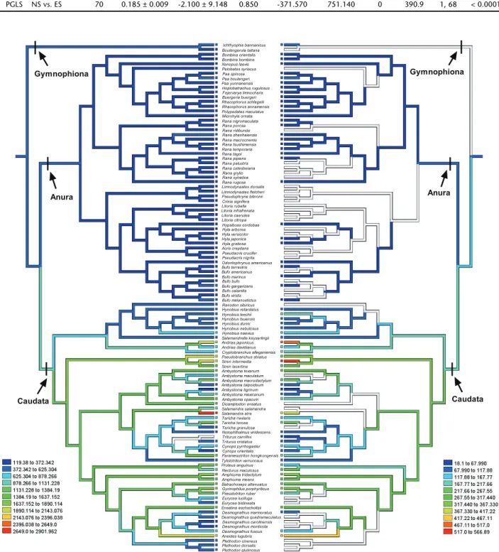
The tests detailed previously were carried out using the topology including all collected amphibian species from Gymnophiona, Caudata and Anura. This topology of species was based on proximate phylogenetic correlation assembled from PYRON & WIENS (2011). We drew the tree and reconstructed the evolutionary history of ES and NS of amphibians by parsi-mony ancestral states in the program Mesquite 2.75 (MADDISON & MADDISON 2011). Because branch lengths lacked divergence time and genetic distance and any other metric proportional to the expected variance for the evolution of each analyzed trait were unavailable, we arbitrarily set the initial branch length to 1, which is appropriate for a speciation model of evolution (MARTINS & GARLAND 1991).
We used ordinary least squares (OLS) and phylogenetic general least squares (PGLS) regressions to estimate the slope for all conventional analyses. These two analyses were imple-mented in R 2.15.3 (R Development Core Team 2013), using the RMS (HARRELL 2012) and Caper (ORME et al. 2012) pack-ages. We used PGLS regression to examine the relationship between NS and ES in amphibians. The PGLS analyses incor-porate phylogenetic information into generalized linear mod-els. They offer a powerful method for analyzing continuous data, and have been applied to estimate the evolutionary model and the relationships among the traits of interest
(BARROS et al. 2011, WARNE & CHARNOV 2008). In PGLS, the strength and type of the phylogenetic signal in the data ma-trix can be accounted for by adjusting branch length trans-formations, which show the degree of phylogenetic correlation in the data. In this study, we used from a maxi-mum likelihood approach to evaluate the phylogenetic ef-fects ( = 0 indicates no phylogenetic effect, and = l indicates the strongest phylogenetic effect equivalent to that expected under the Brownian motion model). We used the Akaike In-formation Criterion (AIC) to estimate merits and drawbacks of the models tested. The best model has the lowest AIC. The model with better ût can be determined by a maximum-like-lihood ratio test in which twice the difference in the natural log of the maximum likelihoods (LnL) of OLS and PGLS mod-els will be distributed approximately as a 2 with degrees of
freedom equal to the difference in the number of parameters estimated in the two models (WARNE & CHARNOV 2008).
RESULTS
Morphological traits of erythrocyte
The erythrocytes of the five anuran species are oval, and their morphological traits are depicted in Table 1. The results of One-way ANOVA indicate that the nine variables of eryth-rocyte morphology were all significantly different among the five species (Table 1). We found that (1) the mean values of EL and ratio of EL/EW and NL/NW were largest in D. melanostictus and smallest in F. limnocharis, the mean value of EW was larger in B. gargarizans than in the other species, the mean value of ES was larger in B. gargarizans and D. melanostictus than in the other species; (2) the mean values of NL and NS were largest in D. melanostictus and smallest in F. limnocharis and M. ornata, the mean value of NW was largest in B. gargarizans and small-est in M. ornata; (3) the mean value of nucleo-cytoplasmic ra-tio was largest in D. melanostictus and R. zhenhaiensis and smallest in M. ornata (Table 1). The variable coefficient was significantly different in NW (F4, 45 = 4.59, p < 0.01, Fig. 1), but not in other erythrocyte morphological traits among the five species (all p > 0.05). The variable coefficient of NW was sig-nificantly larger in D. melanostictus and R. zhenhaiensis than in B. gargarizans, with F. limnocharis and M. ornata in between (Fig. 1).
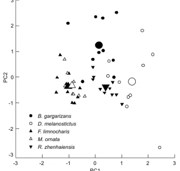
A principal component analysis resolved two compo-nents (eigenvalues ⭓ 1) from nine variables of erythrocyte morphology, accounting for 77.6% of the variation in the origi-nal data (Table 2). The first component (49.1% of variance ex-plained) had high positive loading for EL, NL, NS and ratio of EL/EW. The second axis (28.5% of variance explained) mainly represented EW and ES. Erythrocyte morphology differed sig-nificantly among the five anuran species in their scores on the first axis (F4, 45 = 45.95, p < 0.0001; BG
b, DMª, FLc, MOc, RZb,
Table 1. Descriptive statistics, expressed as mean ± SE and range, for morphological traits of erythrocytes in five anuran species in Lishui, China, and results of one-way ANOVA for each variable of erythrocytes with species as the factor.
Variables B. gargarizans D. melanostictus F. limnocharis M. ornata R. zhenhaiensis Results of statistical analyses
Erythrocyte length (EL, µm) 28.17 ± 0.46 30.02 ± 0.90 23.92 ± 0.22 25.20 ± 0.22 26.96 ± 0.37 F4, 45 = 23.02, p < 0.0001
25.20 – 30.27 26.79 – 36.38 22.56 – 24.82 24.20 – 26.19 25.31 – 29.16 BGab, DMa, FLd, MOcd, RZbc
Erythrocyte width (EW, µm) 20.18 ± 0.50 18.30 ± 0.48 17.71 ± 0.28 18.06 ± 0.21 18.02 ± 0.22 F4, 45 = 7.50, p < 0.001
18.30 – 22.28 15.40 – 21.16 16.65 – 19.70 17.40 – 19.37 17.34 – 19.61 BGa, DMb, FLb, MOb, RZb
Ratio of EL/EW 1.41 ± 0.03 1.66 ± 0.05 1.37 ± 0.03 1.40 ± 0.02 1.51 ± 0.04 F4, 45 = 13.23, p < 0.0001
1.26 – 1.56 1.53 – 1.96 1.25 – 1.50 1.31 – 1.50 1.35 – 1.70 BGbc, DMa, FLc, MObc, RZb
Erythrocyte size (ES, µm2) 447.56 ± 14.87 433.97 ± 21.79 333.18 ± 6.70 358.10 ± 5.99 382.29 ± 4.24 F4, 45 = 15.01, p < 0.0001
363.21 – 516.81 338.67 – 560.78 305.69 – 379.34 333.67 – 387.55 363.56 – 404.21 BGa, DMa, FLb, MOb, RZb
Nucleus length (NL, µm) 10.49 ± 0.28 12.54 ± 0.32 9.20 ± 0.15 9.57 ± 0.15 11.32 ± 0.20 F4, 45 = 34.31, p < 0.0001
8.70 – 11.71 11.00 – 14.74 8.62 – 9.89 8.90 – 10.38 10.28 – 12.47 BGb, DMa, FLc, MOc, RZb
Nucleus width (NW, µm) 6.46 ± 0.19 6.30 ± 0.16 5.88 ± 0.112 5.36 ± 0.09 6.18 ± 0.12 F4, 45 = 9.67, p < 0.0001
5.90 – 7.92 5.67 – 7.50 5.36 – 6.51 4.89 – 5.74 5.71 – 6.82 BGa, DMab, FLbc, MOc, RZab
Ratio of NL/NW 1.65 ± 0.04 2.04 ± 0.05 1.60 ± 0.03 1.82 ± 0.04 1.88 ± 0.05 F4, 45 = 16.07, p < 0.0001
1.44 – 1.81 1.80 – 2.37 1.49 – 1.85 1.57 – 2.02 1.53 – 2.19 BGcd, DMa, FLd, MObc, RZab
Nucleus size (NS, µm2) 53.59 ± 2.74 62.36 ± 2.70 42.62 ± 1.32 40.41 ± 0.94 55.01 ± 1.24 F4, 45 = 22.02, p < 0.0001
40.34 – 69.99 50.57 – 77.05 38.23 – 50.74 36.33 – 44.34 49.30 – 60.38 BGb, DMa, FLc, MOc, RZab
Ratio of NS/ES 0.12 ± 0.01 0.15 ± 0.01 0.13 ± 0.01 0.11 ± 0.00 0.15 ± 0.00 F4, 45 = 5.03, p < 0.01
0.09 – 0.15 0.12 – 0.22 0.11 – 0.17 0.09 – 0.13 0.13 – 0.17 BGab,DMa,FLab, MOb, RZa
BG: B. gargarizans, DM: D. melanostictus, FL: F. limnocharis, MO: M. ornata, RZ: R. zhenhaiensis. Means with different superscripts differ significantly (Tukey’s post hoc test ␣ = 0.05, a > b > c).
Figure 1 The variable coefficients of nucleus width of five species. BG: B. gargarizans, DM: D. melanostictus, FL: F. Limnocharis, MO: M. Ornata, RZ: R. zhenhaiensis. Different superscripts indicate sig-nificant difference (Tukey’s post hoc test, ␣ = 0.05, a > b).
Variability of erythrocyte morphology in amphibians
We assembled published data with our own data on ES, NS for amphibians (Appendix 1). Data from 109 species of amphibians show that mean ES ranged from 119.4 µm2 to
2649 µm2 (N = 108) and the mean NS ranged from 18.1 µm2 to
517 µm2 (N = 71). Our reconstruction of evolutionary changes
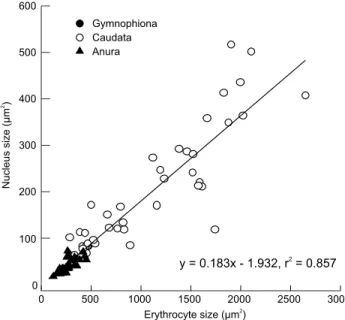
in these variables shows strong positive correlations between NS and ES in amphibians (Fig. 3). The ES and the NS were both significantly different among the three orders of Amphibia (Both p < 0.01). Both traits were greater in Caudata than in Gymnophiona and Anura (Fig. 4). Table 3 summarizes the re-lationships between NS and ES in amphibians according to OLS and PGLS analyses. Mean NS was positively correlated with mean ES in both the OLS and PGLS model (Fig. 5, Table 3). PGLS analysis showed that phylogenetic relationships did not affect NS and ES ( = 0) although there were significant linear relationship between NS and ES (Fig. 5, Table 3).
DISCUSSION
Hematological parameters vary significantly among am-phibian species (ARIKAN et al. 2010, BARAQUET et al. 2013). For example, OLMO & MORESCALCH (1975) documented that inter-specific variation is significant in the volume of erythrocytes and nuclei of seven Plethodontidae (Amphibia: Urodela) spe-cies. In our study, we found species-specificity in nine mor-phological traits of erythrocytes in the five anuran species. In general, variation in the morphological traits of erythrocytes in toads (B. gargarizans and D. melanostictus) was larger than in frogs (F. limnocharis, M. ornata, and R. zhenhaiensis). Further-more, GÜL et al. (2011) found that the number of erythrocytes is also different in toads and frogs. The mean value of erythro-cyte counts was greater in toads (Pseudepidalea viridis and Pelobates syriacus; n = 850530/µl; GÜL et al. 2011) than in frogs
(Hyla arborea, Rana dalmatina and Pelophylax ridibundus; n = 741332/µl; GÜL et al. 2011). The morphological traits of eryth-rocytes were different between toads and frogs and this differ-ence may be attributed to the following three reasons. First, the different habitats of toads and frogs may affect the vari-ability of erythrocyte morphology (ROMANOVA & EGORIKHINA 2006). Toads mainly inhabit terrestrial environments, whereas frogs inhabit semi-aquatic or aquatic environments (GÜL et al. 2011). The terrestrial habitat has selected a series of adaptive structures and mechanisms in frogs that have enabled them to function under conditions of changeable humidity and par-tial oxygen pressure in terrestrial environments (BARAQUET et al. 2013, FOXON 1964, WOJTASZEK & ADAMOWICZ 2003). Second, erythrocyte size may be dependent on the level of metabolism in vertebrates (WOJTASZEK & ADAMOWICZ 2003). Through our field investigation, we found that two toad species (B. gargarizans and D. melanostictus) that crawl slowly and have lower meta-bolic rate consume less energy than the other three species that are agile in their jumping and swimming activity. There-fore, erythrocyte morphology may have evolved to adapt to various levels of activity in vertebrates. Finally, the body size of animals influences erythrocyte size (FRÝDLOVÁ et al. 2012). In our study, the means obtained for the snout-vent length of two toad species (B. gargarizans and D. melanostictus) were greater than the means of the other three frog species (F. limnocharis, M. ornata, and R. zhenhaiensis); this distinction was consistent with erythrocyte size. This finding is logical from a physiological point of view, since smaller erythrocytes have relatively larger surface areas, and therefore, exchange oxygen more efficiently. It is reasonable to expect that erythrocyte size is adjusted to the actual mass-specific metabolic rate that gradu-ally decreases during ontogenetic growth (CLEMENTE et al. 2009, SMITH et al. 2008).
The morphological traits of erythrocytes are variable among individuals of a species. HOTA et al. (2013) found that the erythrocyte profile of M. ornata is variable during the lar-val and adult periods. The coefficient of variation (CV) indi-cated that the level of difference varied among individuals in the same species. Our results showed that the mean values of CV of NW in D. melanostictus and R. Zhenhaiensis were greater than in B. gargarizans (Fig. 1). These differences may be attrib-uted to the different habitats (RUIZ et al. 1983, SALAMAT et al. 2013) and/or variable activity levels (ALLANDER & FRY 2008, SYKES & KLAPHAKE 2008). Moreover, erythrocyte morphology varies with geography in amphibian species. We pooled erythrocyte size data on B. gargarizans from previous studies and our cur-rent study, and found that the erythrocyte profile (EL and EW) differed among three populations from different sampling lo-cations (GUO et al. 2002, ZHOU et al. 2011). The EL and EW of B. gargarizans in Lishui (28°27’N, 119°53’E) were greater than in Chongqing (29°81’N, 106°39’E, GUO et al. 2002), which were greater than in Shuicheng (26.58’N, 104°82’E, ZHOU et al. 2011). However, erythrocyte shape (ratio of EL/EW) showed an
op-Table 2. Loading of the first two axes of a principal component analysis on nine variables of erythrocyte morphology.
Factor loading
PC 1 PC 2
Erythrocyte length (EL) 0.789* 0.403
Erythrocyte width (EW) 0.052 0.974*
Ratio of EL/EW 0.776* -0.354
Erythrocyte size (ES) 0.549 0.789*
Nucleus length (NL) 0.967* -0.184
Nucleus width (NW) 0.592 0.377
Ratio of NL/NW 0.632 -0.490
Nucleus size (NS) 0.924* 0.059
Ratio of NS/ES 0.592 -0.535
Variance explained 49.1% 28.5%
Table 3. Regressions of nuclear sizes (NS) on erythrocyte size (ES) in amphibians based on ordinary least squares (OLS) regression and phylogenetic generalized least squares (PGLS) regression. Significant associations between variables are shown in bold.
Models N Slope Elevation r2 ln likelihood AIC F df p
OLS NS vs. ES 70 0.183 ± 0.009 -1.932 ± 8.638 0.857 -367.534 741.068 – 407.44 1, 68 < 0.0001
PGLS NS vs. ES 70 0.185 ± 0.009 -2.100 ± 9.148 0.850 -371.570 751.140 0 390.9 1, 68 < 0.0001
posite trend in the three populations (Lishui: 1.41; Chongqing: 1.50; Shuicheng: 1.57). These geographic variations in eryth-rocyte morphological traits may be associated with differences in latitude, elevation, or environmental and climatic variables in different sampling locations (GOODMAN et al. 2013).Previous studies have found that morphological variation in the eryth-rocyte traits of amphibians was greater than that in mammals, birds and reptiles (DUELLMAN & TRUEB 1994, GREGORY 2001a, LI et al. 1989, SEVINÇ et al. 2004, WU et al. 1998). Erythrocyte size in animals is generally negatively correlated with the place where the species appears in an evolutionary tree (whether more basal or more apical, indicating a more recent divergence in time).
Howerver, within Amphibia, species of Gymnophiona have larger erythrocytes than the other species of Caudata and Anura (SZARSKI & CZOPEK 1966). Similar results were found in our study, indicating that the ES and NS in Aunra were the smallest among the three orders, but the ES and NS in Caudata were larger than in Gymnophiona (Fig. 4). This may be the result of insuf-ficient data from a limited number of species (only two species in Gymnophiona) collected from previous reports. Likewise, we still could predict that erythrocyte size in Caudata and Gymnophiona evolved to be larger than that in Anura.
PGLS analysis to recover phylogenetic relationships, showed that these did not affect NS and ES, although there were significant linear relationships between NS and ES (Fig. 5, Table 3). Similar results were found in 24 species of sala-manders, which indicate that the more standard relationships between cell size and NS are similarly significant whether phy-logenetically-corrected or not (GREGORY 2003). The increase in erythrocyte size may occur adaptively (e.g., to provide more efficient metabolism), and is correlated with an increase in genome size (GREGORY 2001b). MUELLER et al (2008) demonstrated that positive direct correlations between genome size and NS are significant in the salamander family Plethodontidae. More-over, the “nucleoskeletal” theory emphasizes the need for a balanced ratio of nuclear and cytoplasmic volumes for the maintenance of cell growth and division, and the key impor-tance of cell size to organismal fitness (GREGORY 2003).
ACKNOWLEDGMENTS
Our experimental procedures complied with the current laws on animal welfare and research in China. Funding for this work was supported by the National Science Foundation of China (31270443, 31500308 and 31500329) and the Natu-ral Science Foundation of Zhejiang Province (LY13C030004, LQ15C040002 and LQ16C040001). We thank Rui-Yu Yang for helping to collect the animals.
Figure 5 Ordinary least squares (OLS) regression of nucleus size on erythrocyte size in amphibians. Regression equation and coef-ficient are given in the figure.
LITERATURE CITED
ALLANDER MC, FRY MM (2008) Amphibian haematology.
Veterinary Clinics of North America: Exotic Animal
Practice 11: 463-480. doi: 10.1016/j.cvex. 2008.03.006
ARIKAN H, ATATÜR MK, TOSUNOLU M (2003) A study on the blood cells of the caucasus frog, Pelodytes caucasicus. Zoology in the Middle East30: 43-47. doi: 10.1080/09397140.2003.10637986 ARIKAN H, ALPAGUT-KESKIN N, ÇEVIK IE, ERIOMIO UC (2010) A study on the blood cells of the fire-bellied toad, Bombina bombina L. (Anura: Bombinatoridae). Animal Biology60: 61-68. doi: 10.1163/157075610X12610595764174
ARSERIM SK, MERMER A (2008) Hematology of the Uludað frog, Rana macrocnemis Boulenger, 1885 in Uludað National Park (Bursa, Turkey). Turkish Journal of Fisheries and Aquatic Sciences25: 39-46.
ATATÜR MK, ARÝKAN H, ÇEVIK IE (1999) Erythrocyte sizes of some anurans from Turkey. Turkey Journal of Zoology23: 111-114. ATTADEMO AM, PELTZER PM, LAJMANOVICH RC, CABAGNA-ZENKLUSEN MC, JUNGES CM, BASSO A (2014) Biological endpoints, enzyme activities, and blood cell parameters in two anuran tadpole species in rice agroecosystems of mid-eastern Argentina.
Environmental Monitoring and Assessment186: 635-649.
doi: 10.1007/s10661-013-3404-z
BANERJEE V (1988) Erythrocyte related blood parameters in Bufo melanostictus with reference to sex and body weight.
Environment Ecology Kalyani6: 802-806.
BARAQUET M, GRENAT PR, SALAS NE, MARTINO AL (2013) Intraspecific variation in erythrocyte sizes among populations of Hypsiboas cordobae (Anura: Hylidae). Acta Herpetologica 8: 93-97. doi: 10.13128/Acta_Herpetol-12954
BARNI S, BONCOMPAGNI E, GROSSO A, BERTONE V, FREITAS I, FASOLA M, FENOGLIO C (2007) Evaluation of Rana snk esculenta blood cell response to chemical stressors in the environment during the larval and adult phases. Aquatic Toxicology81: 45-54. doi: 10.1016/j.aquatox.2006.10.012
BARROS FC, HERREL A, KOHLSDORF T (2011) Head shape evolution in Gymnophthalmidae: does habitat use constrain the evolution of cranial design in fossorial lizards? Journal of
Evolutionary Biology24: 2423-2433. doi:
10.1111/j.1420-9101.2011.02372.x
CHOUBEY BG, SHANKAR A, CHOUBEY BJ (1986) Haematological investigation of Himalayan toad Bufo melanostictus Schneider in relation to sex and size. Biological Bulletin of India8: 106-114. CLEMENTE CJ, WITHERS PC, THOMPSON GG (2009) Meta-bolic rate and endurance capacity in Australian vara-nid lizards (Squamata: Varanidae: Varanus). Bio-logical Journal of Linnean Society 97: 664-676. doi: 10.1111/j.1095-8312.2009.01207.x COPPO JA, MUSSART NB, FIORANELLI SA (2005) Blood and urine
physiological values in farm-cultured Rana catesbeiana (Anura: Ranidae) in Argentina. Revista de Biologia53: 545-559. DAS M, MAHAPATRA PK (2012) Blood cell profiles of the tadpoles
of the Dubois’s tree frog, Polypedates teraiensis Dubois, 1986
(Anura: Rhacophoridae). The Scientific World Journal 2012: 701746. doi: 10.1100/2012/701746
DAS M, MAHAPATRA PK (2014) Hematology of wild caught Dubois’s tree frog Polypedates teraiensis, Dubois, 1986 (Anura: Rhacophoridae). The Scientific World Journal 2014: 491415. doi: 10.1155/2014/491415
DICKINSON VM, JARCHOW JL, TRUEBLOOD MH (2002) Hematology and plasma biochemistry reference range values for free-ranging desert tortoises in Arizona. Journal of Wildlife
Diseases38: 143-153. doi: 10.7589/0090-3558-38.1.143
DÖNMEZ F, TOSUNOÐLU M, GÜL Ç (2009) Hematological values in hermaphrodite, Bufo bufo (Linnaeus, 1758). North-Western
Journal of Zoology5: 97-103.
DUELLMAN WE, TRUEB L (1994) Biology of amphibians. New York, McGraw Hill Inc.
FOXON GEH (1964) Blood and respiration, p. 151-209. In: MOORE JA (Ed.). Physiology of the amphibia. New York, Academic Press, XIII+623p.
FRÝDLOVÁ P, HNÍZDO J, CHYLÍKOVÁ L, ŠIMKOVÁ O, CIKÁNOVÁ V, VELENSKÝ P, FRYNTA D (2012) Morphological characteristics of blood cells in monitor lizards: is erythrocyte size linked to actual body size? Integrative Zoology 8: 39-45. doi: 10.1111/ j.1749-4877.2012.00295.x
GONIAKOWSKA-WITALIÑSKA L (1978) Ultrastructural and morphometric study of the lung of the European salamander, Salamandr salamandra L. Cell Tissue Research 191: 343-56. doi: 10.1007/ BF00222429
GOODMAN RM, ECHTERNACHT AC, HALL JC, DENG LD, WELCH JN (2013) Influence of geography and climate on patterns of cell size and body size in the lizard Anolis carolinensis. Integrative
Zoology 8: 184-196. doi: 10.1111/1749-4877.12041
GREGORY TR (2001a) The bigger the C-value, the larger the cell: Genome size and red blood cell size in vertebrates. Blood
Cell, Molecules, and Diseases27: 830-843. doi: 10.1006/
bcmd.2001.0457
GREGORY TR (2001b) Coincidence, coevolution, or causation? DNA content, cell size, and the C-value enigma. Biological Reviews 76: 65-101. doi: 10.1111/j.1469-185X.2000.tb00059.x GREGORY TR (2003) Variation across amphibian species in the
size of the nuclear genome supports a pluralistic, hierarchical approach to the C-value enigma. Biological Journal of the
Linnean Society 79: 329-339. doi:
10.1046/j.1095-8312.2003.00191.x
GRENAT PR, BIONDA CL, SALAS NE, MARTINO AL (2009) Variation in erythrocyte size between juveniles and adults of Odontophrynus americanus. Amphibia-Reptilia30: 141-145. doi: 10.1163/ 156853809787392667
GÜL Ç, TOSUNOÐLU M, ERDOÐAN D, ÖZDAMAR D (2011) Changes in the blood composition of some anurans. Acta Herpetologica 6: 137-147. doi: 10.13128/Acta_Herpetol-9137
HARRELL FE (2012) RMS: regression modeling strategies. R package version 3.4-0. http://CRAN.R-project.org/package=rms [Accessed: 15/04/2014]
HARTMAN FA, LESSLER MA (1964) Erythrocyte measurements in fishes, amphibian, and reptiles. Biological Bulletin126: 83-88. HOTA J, DAS M, MAHAPATRA PK (2013) Blood cell profile of the
developing tadpoles and adults of the ornate frog, Microhyla ornata (Anura: Microhylidae). International Journal of
Zoology2013: 716183. doi: 10.1155/2013/716183
HU ZY, LAI YP, CHEN WJ (2005) Comparison of Blood Cells of Paa spinosa, Rana rugulosa and Rana nigromaculata. Sichuan Journal of Zoology24: 5-9.
LAJMANOVICHA RC, CABAGNA-ZENKLUSEN MC, ATTADEMO AM, JUNGES CM, PELTZER PM, BASSÓ A, LORENZATTI E (2014) Induction of micronuclei and nuclear abnormalities in tadpoles of the common toad (Rhinella arenarum) treated with the herbicides Liberty® and glufosinate-ammonium. Mutation Research/
Genetic Toxicology and Environmental Mutagenesis769:
7-12. doi: 10.1016/j.mrgentox.2014.04.009
LI J, LI G, LOU D, MENG S, YAO J (2009) Microscopic Structures of Peripheral Hematocytes of the Caecilian Ichthyophis bannanicus. Chinese Journal of Zoology 44: 102-107. LI PP, HE GX, ZHANG YH, WANG ZH (1989) The Hematological
obser vation of Chinese giant salamander (Andrias davidianus). Journal of Shaanxi Normal University (Na-tural Science Edition)17: 50-53.
LOPEZ-OLIVERA JR, MONTANE J, MARCO I, SILVESTRE AM, OLER JS, LAVIN S (2003) Effect of venipuncture site on hematologic and serum biochemical parameters in marginated tortoise (Tes-tudo marginata). Journal of Wildlife Diseases39: 830-836. doi: 10.7589/0090-3558-39.4.830
MA DB (2005) Morphological parameter of blood cells in Hynobius leechii Boulenger and Salamandrella keyserlingi.
Journal of Harbin University 10: 123-124. doi: 10.3969/
j.issn.1004-5856.2005.10.032
MA DB, WU WF, WEI H (2003) Morphological parameter of blood ceils in Hynobius leechii Boulenger and Cynops orientalis David. Journal of Harbin University 23: 44-45. doi: 10.3969/j.issn.1004-5856.2005.10.032
MADDISON WP, MADDISON DR (2011) Mesquite: A modular
system for evolutionary analysis. Version 2.75. Available
online at: http://mesquiteproject.org [Accessed: 29/03/2014] MAHAPATRA BB, DAS M, DUTTA SK, MAHAPATRA PK (2012) Hematology of Indian rhacophorid tree frog Polypedates maculatus Gray, 1833 (Anura: Rhacophoridae). Comparative Clinical
Pathology21: 453-460. doi: 10.1007/s00580-010-1118-y
MARTINS E, GARLAND T (1991) Phylogenetic analyses of the correlated evolution of continuous characters: A simulation study. Evolution45: 534-557. doi: 10.2307/2409910 MISIEK L, SZARSKI H (1978) Dimensions of cells in some tissues of six
amphibian species. Acta Biologica Cracoviensia 21: 127-132. MONNICKENDAM MA, BALLS M (1973) Amphibian organ culture.
Experientia29: 1-17. doi: 10.1007/BF01913222
MUELLER RL, GREGORY TR, GREGORY SM, HSIEH A, BOORE JL (2008) Genome size, cell size, and the evolution of enucleated erythrocytes in attenuate salamanders. Zoology111: 218-230. doi: 10.1016/j.zool.2007.07.010
OLMO O, MORESCALCHI A (1975) Evolution of the genome and cell sizes in salamanders. Experientia31: 804-806. doi: 10.1007/BF01938475
ORME D, FRECKLETON R, THOMAS G, PETZOLDT T, FRITZ S, ISAAC N (2012)
Comparative analyses of phylogenetics and evolution in R. R package version 0.5. Available online at: http://CRANR-projectorg/ package=caper [Accessed: 15/04/2014]
PYRON RA, WIENS JJ (2011) Large-scale phylogeny of Amphibia including over 2800 species, and a revised classification of extant frogs, salamanders, and caecilians. Molecular
Phylogenetics and Evolution61: 543-583. doi: 10.1016/
j.ympev.2011.06.012
ROMANOVA EB, EGORIKHINA MN (2006) Changes in hematological parameters of Rana frogs in a transformed urban environment.
Russian Journal of Ecology37: 188-192. doi: 10.1134/
S1067413606030076
ROUF MA (1969) Hemamtology of the leopard frog Rana pipiens.
Copeia4: 682-687. doi: 10.2307/1441793
RUIZ G, ROSENMANN M, VELOSO A (1983) Respiratory and hematological adaptations to high altitude in Telmatobius frogs from the Chilean Andes. Comparative Biochemistry
and Physiology A 76: 109-113. doi:
10.1016/0300-9629(83)90300-6
SALAMAT MA, VAISSI S, FATHIPOUR F, SHARIFI M, PARTO P (2013) Morphological observations on the erythrocyte and erythrocyte size of some Gecko species, Iran. Global Veterinaria 11: 248-251. doi: 10.5829/idosi.gv.2013.11.2.75100
SAMANTARAY K (1985) Studies on hematology of Indian skipper frog, Rana cyanophlyctis Schneider. Comparative Physiology
and Ecology10: 71-74.
SEVINÇ M, UÐURTAÞ ÝH, YILDIRIMHAN HS (2004) Morphological observations on the erythrocyte and erythrocyte size of some Gecko species, Turkey. Asiatic Herpetological Research10: 217-223.
SMITH JG, CHRISTIAN K, GREEN B (2008) Physiological ecology of the mangrove-dwelling varanid Varanus indicus. Physiological and
Biochemical Zoology81: 561-569. doi: 10.1086/590372
SYKES IV, KLAPHAKE E (2008) Reptile hematology. Veterinary
Clinics of North America: Exotic Animal Practice11:
481-500. doi: 10.1016/j.cvex.2008.03.005
SZARSKI H, CZOPEK G (1966) Erythrocyte diameter in some amphibian and reptiles. Bulletin de l’Académie Polonaise des Sciences14: 433-437.
WANG LW (1996) Observation on the Morphology of Blook Ceel and Test of Blood of Hynobius Lecchii. Journal of Shenyang
Teachers College (Natural Science) 14: 53-57.
WOJTASZEK J, ADAMOWICZ A (2003) Haematology of the fire-bellied toad, Bombina bombina L. Comparative Clinical Pathology 12: 129-134.doi: 10.1007/s00580-003-0482-2
WOJTASZEK J, BARANOWSKA M, GLUBIAK M, DZUGAJ A (1997) Circulating blood parameters of the water frog, Rana esculenta L. at pre-wintering stage. Zoologica Poloniae42: 117-126.
WU XB, ZHANG SZ, WU HL (1998) Morphological parameter of blood cells in 16 reptiles species. Chinese Journal of Zoology 33: 29-321. doi: 10.3969/j.issn.0250-3263.1998.01.009 YE XF, ZhANG JJ, YUAN L, WANG XL (2012) Hematologic studies on
Ranodon sibiricus. Journalof Xinjiang Animal Husbandry 11: 33-36. doi: 10.3969/j.issn.1003-4889.2012.11.017 ZHANG QY, LI ZQ, GUI JF (1999) Studies on morphogenesis and
cellular interactions of Rana grylio virus in an infected fish
cell line. Aquaculture 175: 185-197. doi: 10.1016/S0044-8486(99)00041-1
ZHOU QP, LI S, HUANG Q (2010) The observation on the blood cells of Paa yunnanensis by LM. Journal of Northwest
Nor-mal University 46: 87-90. doi:
10.3969/j.issn.1001-988X.2010.04.021
ZHOU QP, ZHOU XL, WEI GB (2011) Observation on the Blood Cells of Bufo Gargarizans of Shuicheng by LM. Journal of
Henan Normal University39: 136-137, 158.
Appendix 1. Erythrocyte size and erythrocyte nuclei size in Amphibia (µm2).
Erythrocyte size Nucleus size Reference
Gymnophiona Caeciliidae
Boulengerula taitana 270.77 – GREGORY (2003)
Ichthyophiidae
Ichthyophis bannanicus 467.72 83.41 LI et al. (2009)
Caudata
Ambystomatidae
Ambystoma macrodactylum 1192.00 247.00 OLMO &MORESCALCH (1975)
Ambystoma maculatum 711.42 – GREGORY (2003)
Ambystoma mexicanum 887.74 84.78 GREGORY (2003)
Ambystoma opacum 1611.00 212.00 OLMO &MORESCALCH (1975)
Ambystoma talpoideum 538 90.00 OLMO &MORESCALCH (1975)
Ambystoma texanum 1462.00 287.00 OLMO &MORESCALCH (1975)
Ambystoma tigrinum 333.25 64.32 GREGORY (2003)
Amphiumidae
Amphiuma means 1383.48 292.00 MONNICKENDAM &BALLS (1973)
Amphiuma tridactylum 1877.09 348.34 HARTMAN &LESSLER (1964)
Cryptobranchidae
Andrias davidianus 821.92 133.52 LI et al. (1989)
Andrias japonicus 2105.00 502.00 OLMO &MORESCALCH (1975)
Cryptobranchus alleganiensis 791.98 168.08 GREGORY (2003)
Dicamptodontidae
Dicamptodonensatus 1182.83 – GREGORY (2003)
Hynobiidae
Hynobius dunni 437.00 111.00 OLMO &MORESCALCH (1975)
Hynobius leechii 501.38 172.56 WANG 1996, MA et al. (2003)
Hynobius naevius 681.00 123.00 OLMO &MORESCALCH (1975)
Hynobius nebulosus 445.79 83.32 GREGORY (2003)
Hynobius retardatus 413.00 83.00 OLMO &MORESCALCH (1975)
Hynobius tsuensis 464.85 84.19 GREGORY (2003)
Ranodon sibiricus 409.98 YE et al. (2012)
Salamandrella keyserlingii 386.75 113.22 MA (2005)
Continues
Submitted: 19 April 2015
Received in revised form: 1 August 2015 Accepted: 29 August 2015
Appendix 1. Continued.
Erythrocyte size Nucleus size Reference
Plethodontidae
Aneides lugubris 1995.00 435.00 OLMO &MORESCALCH (1975)
Batrachoseps attenuatus 1233.00 228.00 OLMO &MORESCALCH (1975)
Desmognathus carolinensis 306.22 – GREGORY (2003)
Desmognathus fuscus 765.00 122.00 OLMO &MORESCALCH (1975)
Desmognathus marmoratus 417.00 77.00 OLMO &MORESCALCH (1975)
Desmognathus monticola 344.05 – GREGORY (2003)
Desmognathus quadramaculatus 585.51 – GREGORY (2003)
Ensatina eschscholtzii 1523.00 281.00 OLMO &MORESCALCH (1975)
Eurycea bislineata 445.95 – GREGORY (2003)
Eurycea lucifuga 628.95 – GREGORY (2003)
Gyrinophilus porphyriticus 1664.00 359.00 OLMO &MORESCALCH (1975)
Plethodon cinereus 431.26 – GREGORY (2003)
Plethodon dorsalis 440.04 – GREGORY (2003)
Plethodon glutinosus 529.89 – GREGORY (2003)
Pseudotriton ruber 1157.00 171.00 OLMO &MORESCALCH (1975)
Proteidae
Necturus maculosus 1119.55 273.13 HARTMAN & LESSLER (1964)
Proteus anguinus 1740.56 118.63 GREGORY (2003)
Salamandridae
Cynops orientalis 286.76 102.54 MA et al. (2003)
Cynops pyrrhogaster 660.05 150.82 GREGORY (2003)
Notophthalmus viridescens 454.58 68.82 GREGORY (2003)
Paramesotriton hongkongensis 1575.00 213.00 OLMO &MORESCALCH (1975)
Salamandra atra 2649.00 407.00 OLMO &MORESCALCH (1975)
Salamandra salamandra 878.89 – GONIAKOWSKA-WITALINSKA (1978)
Taricha granulosa 603.77 – GREGORY (2003)
Taricha rvularis 828.00 119.00 OLMO &MORESCALCH (1975)
Taricha torosa 1518.00 241.00 OLMO &MORESCALCH (1975)
Triturus carnifex – 62.49 GREGORY (2003)
Triturus cristatus 466.53 89.46 GREGORY (2003)
Tylototriton verrucosus 524.00 97.00 OLMO &MORESCALCH (1975)
Sirenidae
Pseudobranchus striatus 2021.00 364.00 OLMO &MORESCALCH (1975)
Siren intermedia 1902.00 517.00 OLMO &MORESCALCH (1975)
Siren lacertina 1587.05 221.44 GREGORY (2003)
Anura Alytidae
Bombinabombina 256.67 60.14 ATATÜR et al. (1999), WOJTASZEK & ADAMOWICZ (2003)
Bombinatoridae
Bombina bombina 201.01 – MISIEK & SZARSKI (1978)
Bombina orientalis 306.31 52.22 GREGORY (2003)
Bufonidae
Bufo americanus 183.59 – GREGORY (2003)
Bufo bufo 220.25 – ATATÜR et al. (1999)
Bufo calamita 201.01 – GREGORY (2003)
Bufo gargarizans 447.56 53.59 Our study
Bufo marinus 192.97 31.81 HARTMAN & LESSLER (1964)
Bufo melanostictus 433.97 62.36 Our study
Bufo terrestris 153.70 23.59 GREGORY (2003)
Bufo viridis 178.29 – ATATÜR et al. (1999)
Appendix 1. Continued.
Erythrocyte size Nucleus size Reference
Dicroglossidae
Fejervarya limnocharis 333.18 42.63 Our study
Hoplobatrachus rugulosus 169.25 29.37 HU et al. (2005)
Paa boulengeri 418.85 71.71 ZHOU et al. (2011)
Paa spinosa 188.02 35.28 HU et al. (2005)
Paa yunnanensis 356.79 56.03 ZHOU et al. (2010)
Hylidae
Acris crepitans 330.46 – GREGORY (2003)
Hyla arborea 200.45 – ATATÜR et al. (1999)
Hyla gratiosa 213.64 26.15 HARTMAN & LESSLER (1964)
Hyla japonica 222.17 34.85 GREGORY (2003)
Hyla versicolor 195.09 – GREGORY (2003)
Hypsiboas cordobae 265.40 40.86 BARAQUET et al. (2013)
Litoria caerulea 197.50 – GREGORY (2003)
Litoria citropa 253.40 – GREGORY (2003)
Litoria infrafrenata 155.43 – S. Young (unpubl. data)
Litoria rubella 144.20 – GREGORY (2003)
Pseudacris crucifer 183.59 – GREGORY (2003)
Pseudacris nigrita 141.37 – GREGORY (2003)
Limnodynastinae
Limnodynastes dorsalis 184.57 – GREGORY (2003)
Limnodynastes fletcheri 138.26 – GREGORY (2003)
Microhylidae
Microhylaornata 358.10 40.41 Our study
Myobatrachinae
Crinia signifera 215.24 – GREGORY (2003)
Pseudophryne bibronii 230.91 – GREGORY (2003)
Odontophrynidae
Odontophrynusamericanus 187.69 23.84 GRENAT et al. (2009)
Pelobatidae
Pelobates syriacus 161.36 – ATATÜR et al. (1999)
Pipidae
Xenopus laevis 119.38 18.10 GREGORY (2003)
Ranidae
Rana catesbeiana 307.91 – COPPO et al. (2005)
Rana grylio 314.84 – ZHANG et al. (1999)
Rana zhenhaiensis 382.29 55.01 Our study
Rana macrocnemis 278.31 36.45 ARSERIM & MERMER (2008)
Rana nigromaculata 275.83 63.38 HU et al. (2005)
Rana palustris 203.48 – GREGORY (2003)
Rana pipiens 206.01 35.94 GREGORY (2003)
Rana porosa 267.99 36.05 GREGORY (2003)
Rana ridibunda 276.65 – ATATÜR et al. (1999)
Rana rugosa 336.35 60.32 GREGORY (2003)
Rana sylvatica 330.46 – GREGORY (2003)
Rana tagoi 273.24 44.77 GREGORY (2003)
Rana temporaria 250.00 25.76 GREGORY (2003)
Rana tsushimensis 247.59 38.05 GREGORY (2003)
Rhacophoridae
Buergeria buergeri 202.16 30.04 GREGORY (2003)
Polypedates maculatus 176.34 32.04 MAHAPATRA et al. (2012)
Rhacophorus annamensis 199.49 31.02 GREGORY (2003)


