From the General Surgery Division and Laboratory of Surgical Anatomy, Hospital das Clínicas, Faculty of Medicine, University of São Paulo.
Received for publication on August 08, 2002.
THE STOPPA PROCEDURE IN INGUINAL HERNIA
REPAIR: TO DRAIN OR NOT TO DRAIN
Aldo Junqueira Rodrigues Jr., Jin Hwan Yoo, Edivaldo Massazo Utiyama and Consuelo Junqueira Rodrigues
RODRIGUES Jr. AJ et al. – The Stoppa procedure in inguinal hernia repair: to drain or not to drain. Rev. Hosp. Clin. Fac. Med. S. Paulo 58(2):97-102, 2003.
OBJECTIVE: The objective of this study is to evaluate the benefits of drainage in the Stoppa procedure for inguinal repair.
PATIENTS AND METHODS: The use of a suction drain was randomized at the end of the surgical intervention in 26 male patients undergoing inguinal hernia repair, divided into 2 groups: Group A, 12 patients undergoing drainage, and group B, 14 patients not undergoing drainage. On the second postoperative day, all patients underwent abdominal pelvic computed tomography scan examination to detect the presence of abdominal fluid collection.
RESULTS: In group A, no patient developed fluid collection in the preperitoneal space, and 1 patient presented with an abscess in the preperitoneal space on the 15th postoperative day.
In group B, 12 patients presented with fluid collections in the preperitoneal space on computed tomography scan evaluation. However, only 3 patients presented minor complications. None of the patients developed a major complication.
CONCLUSION: The use of suction drainage with the Stoppa procedure does not provide any benefit.
DESCRIPTORS: Hernia. Inguinal Hernia. Prosthesis. Mesh. Drainage.
INTRODUCTION
The reconstruction of the posterior barrier of the groin represents one of the major objectives in groin hernia repair. There are 2 primary methods used to achieve this objective: “tissue-repair technique” and “tension-free re-pair”. Recently, tension-free repair has become the gold standard procedure for repairing inguinal hernias. Many techniques have been described by dif-ferent authors1-6. Tension-free repair
involves the use of synthetic pros-thetic materials for rebuilding the pos-terior inguinal wall. The prosthetic materials, now disposable, have a well-tolerated bioreactivity, allow efficient fibroplasia, diminish postoperative
pain, and significantly reduce the re-currence rate and convalescence pe-riod.
The Stoppa procedure, or giant pros-thetic reinforcement of the visceral sac (GPRVS), is performed by wrapping the lower part of the parietal peritoneum with prosthetic mesh. The mesh contrib-utes to a physiological healing process that creates a special bilateral anatomi-cal reinforcement in the inguinal re-gion, which effectively prevents in-guinal hernia recurrence7-11. The
proce-dure’s rationale is based on an elegant surgical and anatomical prosthetic placement that occludes the myopec-tineal ostium of Fruchaud. The GPRVS procedure requires wide dissection of the subfascial preperitoneal space. As a corollary, the GPRVS operation calls for the use of suction drainage. Sometimes this drainage procedure is responsible for longer hospitalization that may be as long as 9.7 days11.
was to analyze the actual benefit of drainage in the GPRVS procedure and to answer the question of whether drainage is better than no drainage.
PATIENTS AND METHODS
Patients
Twenty-six male patients, of ASA I or II surgical risk, were prospectively randomized: Twelve patients pre-sented with recurrent inguinal hernia, and 14 patients presented with bilat-eral groin hernia (Table 1).
All patients underwent general anesthesia, and each surgical interven-tion was performed by the same sur-geon. None of the patients received heparin for prophylaxis of throm-boembolism, since they were encour-aged to walk on the first postoperative day (POD).
Surgical technique
The patient was positioned at the dorsal horizontal decubitus in a mild Trendelenburg position. A single dose of cephalothin was given intrave-nously immediately before the start of the operation. A midline infraumbi-lical incision was performed, and the preperitoneal space was opened. Dis-section was performed in the retropu-bic space of Retzius in front of the bladder as far as the prostate. The dis-section was extended laterally behind the rectus muscles and epigastric ves-sels in the retroinguinal space as far as the iliopsoas fascia. The sac of the in-guinal hernia was identified; when the inguinal hernia was indirect, the sac and the spermatic cord were gently re-tracted, and the spermatic elements were carefully isolated. The prepe-ritoneal cleavage plane was extended to expose the deep aspect of the obtu-rator region below, the iliac vessels lat-erally, and the fascia of the psoas
ma-jor muscle. The direct hernia sacs were inverted with a purse-string suture, and the indirect hernia sac was opened and a finger introduced within it to facili-tate the isolation of the spermatic ele-ments. In 8 patients the indirect her-nia sac was resected, and in 16 patients the parietal peritoneum was inadvert-ently opened and its borders were ap-proximated by continuous suture with polyglycolic acid 2-0 (Table 1). The contents of the spermatic cord were then parietalised . Careful hemostasis was carried out, and a chevron-shaped polypropylene mesh was placed and distended enough to guarantee the most flattened accommodation of the prosthesis in the preperitoneal space. The prosthesis was sutured at the pectineal ligament and the fascia of major psoas muscle with 2-0 poly-propylene stitches to prevent disloca-tion. The dimensions of the mesh were measured in centimeters (Table 1).
Randomization
Once hemostasis in the preperi-toneal space was obtained, the patient was randomly assigned to receive 1 of the 2 treatments: placement or not of a single 4.8 mm drain, which was care-fully placed in the preperitoneal space
in front of the prosthesis and exterior-ized at the right flank.
The 2 treatment groups in this trial were Group A, 14 patients without drainage, and Group B, 12 patients with suction drainage (Table 1).
Postoperative evaluation
On the second POD, all patients underwent an abdominal pelvic com-putated tomography (CT) scan to de-tect the presence of any fluid collec-tion, with particular attention to the consistency and the volume in the preperitoneal space. All patients in Group A were discharged after the CT scan. In Group B, the criterion for tak-ing out the suction drain was a 24-hour drainage less than 50 mL. When this volume was observed, the patient was discharged on the same POD.
All patients were instructed to re-turn to the Division of Surgery if pre-senting with fever, incisional or in-guinal pain, local ecchymosis, hema-toma, or scrotal swelling.
The ambulatory followup was on the 15th POD, when a physical exami-nation was performed to search for ec-chymosis, seromas, hematomas, in-flammation, or infectious signs at the infraumbilical or inguinal regions.
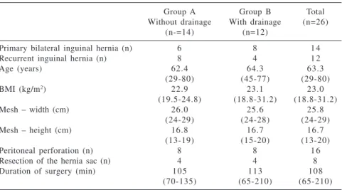
Table 1 - Characteristics of groups A and B.
Group A Group B Total
Without drainage With drainage (n=26)
(n-=14) (n=12)
Primary bilateral inguinal hernia (n) 6 8 1 4
Recurrent inguinal hernia (n) 8 4 1 2
Age (years) 62.4 64.3 63.3
(29-80) (45-77) (29-80)
BMI (kg/m2) 22.9 23.1 23.0
(19.5-24.8) (18.8-31.2) (18.8-31.2)
Mesh – width (cm) 26.0 25.6 25.8
(24-29) (24-28) (24-29)
Mesh – height (cm) 16.8 16.7 16.7
(13-19) (15-20) (13-20)
Peritoneal perforation (n) 8 8 1 6
Resection of the hernia sac (n) 4 4 8
Duration of surgery (min) 1 0 5 1 1 3 1 0 8
(70-135) (65-210) (65-210)
RESULTS
In Group A, 12 out 14 patients (85.7%) presented with preperitoneal space fluid collection as revealed by CT scan on the second POD. From these 12, only 3 (21.4%) exhibited mi-nor clinical manifestations: one showed a scrotal ecchymosis, the other an incisional hematoma, and the lat-ter a seroma. All these complications were treated clinically with good out-comes. In this group the hospital stay ranged from 2 to 7 days (mean of 3.5 days).
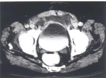
No patient in the Group B pre-sented with any fluid collection on the second POD revealed by CT scan. However, 1 patient had fever and pain in the infraumbilical region without any physical signs of inflammatory process on the 15th POD. This patient underwent an ultrasound scan of the lower abdomen with no findings of fluid collection. We performed an ab-dominal pelvic CT scan, which re-vealed fluid collection in the prepe-ritoneal space (Fig. 1) that was absent in the CT scan performed on the sec-ond POD (Fig. 2). A guided needle
puncture was performed, and about 60 mL of purulent secretion was aspirated; and a wire-guided tubular suction drainage was percutaneously inserted. Drainage was maintained for 4 days, and then the catheter was taken out. The patient recovered well, and no re-currence of inguinal hernia could be detected at the follow-up exam.
DISCUSSION
The Stoppa (GPRVS) procedure utilizes the many advantages of the preperitoneal approach in inguinal hernia repair. It has many advantages, particularly in cases of recurrent or multirecurrent inguinal hernias. We have used GPRVS for inguinal hernia repair since 1983. At present, this op-eration is performed in 22% to 28% of groin hernias referred to our General Surgery Division . A key feature of GPRVS is the application of Pascal’s principle in mesh placement that rein-forces the lower abdominal wall with an elegant anatomical approach that does not disturb groin structures, even in cases that were dissected before. However, the GPRVS procedure re-quires a very extensive dissection of the preperitoneal space for the inser-tion and wrapping of the visceral sac in a large bilateral mesh prosthesis.
Recent publications about the GPRVS have revealed that there still are authors who routinely use drainage with this procedure15,17; however, some
others do not use drainage12,16, and
some eventually use drainage13,20.
When we looked for potential compli-cations reported in these publicompli-cations that might have arisen from fluid col-lections (seroma, hematoma, and infec-tion), we found the following compli-cation rate: 1) routine drainage: 3%15
and 9%17; 2) use of suction drain
ac-cording to intraoperative parameters: 10.2%20 and 24.6%13; and 3) no
drain-age: 14%16 and 22.7%12. From these Figure 1 - Patient on the 15th POD. Abdominal pelvic CT scan showed an abscess in the
preperitoneal space (arrow).
data, it appears that the use of a suc-tion drain would bring some benefit. However, considering that Coda et al.13
reported a 24.6% rate of hematoma and seroma, even with the use of suction drainage in 83% of the patients in their study, it appeared that there was some controversy about the benefit of the drainage in GPRVS. To the best of our knowledge, this issue has not been studied before.
We have been using the procedure for repair of all inguinal hernias poten-tially disposed to recurrence, includ-ing those of obese patients with ab-dominal distension and patients with chronic bronchitis. However, for the present study, we included for prospec-tive randomization 2 categories of pa-tients with groin hernias: 1) recurrent or multirecurrent inguinal hernias, and 2) patients over 50 years with bilateral inguinal hernias. This protocol was aimed towards answering the question about the necessity of drainage in GPRVS, minimizing as much as possi-ble patient comorbidities that could interfere with the interpretation of the outcome. It is clear that obesity inter-feres with the dissection. In this study, the patients had a body mass index (BMI) ranging from 18.81 to 31.20 kg/ m2 (mean = 23.0 kg/m2). On the other
hand, by considering the anthropomet-ric parameters and the size of the pros-thesis placed in preperitoneal space, we found that Groups A and B were homogeneous.
We performed the Stoppa proce-dure with special attention to the origi-nal description7-10. The choice of
polypropylene mesh is a consequence of availability in our country. At the beginning of our experience, we used 4 drains in the preperitoneal space. Further we believed that 1 suction drain was sufficient by assuming that the preperitoneal space created by the surgical dissection represented only 1 bilateral continuous space; in fact, this strategy worked effectively.
Aspirative drainage in GPRVS was responsible for 2 to 7 days of postop-erative hospital stay with a mean of 3.5 days, which is compatible to the find-ings of others11,14,16,20. Of the patients
who did not undergo drainage, 85.7% presented with preperitoneal fluid col-lection revealed by CT scan on the sec-ond POD, but this was diagnosed clini-cally in only 21.4%, with minimal clinical evidence that did not require any specific treatment. This finding suggests that it is safe to not drain sys-tematically after the GPRVS procedure. This concept is strongly supported when we consider that the use of drain-age did not prevent the collection of pus after the second POD in 1 patient. Moreover, the clinical signs of the in-flammatory process are more reliable than the potential benefit that drainage in all patients might bring. The success-ful percutaneous puncture and drain-age of fluid collection in 1 patient demonstrated that the presence of post-operative infectious fluid collection following GPRVS does not require that the mesh be taken off.
Thus, the results presented from Group A on clinical evaluation are similar to those reported by Solorzano
et al.16 and Beets et al.12 who also did
not use drainage systematically in GPRVS. Solorzano et al.16 reported
14% hematoma and infectious com-plications while Beets et al.12 reported
22.6% seroma, hydrocele, hematoma, and infectious complications.
Our results justify the concept that the GPRVS procedure is not an opera-tion that routinely requires drainage. We are convinced that, for general use, precise case selection, and careful at-tention to anatomical and surgical principles are the foundations for every surgical procedure, including GPRVS. We can safely propose that the use of aspirative drainage in GPRVS must be used in situations without good hemostasis. Schimitz et al.21 reported
that in Bassini-Lotheissen inguinal hernia repair, patients receiving low-dose subcutaneous heparin have a high rate of hemorrhagic complications, with 22% hematomas and 13.3% ec-chymosis. Similar results were also re-ported by Mumme et al.22, including
increased postoperative hematomas and subsequent increased surgical reintervention with anticoagulation in inguinal hernia surgery.
Finally, our results corroborate the unquestionable value of the GPRVS procedure as an efficient inguinal her-nia repair technique that reduces the recurrence rate. The GPRVS procedure is not only the last weapon of defense, but is actually a good weapon. By as-suming this posture, we think that we are safely decreasing postoperative stay, and as a consequence, improving the cost effectiveness of the procedure.
RESUMO
RODRIGUES Jr. AJ e col. – Hernioplastia inguinal segundo procedimento de Stoppa: drenar ou não drenar. Rev. Hosp. Clin. Fac. Med. S. Paulo 58(2):97-102, 2003.
OBJETIVO: O objetivo do
pre-sente estudo é avaliar os benefícios da drenagem no procedimento de Stoppa no tratamento da hérnia inguinal.
PACIENTES E MÉTODOS: O
Grupo A, com 12 pacientes submetidos à drenagem e Grupo B, com 14 paci-entes não submetidos à drenagem. No segundo dia de pós-operatório, todos os pacientes foram submetidos à tomografia computadorizada de abdo-me para a verificação de coleções ab-dominais.
RESULTADOS: No Grupo A,
ne-nhum paciente apresentou coleção no espaço pré-peritonial. Por outro lado, um paciente desenvolveu abcesso no espaço pré-peritonial no décimo quin-to dia de pós-operatório. No Grupo B, 12 pacientes apresentaram coleção pré-peritonial à tomografia. Entretanto, so-mente três apresentaram complicações menores. Nenhum paciente apresentou
complicação maior.
CONCLUSÃO: O uso de drenagem
de aspiração contínua no proce-dimento de Stoppa não traz nenhum benefício.
DESCRITORES: Hérnia. Hérnia Inguinal. Prótese. Tela. Drenagem.
REFERENCES
1 . GILBERT AI - Sutureless repair of inguinal hernia. Am J Surg 1992; 163:331-335.
2 . GILBERT AI, GRAHAM MF, VOIGT WJ - A bilayer patch device for inguinal hernia repair. Hernia 1999; 3:161-166. 3 . LICHTENSTEIN IL, SHULMAN AG - Ambulatory outpatient
hernia surgery. Including a new concept, introducing tension-free repair. Int Surg 1986; 71:1-4.
4 . LICHTENSTEIN IL, SHULMAN AG, AMID PK et al. - The tension-free hernioplasty. Am J Surg 1989;157:188-193. 5 . RUTKOW IM, ROBBINS AW - Mesh plug hernia repair: A
follow-up report. Surgery 1995; 117:597-598.
6 . TRABUCCO EE, TRABUCCO AF.- Flat plug and mesh hernioplasty in the “inguinal box”: description of the surgical technique. Hernia 1998; 2:133-138.
7 . STOPPA R - Prosthetic repair via the open abdomen. In: CHEVREL JP - Hernias and surgery of the abdominal wall. Berlin, Springer, 1998. p. 216-223.
8 . STOPPA R, ABOURACHID H, DUCLAYE C et al.- Plastie des hernies de l’aine. L’interposition sans fixation de tulle de Dacron par voie médiane sous-péritonéale. Nouv Presse Pr Med 1973;2:1949-1951.
9 . STOPPA R, PETIT J, HENRY X - Unsutured Dacron prosthesis in groin hernias. Int Surg 1975;60:411-412.
10. STOPPA R, WARLAMOUNT CR - The pre-peritoneal approach and prosthetic repair of groin hernia. In: NYHUS LN, CONDON R - Hernia. 3rd ed. Philadelphia, Lippincott, 1989.
p. 199-255.
11. STOPPA R, WARLAUMONT CR, VERHAEGHE PJ et al. -Prosthetic repair in the treatment of groin hernias. Int Surg 1986; 71:154-158.
12. BEETS GL, VAN GELDERE D, BAETEN CGMI et al. - Long-term results of giant prosthetic reinforcement of the visceral sac for complex recurrent inguinal hernia. Br J Surg 1996; 83:203-206.
14. MALAZGIRT Z, OZKAN K, DERVISOGLU A et al. - Comparison of Stoppa and Lichtenstein techniques in the repair of bilateral inguinal hernias. Hernia 2000; 4:264-267.
15. PRICOLO R, PARZIALE A, RIZZITELLI E et al. - La riparazione delle ernie inguinali e/o crurali com protesi gigante in mersilene in posizione preperitoneale. Minerva Chir 1993; 48 :1007-1010.
16. SOLORZANO CC, MINTER RM, CHILDERS TC et al. -Prospective evaluation of the giant prosthetic reinforcement of the visceral sac for recurrent and complex bilateral inguinal hernias. Am J Surg 1999; 177:19-22.
17. THAPAR V, RAO P, PRABHU R et al. - Giant prosthesis for reinforcement of visceral sac for complex bilateral and recurrent inguinal hernias: A prospective evaluation. J Postgrad Med 2000; 46:80-82.
18. VAN DAMME JPJ - A preperitoneal approach in the prosthetic repair of inguinal hernia. Int Surg 1985; 70:223-226. 19. WANTZ GE - Giant prosthetic reinforcement of the visceral sac.
Surg Gynecol Obstet 1989; 169:408-417.
20. CHAMPAULT GG, RIZK N, CATHELINE JM et al. - Inguinal hernia repair. Totally preperitoneal laparoscopic approach versus Stoppa operation: Randomized trial of 100 cases. Surg Laparosc Endosc 1997; 7:445-450.
21. SCHMITZ R, KANSY M, MOSER KH et al. - Effect of low-dose subcutaneous heparin on postoperative wound hematomas: Randomized clinical trial on hospitalized inguinal hernia patients in Germany. World J Surg 1995; 19:416-419.