Polypropylene mesh vs. Site-specific repair in the treatment of
Polypropylene mesh vs. Site-specific repair in the treatment of
Polypropylene mesh vs. Site-specific repair in the treatment of
Polypropylene mesh vs. Site-specific repair in the treatment of
Polypropylene mesh vs. Site-specific repair in the treatment of
anterior vaginal wall prolapse: preliminary results of a
anterior vaginal wall prolapse: preliminary results of a
anterior vaginal wall prolapse: preliminary results of a
anterior vaginal wall prolapse: preliminary results of a
anterior vaginal wall prolapse: preliminary results of a
randomized clinical trial
randomized clinical trial
randomized clinical trial
randomized clinical trial
randomized clinical trial
Tela de polipropileno versus correção sítio-especifica no tratamento do prolapso
Tela de polipropileno versus correção sítio-especifica no tratamento do prolapso
Tela de polipropileno versus correção sítio-especifica no tratamento do prolapso
Tela de polipropileno versus correção sítio-especifica no tratamento do prolapso
Tela de polipropileno versus correção sítio-especifica no tratamento do prolapso
de parede vaginal anterior: resultados preliminares de ensaio clínico randômico
de parede vaginal anterior: resultados preliminares de ensaio clínico randômico
de parede vaginal anterior: resultados preliminares de ensaio clínico randômico
de parede vaginal anterior: resultados preliminares de ensaio clínico randômico
de parede vaginal anterior: resultados preliminares de ensaio clínico randômico
JACQUELINE LEME LUNARDELLI1; ANTONIO PEDRO FLORES AUGE2; NUCÉLIO LUIZDE BARROS MOREIRA LEMOS3; SILVIADA SILVA CARRAMÃO3; ANDRÉ
LIMADE OLIVEIRA3; ELIANA DUARTE4; TSUTOMU AOKI5
A B S T R A C T A B S T R A C T A B S T R A C T A B S T R A C T A B S T R A C T
Objective Objective Objective Objective
Objective: Pelvic organ prolapse is a disorder caused by the imbalance between the forces responsible for supporting the pelvic organs in their normal position and those that tend to expel them from the pelvis. Anterior vaginal wall prolapse, known as cystocele, is the most common form of prolapse and can result from lesions in different topographies of the endopelvic fascia. Currently, a woman has an 11% risk of being submitted to a surgical procedure to correct pelvic floor disorder, and a 29% chance of being reoperated due to failure in the first surgery. Methods:Methods:Methods:Methods:Methods: A prospective randomized study was conducted to compare the use of polypropylene mesh with site-specific repair in the surgical treatment of anterior vaginal prolapse. Thirty-two patients aged between 50 and 75 years, who had previous vaginal prolapse at stage III or IV, or prolapse recurrence, were operated. Mean follow-up was 8.5 months. Results:Results:Results:Results: The results demonstrate the superiority of the anatomical outcomes with the use of polypropyleneResults: mesh over site-specific repair. Regarding surgical morbidity, shorter operative time was observed for the mesh group. Conclusion:Conclusion:Conclusion:Conclusion:Conclusion: The results observed in this study indicate the superiority of anatomical results obtained with the use of polypropylene mesh over site-specific repair.
Key words: Key words: Key words: Key words:
Key words: Uterine Prolapse. Pelvic Floor. Surgical Mesh. Gynecologic Surgical Procedures.
Study conducted at the Department of Gynecology and Obstetrics of Irmandade da Santa Casa de Misericórdia de São Paulo, São Paulo, SP Brazil. 1. Master in Gynecology and Assistant Professor - Department of Gynecology and Obstetrics (DGO) of Irmandade da Santa Casa de Misericórdia de São Paulo (ISCMSP), São Paulo, SP Brazil; 2. Associate Professor - DGO of ISCMSP; 3. PhD and Assistant Professor - DGO of ISCMSP; 4. Post-Graduation student of Ciências Médicas Faculty of ISCMSP; 5. Adjunct Professor and Director - DGO of ISCMSP.
INTRODUCTION
INTRODUCTION
INTRODUCTION
INTRODUCTION
INTRODUCTION
P
elvic organ prolapse (POP) is defined as the partial or total permanent displacement of any pelvic segment or organ from its normal location, including the procidentia of the vaginal walls (cystocele, rectocele, enterocele) or the uterus. It is a disorder caused by the imbalance between the forces responsible for supporting the pelvic organs in their normal position and those which tend to push them out of the pelvis.Pelvic floor defects constitute an epidemic which goes unnoticed by many1. Studies show that in
the United States some 300,000 women a year undergo surgery for the correction of prolapse and urinary incontinence2.
Currently, a woman has an 11% risk of undergoing some surgical procedure for the repair of pelvic floor disorders, and a 29% chance of repeat surgery due to failure in the first operation. Among the various prolapse sites, the anterior vaginal wall is the most frequent and the primary site of recurrence4. A number of surgical techniques
for the correction of anterior vaginal prolapse (AVP), widely known as cystocele, have been described. Classically, for many years, anterior colporrhaphy, described by Kelly-Kennedy in 1913, was performed for the correction of stress urinary incontinence (SUI) and cystocele.
The correction of AVP through anterior colporrhaphy without the utilization of implants shows a high recurrence rate, which may reach as much as 40%4-6.
Thus, the objective of this study was to compare the anatomical outcomes obtained with a polypropylene mesh versus the site-specific operation for the correction of ante-rior vaginal wall prolapse.
METHODS
METHODS
METHODS
METHODS
METHODS
Division of the Obstetrics and Gynecology Department (DOGI) of ISCMSP between June 2006 and May 2008.
The study was randomized and comprised 32 women with AVP who were seen at the clinic of that hospi-tal. These women were allocated into two groups: Mesh Group – 16 patients who underwent AVP repair with a synthetic monofilament polypropylene mesh (Nazca TC, Promedon, Córdoba, Argentina), and No-Mesh Group – 16 patients who underwent site-specific surgical repair of AVP, without the use of a synthetic mesh.
Group allocation was performed through a randomization table by a third party not involved in the study, who placed the results obtained inside sealed enve-lopes, which were opened upon the patients’ admission.
Patients with ages ranging between 50 and 75 years and diagnosed with AVP stage III or IV, or recurrent anterior vaginal prolapse were included. The study excluded pregnant women, mothers in the puerperal period and up to six months post partum, patients with a history of use of implants in reconstructive or anti-incontinence pelvic procedures, patients with blood coagulation disorders, kidney failure and/or upper urinary tract obstruction, urethral diverticulum or a history of pelvic irradiation. The patients’ history was taken and they underwent a general physical and a gynecological examination.
Pelvic organ support was evaluated according to the guidelines of the International Continence Society (ICS), following the POP-Q7 system through the Pelvic Organ
Prolapse Quantification Index (POP-Q-I), Absolute and Relative8-10. For the screening of occult stress urinary
incontinence, all patients, whether symptomatic or not, were submitted to a urodynamic study both in the dorsal recumbent and the semi-sitting position, with prolapse reduction using a Cheron forceps.
Operative time (minutes), intraoperative blood loss (mL) and intra- and postoperative complications were recorded. Operative time was measured by means of a chronometer, but only the duration of the actual site-specific operation or surgical mesh repair was used for comparison; the length of concurrent procedures was not taken into consideration. Blood loss was measured through a disposable plastic surgical aspirator, coupled with a collecting bottle graduated in 20 mL increments.
Antibiotic prophylaxis was done by using 2.0 g cefazolin and 1.0 g metronidazole upon anesthetic induction. The bladder catheter was removed after 24 hours. Patients were instructed to avoid physical strain for 30 days and refrain from sexual activity for 60 days after the procedure. The patients’ pelvic organ support was re-evaluated postoperatively as previously described, and recorded at one, three, six and 12 months.
Concurrent surgical procedures such as transvaginal hysterectomy (HV), sacrospinous fixation (FSE), colpoperineoplasty (CPP), rectocele correction and McCall culdoplasty were performed as indicated, depending on the preoperative findings, and recorded.
The patients in the no-mesh group who had a preoperative diagnosis of SUI underwent the placement of
a suburethral transobturator sling (Safyre T®, Promedon®,
Córdoba, Argentina) through the same incision made for AVP correction. In the patients of the mesh group who presented with SUI, the same mesh was used for both AVP and SUI correction (Nazca TC®). All data were recorded in
specific study protocols.
The mesh group patients underwent AVP repair with the utilization of a synthetic monofilament polypropylene mesh (Nazca TC®, PromedonÒ Ltda, Córdoba, Argentina).
The patients were placed in the lithotomy position and submitted to bladder catheterization. Next, a solution of 250 mL of saline (NaCl 0.9%) and 1 mL of adrenaline 1% was infiltrated into the vaginal wall to aid dissection and hemostasis.
A median longitudinal incision was made on the anterior vaginal wall, from 1.5 cm below the urethral meatus at the level of the pubourethral ligament insertion down to the uterine cervix. From that incision, the dissection was extended to the ischio-pubic ramus, bilaterally. Two 1.0 cm suprapubic incisions, 2.5 cm lateral and 3.0 cm above the clitoris were made for the subsequent passage of the pre-pubic needles.
The synthetic mesh composed of monofilament polypropylene with 6 mm macropores at center was designed for the correction of AVP alone or associated with SUI (Nazca TCÒ). The kit contains three needles: one for the pre-pubic approach and two helical needles for the inferior transobturator approach. The mesh has four anchoring points: two pre-pubic arms and two transobturator arms. It has silicone at the ends, which enables the attachment of the respective needles.
The pre-pubic needle was inserted vaginally, directed to the subcutaneous tissue of the suprapubic incisions previously made, and guided along the proper path by the surgeon’s index finger after paraurethral tunneling. The needle and the polypropylene mesh were connected and the upper arms of the mesh were pulled through the suprapubic incisions towards the mid-urethra.
The transobturator needles were inserted 2 cm laterally and 3 cm inferiorly in relation to the conventional transobturator approach, the tip of the needle pointing towards the ischial spine so as to perforate the obturator membrane at the level of the tendinous arch of the endopelvic fascia, in its proximal third. Following that, the lower portion of the mesh was attached to the pericervical ring with nylon 0 (Mononylon®, Ethicon®), and the lower
arms of the mesh were pulled, thereby correcting the pubocervical fascia defects.
Following hemostasis reassessment, the vaginal wall was closed through the overlapping technique (“double-breasting” closure) with interrupted 2-0 polyglactin (Vicryl®,
Ethicon®) suture, with the purpose of preventing resection
The no-mesh patients underwent site-specific surgical AVP repair without a synthetic mesh. They were placed in the lithotomy position and submitted to bladder catheterization. Next, the vaginal wall was infiltrated with 250 mL saline (NaCl 0.9%) and 1 mL 1% adrenaline to make dissection and hemostasis easier. A median incision was made on the anterior vaginal wall, from 1.5 cm below the urethral meatus at the level of the pubourethral ligament insertion down to the uterine cervix.
Starting from that incision, a lateral dissection was extended as far as the ischio-pubic ramus, bilaterally. It was possible, then, to identify the anterior vaginal wall defects for the site-specific repair.
Once a lateral defect was observed, interrupted stitches 1 cm apart were passed for the reinsertion of the pubocervical fascia into the tendinous arch, from the most anterior portion of the ischio-pubic ramus to the ischial spine. The repair of a central defect, on the other hand, consisted of the plication of the pubocervical fascia in the midline. The transverse defect was repaired through the reinsertion of the pubocervical fascia into the pericervical ring. The suture was multifilament polyethylene (Ethibond® 1).
The vaginal wall was closed through the “double-breasting” technique as described for the mesh group, and the Foley catheter was left in place in the immediate postoperative period and remained for 24 hours.
The collected data were input to the spreadsheet program Excel of the Office package by Microsoft® (Excel
2003). The calculations were made by the SigmaStat program of Jandel Corporation® (SigmaStat®;1995).
The Mann-Whitney test was used in the comparison between the groups for age, BMI, obstetric history, preoperative POP-Q-I, postoperative POP-Q-I, operative time (minutes) and blood loss (mL). Two tables showing the frequency of previous and concurrent surgical procedures for each group were produced.
The risk of rejecting the null hypothesis was established at α < 0.05 or 5%.
Sample size was calculated on the basis of the standard deviation for point Ba10 of 0.7 cm. Calculations
were made based on the formula for ideal sample size of Student’s t-test, considering a=5%, a two-way analysis,
90% statistical power to detect a 1-cm difference between the groups, and an estimated noncompliance rate of 30%. The Minitab® 15.1.1.0 program (Minitab® Inc. EUA) was
used to estimate sample size.
RESULTS
RESULTS
RESULTS
RESULTS
RESULTS
Thirty-two patients were evaluated with a mean follow-up of nine months (mesh group = 9.0; no-mesh group = 7.9). There was no significant difference between the groups. No patient was lost during follow-up, thus noncompliance rate was zero.
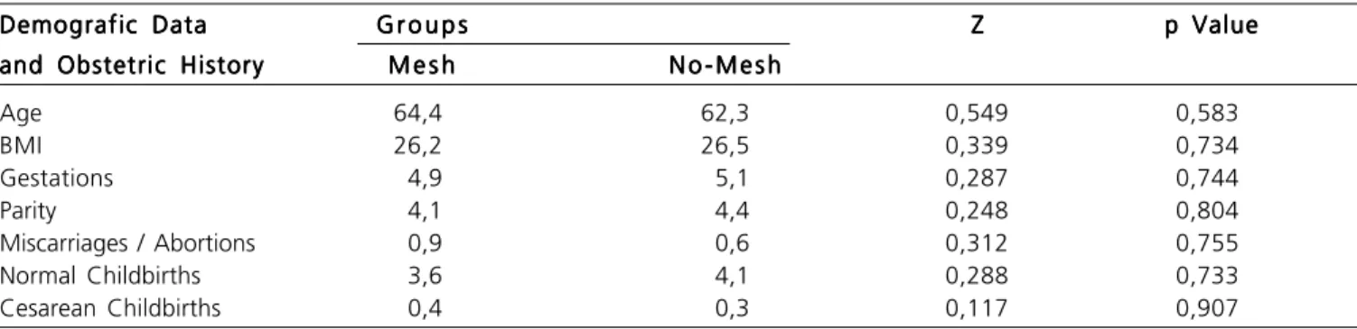
Patient age ranged from 54 to 76 years (mean = 63.3; median = 62.5) and body mass index (BMI) ranged from 21.4 to 35.7 (mean = 26.4; median = 26.1). The groups were homogeneous with regard to anthropometrics and obstetric history (Table 1). Nineteen previous operations had been performed on the study patients, 10 (52.6%) in the mesh group (six colpoperineoplasties, three abdominal hysterectomies, one vaginal hysterectomy) and nine (47.4%) in the no-mesh group (five colpoperineoplasties, three abdominal hysterectomies and one vaginal hysterectomy).
The concomitant surgical procedures performed on the patients of both groups are demonstrated in table 2. No intraoperative complications occurred.
Two patients in the mesh group and seven from the no-mesh group exhibited preoperative SUI. One patient from the mesh group and one from the no-mesh group presented with de novo SUI. The patients whose SUI treatment failed were referred to pelvic floor physical therapy and are now cured.
There occurred one case of mesh erosion (6.25%), which was located on the lateral left side of the anterior vaginal wall. The lesion was treated with topical estrogen and partial exeresis of the mesh, under local anesthesia.
The mean operation time for the mesh group was 56.1 minutes, and 80.9 minutes for the no-mesh group (p=0.002). Intraoperative blood loss was 76.3 mL in the mesh group and 126.9 mL in the no-mesh group, a non-significant statistical difference (p=0.260).
Table 1 Table 1Table 1 Table 1
Table 1 - Comparison of means for demographic data and obstetric history between the mesh and no-mesh groups of patients.
Demografic Data Demografic DataDemografic Data Demografic Data
Demografic Data G r o u p sG r o u p sG r o u p sG r o u p sG r o u p s ZZZZZ p Valuep Valuep Valuep Valuep Value
and Obstetric History and Obstetric Historyand Obstetric History and Obstetric History
and Obstetric History M e s hM e s hM e s hM e s hM e s h N o - M e s hN o - M e s hN o - M e s hN o - M e s hN o - M e s h
Age 64,4 62,3 0,549 0,583
BMI 26,2 26,5 0,339 0,734
Gestations 4,9 5,1 0,287 0,744
Parity 4,1 4,4 0,248 0,804
Miscarriages / Abortions 0,9 0,6 0,312 0,755
Normal Childbirths 3,6 4,1 0,288 0,733
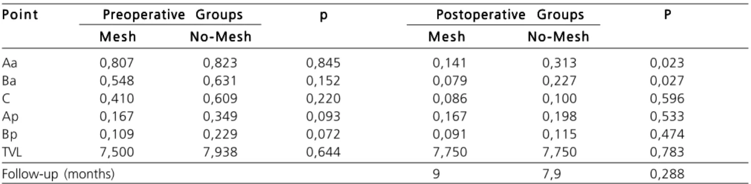
The Pelvic Organ Prolapse Quantification Index (POP-Q-I) for points Aa, Ba, C, Bp and Ap was applied as proposed and validated in other studies9,11. POP-Q-I quantifies
prolapse as a continuous variable, “zero” being the total absence of prolapse, and “one”, the maximum extent of prolapse for a given point (Table 3).
DISCUSSION
DISCUSSION
DISCUSSION
DISCUSSION
DISCUSSION
The correction of AVP represents one of the ma-jor challenges in the reconstructive surgery of the pelvic floor in terms of success and anatomical support durability. Numerous studies for the correction of anterior vaginal wall defects were proposed in the last century, both by the ab-dominal and the vaginal route.
Despite the better understanding of anatomy and function and the advancement in surgical techniques, success rates in the long-term are still variable. The literature shows that these rates range between 37% and 100%11 in
relation to anterior colporrhaphy.
The importance of paravaginal defects in the prolapses of the anterior compartment was described by
White in 191212. Richardson et al 13 demonstrated that
patients may present with a combination of two or more types of defects; therefore, it is possible to understand one of the reasons for the high recurrence rate in anterior colporrhaphy.
Inumerous studies on the use of nonabsorbable synthetic meshes in human models have been reported in the literature for the repair of posterior vaginal wall14 and
anterior vaginal wall procidentia15,16, vaginal vault
prolapse17, total genital prolapse 18 and stress urinary
incontinence19.
The different surgical procedures for AVP correction report recurrence rates of 3% to 70% following anterior colporrhaphy20-22 and 5% to 50% after
paravaginal repair via vaginal approach. In another study23
with 10-year follow-up, it was found that the use of synthetic meshes for the correction of pelvic organ prolapse revealed satisfaction rates of 68% (follow-up of 6 months to 3 years), 73% (3 – 6 years) and 74% (over 6 years)24.
In a systematic review of the literature on AVP repair, considering levels of evidence 1 and 2, other authors25
noted that the use of meshes offers anatomic and surgical
Table 2 Table 2 Table 2
Table 2 Table 2 – Frequency of concurrent surgical procedures in the mesh and no-mesh groups (CPP: colpoperineoplasty; SSF: sacrospinous fixation; USF: uterosacral fixation; VH: vaginal hysterectomy; Safyre T: suburethral sling).
Concurrent Operations Concurrent OperationsConcurrent Operations
Concurrent OperationsConcurrent Operations G r u p o sG r u p o sG r u p o sG r u p o sG r u p o s T o t a lT o t a lT o t a lT o t a lT o t a l
M e s h M e s h M e s h M e s h
M e s h N o - M e s hN o - M e s hN o - M e s hN o - M e s hN o - M e s h
None 13 0 13
CPP 0 13 13
SSF 1 0 1
USF 1 0 1
VH 1 7 8
Mc Call 1 3 4
Paravaginal 0 11 11
Retocele 1 3 4
Safyre T 0 7 7
Total operations * 18 44 62
* more than one operation per patient.
Table 3 Table 3Table 3
Table 3Table 3 - Comparison of Pelvic Organ Prolapse Quantification Index (POP-Q-I) preoperative and postoperative between the mesh and no-mesh groups.
P o i n t P o i n tP o i n t
P o i n tP o i n t Preoperative GroupsPreoperative GroupsPreoperative GroupsPreoperative GroupsPreoperative Groups ppppp Postoperative GroupsPostoperative GroupsPostoperative GroupsPostoperative GroupsPostoperative Groups PPPPP
M e s h M e s h M e s h M e s h
M e s h N o - M e s hN o - M e s hN o - M e s hN o - M e s hN o - M e s h M e s hM e s hM e s hM e s hM e s h N o - M e s hN o - M e s hN o - M e s hN o - M e s hN o - M e s h
Aa 0,807 0,823 0,845 0,141 0,313 0,023
Ba 0,548 0,631 0,152 0,079 0,227 0,027
C 0,410 0,609 0,220 0,086 0,100 0,596
Ap 0,167 0,349 0,093 0,167 0,198 0,533
Bp 0,109 0,229 0,072 0,091 0,115 0,474
TVL 7,500 7,938 0,644 7,750 7,750 0,783
Follow-up (months) 9 7,9 0,288
outcomes which are superior to those obtained with traditional colporrhaphy, although this evidence is still limited to only a few clinical studies.
In a randomized study with 83 patients, Weber et al22 compared the repair of AVP through anterior
colporrhaphy techniques, paravaginal defect repair and the use of absorbable synthetic polyglactin 910 mesh (Vicryl®,
Ethicon, USA). With mean follow-up of two years, they found objective success rates of 30%, 46% and 42 % respectively. Vaginal tissue erosion occurred in only 2.9% of cases, which required the surgical removal of the material.
Meanwhile, Sand et al25 compared AVP repair by
traditional colporrhaphy and the use of a mesh similar to that of the above-mentioned study. Success rate in the mesh group was significantly higher than in the colporrhaphy group: 75% and 57%, respectively.
The inconsistent results obtained with absorbable meshes have discouraged its use, and type 1 nonabsorbable meshes (macroporous, monofilament) have been more widely employed.
The first to report on the use of nonabsorbable synthetic meshes for AVP repair was Julian, in 199615. In a
retrospective study, 24 women having recurrent AVP were randomly allocated; 12 of them underwent anterior colporrhaphy and 12 underwent both anterior colporrhaphy and a MarlexÒ (Bard, USA) mesh placement. On 24-month follow-up, success rates of 66% and 100% were observed in the groups, respectively.
A number of factors contribute to the heterogeneity of the results observed in the literature. Among those factors are different designs (many of the studies are retrospective), different surgical techniques, differences in sampling and, especially, the criteria of failure and recurrence adopted by each author.
In the present study, outcomes were not evaluated in terms of success or recurrence. The only instrument for comparison was the Pelvic Organ Prolapse Quantification Index (POPQ-I), which was proposed7 and
validated in a prospective, unicenter study10.
POP-Q-I quantifies prolapse directly, in a continuous manner. Thus, only the anatomical outcomes are compared, through means and dispersion values (Mann-Whitney test), without considering success or recurrence rates, since no consensus exists on those concepts in the literature.
This system quantifies the prolapse between zero and one for each point, where “zero” indicates no prolapse and “one” corresponds to the maximum extent of prolapse. In the present study, statistically significant results were obtained for points Aa and Ba, which are related to anterior vaginal wall prolapse. It was found that the mean prolapse rate was significantly lower for the mesh group (7.9%) compared with the no-mesh group (22.7%). These figures show the superiority of the
anatomical outcomes obtained with the use of implants over site-specific repair.
The transobturator route offers the benefit of the subvesical surgical plane, with lower risk of bladder, bowel or large vessel lesion26. Englin et al27 were the first
to propose AVP repair via transobturator using a mesh, and obtained a recurrence rate of only 3% in 18 months, with a 5% incidence of erosions. This good result was observed in the present study as well, and translates into absence of visceral or vascular lesion as well as shorter operative time.
One of the major disadvantages to using synthetic meshes derives from the erosion and extrusion rates of the material. This type of complication is described in the literature and occurs in 2.8% to 17.3% of cases10, 28-31. One
case of erosion was observed in 16 patients (6.25%). However, the sample employed is not adequate for the evaluation of erosion rates, considering that the variable of interest consists of the anatomical outcomes as measured by the POP-Q-I.
One of the advantages to using mesh kits in repairing pelvic organ prolapses is the less invasive character of the procedure, which results in decreased operative time and blood loss28. In the present study, a difference was
observed in operative time, which was significantly shorter for the mesh group (56.1 min) compared with the no-mesh group (80.9 min). Regarding intraoperative blood loss, no statistically significant difference was found between the groups.
Recent research studies on the biomechanism and composition of the connective tissue of the pelvic floor provide a new insight into the matter of pelvic organ stability, as well as the postoperative healing process32.
Drawing on these concepts, some authors state that the reconstruction of the pelvic floor will be better accomplished if synthetic meshes are used, instead of having only the native tissue as support33. The results
presented herein corroborate that statement, since the anatomical outcomes for the mesh group patients were found to be statistically superior to those for the no-mesh group.
The challenge in any pelvic reconstructive procedure is to provide a supporting structure while the normal anatomic condition of the surrounding structures is restored. Even though there are several materials which could provide that support, few have the necessary properties to restore the qualities of the living tissue. More randomized prospective clinical trials, preferably multicenter, are needed before recommending the utilization of implants in daily practice.
R E S U M O R E S U M O R E S U M O R E S U M O R E S U M O
Objetivo Objetivo Objetivo Objetivo
Objetivo: O prolapso de órgãos pélvicos é enfermidade decorrente do desequilíbrio entre as forças encarregadas de manter os órgãos pélvicos em sua posição normal, e aquelas que tendem a expeli-los para fora da pelve. O prolapso de parede vaginal anterior é a mais freqüente forma de prolapso e pode ser ocasionado por lesões em diferentes topografias da fáscia endopélvica. O objetivo deste estudo foi comparar o uso de tela de polipropileno e correção sitio-específica no tratamento cirúrgico do prolapso vaginal anterior. MétodosMétodosMétodosMétodosMétodos: Estudo prospectivo randômico comparativo em que foram operadas 32 pacientes com idades entre 50 e 75 anos, que apresentavam prolapso vaginal anterior estádio III ou IV, ou recidivado. A estática pélvica foi avaliada segundo as recomendações da International Continence Society (ICS), o sistema POP-Q e pelo Índice de Quantificação de Prolapso (POP-Q-I) Absoluto e Relativo. Para o rastreamento da incontinência urinária de esforço oculta todas as pacientes, sintomáticas ou não, foram submetidas a estudo urodinâmico em posição semi-ginecológica e semi-sentada, com redução do prolapso com pinça de Cheron. Registrou-se o tempo cirúrgico, o volume de sangramento intra-operatório e as complicações intra e pós-operatórias. O tempo de seguimento médio do estudo foi de 8,5 meses. ResultadosResultadosResultadosResultadosResultados: Em relação aos resultados anatômicos ocorreu melhores resultados com a utilização de tela de polipropileno sobre o reparo sitio-específico. Em relação à morbidade cirúrgica, observou-se menor tempo cirúrgico no grupo em que utilizou-se tela. ConclusãoConclusãoConclusãoConclusãoConclusão: Houve superioridade dos resultados anatômicos obtidos com a utilização de tela de polipropileno sobre o reparo sitio-específico.
Descritores Descritores Descritores Descritores
Descritores: Prolapso Uterino. Soalho Pélvico. Telas Cirúrgicas. Procedimentos Cirúrgicos Ginecológicos.
REFERENCES
REFERENCES
REFERENCES
REFERENCES
REFERENCES
1. DeLancey JO. The hidden epidemics of pelvic floor dysfunction: achievable goals for improved prevention and treatment. Am J Obstet Gynecol. 2005; 192(5):1488-95.
2. Boyles SH, Weber AM, Meyn L. Procedures for urinary incontinence in the United States, 1979-1997. Am J Obstet Gynecol. 2003; 189(1):70-5.
3. Olsen AL, Smith VJ, Bergstrom JO, Colling JC, Clark AL. Epidemiology of surgically managed pelvic organ prolapse and urinary incontinence. Obstet Gynecol. 1997; 89(4):501-6.
4. Shull BL, Capen CV, Riggs MW, Kuehl TJ. Preoperative and postoperative analysis of site-specific pelvic support defects in 81 women treated with sacrospinous ligament suspension and pelvic reconstruction. Am J Obstet Gynecol. 1992; 166(6 Pt 1):1764-8; discussion 1768-71.
5. Kohli N, Sze EHM, Roat TW, Karram MM. Incidence of recurrent cystocele after anterior colporrhaphy with or without concomitant transvaginal needle suspension. Am J Obstet Gynecol. 1996; 175(6):1476-80; discussion 1480-2.
6. Benson JT, Lucente V, McClellan E. Vaginal versus abdominal reconstructive surgery for the treatment of pelvic support defects: a prospective randomized study with long-term outcome evaluation. Am J Obstet Gynecol. 1996; 175(6):1418-21; discussion 1421-2. 7. Bump RC, Mattiasson A, Bo K, Brubaker LP, DeLancey JO, Klarskov
P et al. The standartization of terminology of female pelvic organ prolapse and pelvic floor dysfunction. Am J Obstet Gynecol. 1996; 175(1):10-7.
8. de Barros Moreira Lemos NL, Flores Auge AP, Lunardelli JL, Brites Frade A, Frade CL, de Oliveira AL et al. Optimizing pelvic organ prolapse research. Int Urogynecol J Pelvic Floor Dysfunct. 2007; 18(6):609-11. Epub 2006 Sep 26.
9. De Tayrac R, Gervaise A, Fernandez H. Cystocele repair with a fixation-free prosthetic polypropylene mesh. Int Urogynecol J. 2001;12(Suppl 3):S92.
10. Lemos NLBM, Auge APF, Lunardelli JL, Carramão SS, Faria ALA, Aoki T. Validation of the Pelvic Organ Prolapse Quantification Index (POP-Q-I): a novel interpretation of the POP-Q system for optimization of POP research. Int Urogynecol J. 2008; 19(7):995-7. Epub 2008 Jan 24.
11. Maher C, Baessler K. Surgical management of anterior vaginal wall prolapse: an evidence based literature review. Int Urogynecol J Pelvic Floor Dysfunct. 2006; 17(2):195-201. Epub 2005 May 25. 12. White GR. An anatomic operation for the cure of cystocele. Am J
Obstet Dis Women Child. 1912; 65:286–90.
13. Richardson AC, Lyon JB, Williams NL. A new look at pelvic relaxation. Am J Obstet Gynecol. 1976; 126(5):568-73.
14. Kohli N, Miklos JR. Dermal graft-augmented rectocele repair. Int Urogynecol J Pelvic Floor Dysfunct. 2003; 14(2):146-9. Epub 2003 Feb 13.
15. Julian TM. The efficacy of Marlex mesh in the repair of severe, recurrent vaginal prolapse of the anterior midvaginal wall. Am J Obstet Gynecol.1996; 175(6):1472-5.
16. Salomon LJ, Detchev R, Barranger E, Cortez A, Callard P, Darai E. Treatment of anterior vaginal wall prolapse with porcine skin collagen implant by transobturator route: preliminary results. Eur Urol. 2004; 45(2):219-25.
17. Fox SD, Stanton SL. Vault prolapse and rectocele: assessment of repair using sacrocolpopexy with mesh interposition. BJOG. 2000; 107(11):1371-5.
18. Nicita G. A new operation for genitourinary prolapse. J Urol. 1998; 160(3 Pt 1):741-5.
19. Dik P, Klijn AJ, van Gool JD, de Jong TP. Transvaginal sling suspension of the bladder neck in female patients with neurogenic sphincter incontinence. J Urol. 2003; 170(2 Pt 1):580-2; discussion 581-2. 20. Porges RF, Smilen SW. Long-term analysis of the surgical
management of pelvic support defects. Am J Obstet Gynecol. 1994; 171(6):1518-26; discussion 1526-8.
21. Flood CG, Drutz HP, Waja L. Anterior colporraphy reinforced with Marlex mesh for the treatment of cystoceles. Int Urogynecol J Pelvic Floor Dysfunct. 1998; 9(4):200–4.
22. Weber AM, Walters MD, Piedmonte MR, Ballard LA. Anterior colporrhaphy: a randomized trial of three surgical techniques. Am J Obstet Gynecol. 2001; 185(6):1299-304; discussion 1304-6. 23. Sullivan ES, Longaker CJ, Lee PYH. Total pelvic mesh repair: a
ten-year experience. Dis Colon Rectum. 2001; 44(6):857-63. 24. Maher C, Baessler K, Glazener CM, Adams EJ, Hagen S. Surgical
management of pelvic organ prolapse in women: a short version Cochrane review. Neurourol Urodyn. 2008; 27(1):3-12.
25. Sand PK, Koduri S, Lobel RW, Winkler HA, Tomezsko J, Culligan PJ et al. Prospective randomized trial of polyglactin 910 mesh to prevent recurrence of cystoceles and rectoceles. Am J Obstet Gynecol 2001; 184(7):1357-62; discussion 1362-4.
26. Dargent D, Bretones S, Mellier G. [Insertion of a sub-urethral sling through the obturating membrane for treatment of female urinary incontinence]. Gynecol Obstet Fertil. 2002; 30(7-8):576-82. French. 27. Eglin G, Ska JM, Serres X. [Transobturator subvesical mesh tolerance and short term results of a 103 cases continuous series]. Gynecol Obstet Fertil. 2003; 31(1):14-9. French.
28. Birch C. The use of prosthetics in pelvic reconstructive surgery. Best Pract Res Clin Obstet Gynaecol. 2005; 19(1):979–91. Epub 2005 Sep 26.
30. Gauruder-Burmester A, Koutouzidou P, Rohne J, Gronewold R, Tunn R. Follow-up after polypropylene mesh repair of anterior and posterior compartments in patients with recurrent prolapse. Int Urogynecol J Pelvic Floor Dysfunct. 2007; 18(9):1059-64. Epub 2007 Jan 12.
31. Hiltunen R, Nieminen K, Takala,T, Heiskanen E, Merikari M, Niemi K, Heinonen PK. Low-weight polypropylene mesh for anterior va-ginal wall prolapse: a randomized conttolled trial. Obstet Gynecol. 2007; 110(2 Pt 2):455-62.
32. Farrell SA, Dempsey T, Geldenhuys L. Histologic examination of “fascia” used in colporraphy. Obstet Gynecol. 2001; 98(5 Pt 1):794-8. 33. Barber MD, Visco AG, Weidner AC, Amudsen CL, Bump R.Bilateral
uterosacral ligament vaginal vault suspension with site-specific endopelvic fascia defect repair for the treatment of pelvic organ prolapse. Am J Obstet Gynecol. 2000; 183(6):1402–10; discussion 1410-1.
Received in 10/10/2008
Accepted for publication in 20/12/2008 Conflict of interest: None
Financial source: None
How to cite: How to cite:How to cite: How to cite: How to cite:
Lunardelli JL, Auge APF, Lemos NLB, Carramão SS, Oliveira AL, Duarte E, Aoki T. Polypropylene mesh vs. site-specific repair in the treatment of anterior vaginal wall prolapse: preliminary results of a randomized clinical trial. Rev Col Bras Cir. [periódico na Internet] 2009; 36(3). Disponível em URL: http://www.scielo.br/rcbc