Article
J. Braz. Chem. Soc., Vol. 23, No. 3, 461-467, 2012. Printed in Brazil - ©2012 Sociedade Brasileira de Química 0103 - 5053 $6.00+0.00
A
*e-mail: [email protected]
Short-Term Toxicity Test: Monitoring Klebsiella oxytoca Bacterium Respiration
using a Flow Injection Analysis/Conductometric System
José R. Guimarães,*, a Carolina R. T. Faraha and Pedro S. Fadinib
aFaculdade de Engenharia Civil, Arquitetura e Urbanismo,Universidade Estadual de Campinas,
CP 6021, 13083-852 Campinas-SP, Brazil
bDepartamento de Química, Universidade Federal de São Carlos, CP 676,
13565-905 São Carlos-SP, Brazil
Neste estudo, foi desenvolvido um teste de toxicidade rápido, utilizando-se a bactéria Klebsiella
oxytoca como organismo-teste. Ensaios com Escherichia coli foram usados como referência.
A inibição do crescimento bacteriano foi avaliada por um sistema de análise por injeção em fluxo (FIA) com detecção condutométrica do CO2 produzido durante o processo respiratório. Os
resultados foram expressos em termos de CE50 (concentração efetiva). A bactéria K. oxytoca foi
mais resistente que a E. coli. A ordem de sensibilidade da K. oxytoca em relação aos metais foi Hg2+ > Cd2+ > Cu2+ e para E. coli, Hg2+ > Cu2+ > Cd2+. Para o detergente Laborhex 2 (princípio ativo:
digluconato de clorexidina), o CE50 foi 1,55 ± 0,32 mg L
-1 e 0,32 ± 0,10 mg L-1 para K. oxytoca
e E. coli, respectivamente, enquanto que para o detergente Riodeine Degermant (princípio ativo:
iodeto de polivinilpirrolidona-PVP-I), ambas as bactérias apresentaram sensibilidades bem semelhantes, 11,0 ± 1,7 mg L-1 e 12,0 ± 2,0 mg L-1, respectivamente.
In this study, the Klebsiella oxytoca bacterium was used as a test organism in short-term toxicity evaluations, and Escherichia coli was used as reference. The inhibition of bacterial growth was quantified by flow injection analysis (FIA) via conductometric measurements of the CO2 produced
during respiration. The results were expressed as effective concentration (EC50)values. K. oxytoca
was more resistant than E. coli in respect to growth inhibition. The metal sensitivity order for
K. oxytoca was found to be Hg2+ > Cd2+ > Cu2+ and Hg2+ > Cu2+ > Cd2+ for E. coli. The sensitivity
to the Laborhex 2 detergent (active ingredient: chlorhexidine digluconate) was 1.55 ± 0.32 mg L–1
and 0.32 ± 0.10 mg L–1 for K. oxytoca and E. coli, respectively. The bacteria showed comparable
sensitivities to the Riodeine Degermant detergent (active ingredient: polyvinyl pyrrolidone-iodine-PVP-I), of 11.0 ± 1.7 mg L–1 and 12.0 ± 2.0 mg L–1, for K. oxytoca and E. coli, respectively.
Keywords: short-term toxicity test, Klebsiella oxytoca, Escherichia coli, heavy metals, hospital detergents
Introduction
The toxicity of a compound is usually defined in terms of the biological response of a particular organism to a toxin, such that toxicity reflects the harmful effects on an organism upon exposure to a given concentration of a chemical agent for a given period of time. In a toxicity test, organisms are used to identify the minimum concentration of a chemical agent that results in disturbance, which determines the level at which exposure becomes harmful.
The tests may be used to evaluate the toxicity of chemical products available in the market, to appraise the quality of surface waters, to monitor and verify the efficiency of systems that treat wastewaters and effluents,1,2
to evaluate the effects of industrial effluent discharge into surface waters or in wastewater treatment plants,3,4 or to
evaluate the deleterious actions of industrial products.5 The
tests can also evaluate the sensitivity of an organism to a substance at various stages of life.6
terms of mortality or the onset of symptoms that precede mortality or that occur within the brief period of exposure. Chronic tests evaluate harmful effects on one or more biological functions of the organism, such as reproduction, growth, or behavior, within a period of exposure on the timescale of the full life cycle.7
Bioassays that use bacteria as test organisms are classified as short-term toxicity tests. They have certain advantages because the biochemical cycles of bacteria are as complex as the cycles of larger organisms, while the short life cycle displays a swift response to changes in environmental conditions.8 Such bioassays are performed
in small sample volumes, they are reproducible, they are simple to perform, and they are less expensive than other toxicity tests.9
Parvez et al.9 classified tests that use bacteria into assay
categories that involve monitoring of population growth, substrate consumption, respiration, adenosine tri-phosphate (ATP) luminescence, or bioluminescence inhibition. Bioluminescence inhibition tests employ several bacterial species: Vibrio fischeri, Photobacterium phosphoreum, Vibrio harveyi, or Pseudomonas fluorescens. Among all biochemical processes that take place within cells and bacteria, respiration is the major process that controls the growth of microbial cultures.
In the specialized literature there are studies showing the use of the bacterium Escherichia coli as a test organism to evaluate the toxicity of metals, antibiotics, organic compounds, textile effluent, sediment and fuel.10-14
Like Escherichia coli, Klebsiella oxytoca bacterium belongs to the enterobacter family. It may cause infections of the urinary, respiratory and gastrointestinal tracts. The
K. oxytoca bacterium is one of the sources of hospital infection and can be detected in distilled water containers, resuscitation apparatus and hand-washing scrubbers. According to Reiss et al.,15 this bacterium is resistant to
disinfectants, probably mediated by capsule formation.Due to its resistance, this bacterium can be used as an alternative organism in short-term toxicity tests.
The aim of this study was to evaluate a short-term toxicity test using the Klebsiella oxytoca bacterium as test organism. Bacteria respiration was monitored using a flow injection analysis (FIA) system with conductometric detection.
Experimental
Chemical species evaluated
This work evaluated the following potentially toxic substances: antibiotic tetracycline, the toxic metals Hg2+,
Cd2+, and Cu2+, plus two hospital detergents (Laborhex 2
and Riodeine Degermant).
A 250 mg L–1 tetracycline solution was prepared by
dissolving the contents of one EMS brand capsule of the antibiotic in deionized water. This solution was stored as aliquots in Eppendorf tubes, protected from light, and frozen until use in an assay. The concentrations ranged from 0.008 to 1 mg L–1 tetracycline. This reference antibiotic was used
in the sensitivity testing successive bacteria generations. Metal chloride Merck reference solutions were diluted in calibrated volumetric flasks to concentrations of 1 g L–1.
The concentration ranged from 1 to 100 µg L–1 for Hg2+,
from 0.25 to 100 mg L–1 for Cd2+, and from 1 to 200 mg L–1
for Cu2+.
The detergents were diluted with consideration for the active ingredient. Laborhex 2 contained 20.0 g L–1
chlorhexidine digluconate, so the assay concentrations ranged from 0.1 to 5 mg L–1. The Riodeine Degermant
contained 100.0 g L–1 polyvinyl pyrrolidone-iodine
(PVP-I), and the assay concentrations ranged from 5 to 15 mg L–1.
Test organisms
Bacteria K. oxytoca (CIP 79.32) and E. coli (ATCC 25922) were acquired from the André Tosello Tropical Foundation for Research and Technology in Brazil.
Culture medium for bacteria growth
The culture medium was prepared by adding 1.6 g KH2PO4 (Synth), 1.6 g K2HPO4 (Merck), 1.0 g NaCl
(Synth), 4.0 g (NH4)2SO4 (Merck), 0.1 g MgSO4 (Merck),
and 0.5 g citric acid (Merck) per liter of water, and the pH was adjusted to 7.2 using a 4 mol L–1 NaOH solution
(Merck). The solution was boiled and cooled to 90 °C, and 2.5 g glucose (Ecibra) was added. This procedure was described by Dowards and Barisas,16 and adapted by
Jardim et al.,12 although the quantity of magnesium sulfate
used was adjusted from 0.7 to 0.1 mg L–1.
Toxicity test procedure
The culture medium was inoculated with the bacteria and left in an oven at 37 °C until the bacterial suspension became cloudy. It was then placed in a water bath at 37 °C, and the CO2 was monitored until its concentration reached
0.50 mmol L-1. One-hundred milliliter aliquots were
No test chemicals were added to the control flask. An analytical curve was constructed from the reference solutions with concentrations of 0.25, 0.50, 1.00, 2.00, and 4.00 mmol L–1 for each assay. Culture medium without
bacteria was used as the blank. The cultures were monitored every 20 min with the following order of analysis: control, blank, contaminated cultures, and finally the control once again, until the analytical signal of the control reached the same intensity as the signal of the reference solution with the highest concentration, which was 4 mmol L–1.
CO2 determination using the FIA system
The FIA system used here has been described previously in Jardim et al.,12 and is used for short-term toxicity tests,
which monitored bacterial growth (E. coli) by measuring the CO2 production from the microorganism’s respiration
process.
The FIA system used in the toxicity assay was composed of a peristaltic pump, a samples and standards injector, a diffusion cell, a conductivity cell, reagent delivery tubes, ionic exchange resins, a conductivity meter, a recorder, and a water bath. A schematic diagram is shown in Figure 1.
The analytical procedure consisted of the following steps: a 100 µL aliquot was delivered to a sampling ring and was manually injected into a 0.2 mol L–1 sulfuric acid carrier
flow. According to equation 1, the displaced equilibrium favored formation of CO2 and H2O.
(1)
A fraction of the carbon dioxide gas formed permeated into the diffusion cell through a Teflon® membrane and
into the flow of deionized water, which was continually monitored for conductivity. In this water flow, acid hydrolysis of CO2 occurred, which increased the
conductivity in proportion to the concentration of the carbonic species present in the original sample.
Calculation of the CO2 concentration and effective
concentration (EC)
Short-term toxicity tests using K. oxytoca and E. coli
bacteria measured the inhibition of microbial respiration (CO2) by the test compounds. The CO2 concentration in
all flasks was obtained by interpolation of the analytical curve.13
Bacterial growth was verified by the increase in CO2
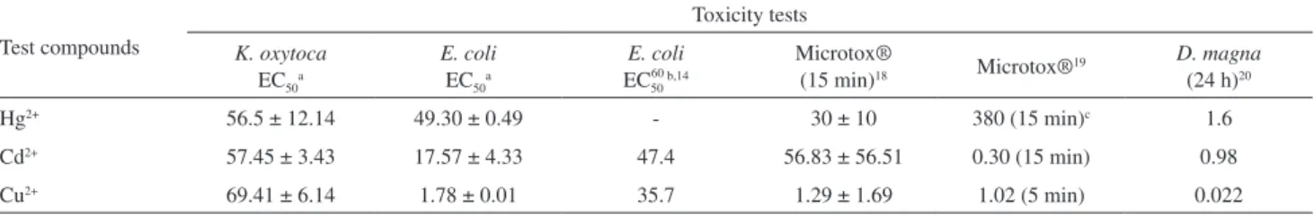
concentration in the control (bacterial suspension without the toxic agent). Figure 3 shows the bacterial growth of the control and the suspensions with increasing concentration of the metal Cd2+.
The effective concentration (EC50) was obtained from
the chart of percent inhibition versus concentration of the test compound over an exposure time, which was determined by the bacterial growth of the control in the CO2 concentration range 0.5-4 mmol L–1 (Figure 3).
Equation 2 provides an expression for the effective concentration,
(2)
where C is the difference between the final and initial CO2 concentrations in the control, and A is the difference
between the final and initial CO2 concentrations in the
sample.
Results and Discussion
The bacterial growth times in the controls, within the 0.5-4 mmol L–1 concentration range, differed for each
species. Table 1 lists the number of assays performed and the average time it took the bacteria to double.
According to the CO2 concentration, K. oxytoca
grew faster than E. coli with duplication times of 29.5 ± 3.4 min and 33.9 ± 3.2 min, respectively. The F-test for the comparison of standard deviations showed that the variances of the two cases did not differ significantly; however, the significance test (t-test) for the comparison of
Figure 1. Diagram of the FIA/conductometric system used to determine the CO2 concentration in toxicity assays.
Table 1. Number of assays performed and the bacteria doubling time
Bacteria Number of assays
performed
Doubling time / min
K. oxytoca E. coli
37 30
two experimental means pointed a significant difference at p = 0.05 (Miller and Miller17).
The assay toxicity method described here was optimized by varying the concentrations of the test compounds to identify concentrations that would be representative of an inhibition interval between 0 and 100%. The assays were performed at least in duplicate.
The tetracycline assays provided a reference assay for comparison. The bacterium may undergo genetic mutations due to several factors. One such common mutation is caused by transfer of the original strain to prolong its use. Each transfer represents a new generation and, after several transfers, the bacteria may display different effective concentrations (EC50). Prior to conducting the
assay with a test compound, an assay must be performed using a solution in which the inhibition concentration or the EC50 is known, and which acts as a control for the
bacterial activity. All toxicity tests were accompanied by such an activity control assay, performed by growing the strain in media containing 0.1 mg L–1 tetracycline to
verify the bacterial activity. This concentration provided inhibition of around 61% relative to the control. The EC50 values remained constant throughout the period of
experiments showing that no significant variations in the characteristics of both bacteria occurred.
The results demonstrated that both bacterial species had similar responses to the reference antibiotic (EC50 = 0.08 ± 0.01 mg L
-1). No effects were noted up to
concentrations of 0.02 mg L–1. Inhibition began beyond
a concentration of 0.05 mg L–1 and was complete after
0.5 mg L–1.
The sensitivity of the assay to a commercial antibiotic indicates the assays’ potential for applications in healthcare. For example, antibiograms may be used to determine the susceptibility of a contaminated material of biological origin to a range of antibiotics.
Comparison among metals
Figure 2 shows the behavior of the bacteria in the presence of Hg2+. No effects on growth were observed
relative to the control in either species in the presence of up to 25 µg L–1 Hg2+. An inhibition response was observed
starting at 50 µg L–1 and became significant for E. coli,
with 70-80% inhibition. K. oxytoca inhibition levels were in the range 40-50%. E. coli proved to be more sensitive than K. oxytoca to this compound.
In the presence of Cd2+, the inhibition effects on the
bacteria differed at a concentration of 2.5 mg L–1, which was
the lowest tested. The K. oxytoca (Figure 3) response was comparable to the response of the control, whereas E. coli
showed slight inhibition (Figure not shown). The EC50 of
Cd2+ for K. oxytoca (Figure 4) was 3 times greater than that
for E. coli (Table 2). Thus, it was possible to conclude that cadmium showed higher toxicity to E. coli, when compared to K. oxytoca bacteria.
Figure 2. Bacterial growth of K. oxytoca and E. coli in the presence of Hg2+.
E. coli proved to be far more sensitive than K. oxytoca
to Cu2+. The compound displayed growth inhibition starting
at 0.25 mg L–1 for E. coli, and exposure to 10 mg L–1 Cu2+
resulted in K. oxytoca inhibition comparable to that of the control. The EC50 for Cu2+ for K. oxytoca was 70 times
greater than that for E. coli (Table 2).
A thorough evaluation of the toxic effects of a test compound requires that toxicity be tested for an array of organisms that are representative of the different trophic levels. The sensitivities of E. coli and K. oxytoca were compared with previous reports of the sensitivities of other organisms to the same toxic agents, as shown in Table 2. The organisms assayed in the literature were E. coli, Vibrio fischeri or Microtox®, and the microcrustacean
Daphnia magna.
Toxic agents act differently on microorganisms. To estimate safe environmental concentration limits, several tests must be performed simultaneously. The assays must also be performed under different conditions. Among the organisms listed in Table 2, D. magna was more sensitive than the bacteria towards metals.
Several conclusions could be drawn with respect to the bacteria used in this study and submitted to the same environmental conditions. K. oxytoca was more resistant
than E. coli to all test compounds. The order of sensitivity was Hg2+ > Cd2+ > Cu2+ for K. oxytoca and Hg2+ > Cu2+ >
Cd2+ for E. coli.
Organisms at the same trophic level may display different sensitivities toward a specific compound, as suggested by the inverted sensitivities to Cd2+ and Hg2+.
The original microorganisms may have had contact with different metals and may have adapted prior to being used in this assay. Different laboratories may obtain variable results, as pointed by Cotman et al.,21 who described an
interlaboratory trial using D. magna tests for wastewater matrices. The coefficient of variation was as high as 62.9%.
Hg2+ was the most toxic agent among the metals tested
for the majority of organisms shown in Table 2. Cu2+ was
the second most toxic (with the exception of toxicity toward
K. oxytoca and one Microtox test), followed by Cd2+.
It is important to point out that in the present study the results were obtained from total concentration of metals and the activity or bioavailability of the test compounds in the culture medium was not tested. However, Jardim et al.,12
and Gimenez and co-workers,14 have previously verified the
activity of Cu2+, Cd2+, and Hg2+ in the culture medium in a
toxicity test similar to this study. They concluded that the toxicity is reduced in the presence of the culture medium.
Toxicity data for tests with pure substances performed in the laboratory are important for the evaluation of environmental risks and also for setting water quality criteria. Aquatic organisms are not normally exposed to isolated substances. Rather, they are exposed to mixtures.
The interactions between organic compounds and toxic metals can change the bioavailability of a metal and reduce its toxicity. The toxicity of metals is more closely correlated with the free ion concentration than with the total metal concentration or the concentration of the complexed forms.22
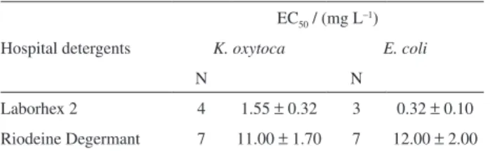
Comparison among active ingredients in hospital detergents
Klebsiella spp. bacteria are an important nosocomial pathogen. The incidence of Klebsiella infection detected in immunocompromised hospitalized patients in hospitals
Figure 4. Effective concentration (EC) of Cd2+ for K. oxytoca.
Table 2. Comparison of the EC50 and IC50 of 6 toxicity tests for Hg2+ (µg L-1), Cd2+ (mg L–1), and Cu2+ (mg L–1) reported in the literature
Test compounds
Toxicity tests
K. oxytoca EC50a
E. coli EC50a
E. coli EC5060 b,14
Microtox®
(15 min)18 Microtox®19
D. magna (24 h)20
Hg2+ 56.5 ± 12.14 49.30 ± 0.49 - 30 ± 10 380 (15 min)c 1.6
Cd2+ 57.45 ± 3.43 17.57 ± 4.33 47.4 56.83 ± 56.51 0.30 (15 min) 0.98
Cu2+ 69.41 ± 6.14 1.78 ± 0.01 35.7 1.29 ± 1.69 1.02 (5 min) 0.022
is around 5-7%.23 The main cause of hospital infections is
the incorrect asepsis of equipment and the hands of hospital staff. To prevent Klebsiella oxytoca outbreaks in hospitals, the efficiency of detergents and disinfectants need to be evaluated. This can be performed using quick tests, as described in this study.
Laborhex and Riodeine detergents are indicated as disinfectant for the hands and arms of the surgical and laboratorial team and for pre-surgical preparing of patients skin.
Concerning to laborhex detergent, it was verified that 0.50 mg L-1 of the compound was the highest concentration
used in the test for E. coli, causing a high inhibition; this same concentration was the lowest used in the test with K. oxytoca. Table 3 shows the EC50 values obtained for both
bacteria. The EC50 was five times higher for K. oxytoca
than E. coli. So, the E. coli was more sensitive to laborhex detergent and, therefore, K. oxytoca was more suitable than
E. coli for evaluating the efficiency of detergents based on chlorhexidine. As shown in Table 3, both species presented similar sensitivities to Riodeine, once the EC50 values were
statistically equal.
Laborhex 2 showed bactericidal action toward E. coli that was superior to the action of Riodeine, whereas
K. oxytoca was more resistant than E. coli to Laborhex 2.
K. oxytoca resistance against the disinfectant was probably aided by the formation of capsules visible as mucold colonies, as reported by Reiss et al.15
The detergent test compound results showed the potential for application of the toxicity assay developed here. The efficiency of the detergents was evaluated from the standpoint of their quality as sanitary products.24
Conclusions
The K. oxytoca appeared to be a good test organism because the EC50 values obtained in the toxicity
assays showed low standard deviations, which denotes reproducibility.
The bacteria proved to be highly sensitive to tetracycline. This drug may be used as a reference to confirm that a strain has retained its characteristics during storage and handling.
For all the metals (Hg2+, Cd2+, and Cu2+) analyzed and
both detergents (Laborhex 2 and Riodeine Degermant),
K. oxytoca proved to be more resistant than E. coli., indicating that Klebsiella as a more reliable organism for efficiency assays of detergents used in hospitals than
E. coli.
Finally, the toxicity test proposed in this study may be used as a complementary test when a battery of toxicity tests are required to characterize both pure substances and mixed compounds.
Acknowledgments
The authors would like to thank the São Paulo Research Foundation (FAPESP) and the Coordenação de Aperfeiçoamento de Pessoal de Nível Superior (CAPES).
References
1. Arslan-Alaton, I.; Eremektar, G.; Germirli-Babuna, F.; Insel, G.; Selcuk, H.; Ozerkan, B.; Teksoy, S.; Water Sci. Technol.
2005, 52, 309.
2. Jamroz, T.; Ledakowicz, S.; Miller, J. S.; Sencio, B.; Environ. Toxicol. 2003, 18, 187.
3. Tisler, T.; Zagorc-Koncan, J.; Water Sci. Technol. 1994, 30, 107. 4. Mamais, D.; Noutsopoulos, C.; Stasinakis, A. S.; Kouris, N.;
Andreadakis, A. D.; Water Environ. Res. 2008, 80, 484. 5. Ezemonye, L. I. N.; Ogeleka, D. F.; Okieimen, F. E; Chem.
Ecol. 2007, 23, 131.
6. Ferrer, L.; Andrade, S.; Austeasuain, R.; Marcovecchio, J.; Ecotoxicol. Environ. Saf. 2006, 65, 209.
7. Conselho Nacional do Meio Ambiente (CONAMA); Resolução n. 357 de 15 de março de 2005, Brasília, DF, Brasil, 2005. 8. Bitton, G.; Crit.Rev. Environ. Control. 1983, 13, 51. 9. Parvez, S.; Venkataraman, C.; Mukherji, S.; Environ. Int. 2006,
32, 265.
10. Moraes, S. G.; Freire, R. S.; Durán, N.; Chemosphere2000, 40, 369.
11. Jardim, W. F., Moraes, S. G., Takiyama, M. M. K.; Water Res.
1997, 31, 1728.
12. Jardim, W. F.; Pasquini, C.; Guimarães, J. R.; Faria, L. C.; Water Res. 1990, 24, 351.
13. Guimarães, J. R.; Jardim, W. F.; Quim. Nova1993, 16, 28. 14. Jardim, W. F.; Canela, M. C.; Gimenez, S. M. N.; Moraes, S. G.;
Chem. Speciation Bioavailability1993, 5, 97.
15. Reiss, I.; Borkhardt, A.; Füssle, R.; Sziegoleit, A.; Gortner, L.; The Lancet2000, 356, 310.
16. Dowards, E. J.; Barisas, B. G.; Aquat. Toxicol. 1984, 4, 129. 17. Miller, J. N.; Miller, J. C.; Statistics and Chemometrics for
Analytical Chemistry, 4th ed.; Prentice Hall, New Jersey, USA,
2000.
Table 3. Comparison of the EC50 for hospital detergents
Hospital detergents
EC50 / (mg L–1)
K. oxytoca E. coli
N N
Laborhex 2 4 1.55 ± 0.32 3 0.32 ± 0.10
18. Greene, J. C.; Miller, W. E.; Debacon, M. K.; Long, M.; Bartels, C. L; Arch.Environ. Contam. Toxicol.1985, 14, 659. 19. Sillanpãã, M.; Oikari, A.; Chemosphere1996, 32, 1485. 20. Sorvari, J.; Sillanpãã, M.; Chemosphere 1996, 33, 1119. 21. Cotman, M.; Drolc, A.; Milenko, R.; Tisler, T.; Int. J. Environ.
Pollut.2007, 31, 13.
22. Kim, K. T.; Lee, Y. G.; Kim, S. D.; Environ. Int. 2006, 32, 487. 23. Brisse, S.; Milatovic, D.; Fluit, A. C.; Verhoef, J.; Schmitz, F.-J.;
Eur. J. Clin. Microbiol. Infect. Dis.2000, 19, 64.
24. Santos, A. A.; Nascimento, A. R.; Kondo, M. M.; Grassi, M. T.; Annals of the Brazilian Chemical Society, 23th Meeting, Poços
de Caldas, MG, Brasil, 2000.
Submitted: March 23, 2011
Published online: January 17, 2012