vol. 42, n. 1, jan./mar., 2006
A study of the characteristics and
in vitro
permeation properties of CMC/
chitosan microparticles as a skin delivery system for vitamin E
Juliana Bucchi Alencastre
1, Maria Vitoria Lopes Badra
Bentley
1, Fabiola Silva Garcia
1, Maria de
Moragas
2, Joseph Luis Viladot
2, Juliana Maldonado
Marchetti
1*1Departamento de Ciências Farmacêuticas, Universidade de São Paulo, 2Cognis Iberia (Primacare),
Polígono Industrial San Vicente, Spain
Carboxymethylcellulose (CMC)/chitosan microparticles containing vitamin E were prepared by a complex coacervation method and their potential use as a topical delivery system was evaluated. Morphology, particle size distribution, encapsulation yield, physical and chemical stability, in vitro release and permeation through skin were studied. The microparticles appeared to be spherical, with a homogeneous surface and were not aggregated. Mean diameters ranged from 2.7 to 7.6 μm and the encapsulation yield was 81%. Chemical stability studies indicated a protection of encapsulated vitamin E, of 8.1% for O/W and of 10.83% for W/O emulsions, following storage at 45 °C for 60 days. Forty-eight% of vitamin E, determined by HPLC, were released within 24 hours.
In vitro permeation and retention studies showed a higher penetration rate of vitamin E in the free and encapsulated forms, from the W/O emulsion. The carriers studied seem to be promising systems for topical administration.
INTRODUCTION
One of the biggest challenges to Pharmaceutical Sciences over the last 40 years has been finding new ways to extend the releasing capacity of active compounds from delivery systems (Kydoneus, Berner, 1987).
Cutaneous administration, leading to the release of active substances on the epidermis, coupled to their maintenance at the site of action, is a new research area. Improved delivery systems maximizing the time of permanence of active compounds on the skin by minimizing transdermal absorption may play an important role in the success of this therapy (Embril, Nacht, 1996).
A c t i v e c o m p o u n d s s u c h a s U V f i l t e r s a n d antiseptics should remain on the skin surface to effectively perform their functions. Other active substances such as antioxidants, whitening and antiaging agents can be effective on the epidermis or dermis (Morganti et al., 2001). In recent years, microparticles have aroused increasing interest as drug delivery systems. Many authors reported that m i c r o p a r t i c l e s c o u l d b e u s e d t o d e l i v e r a c t i v e compounds into the skin in greater amounts than do conventional topical formulations, with improved localization at the desired site of action. Natural or synthetic polymers can be used for the obtention of microparticles. Among the polymers available for
*Correspondence:
J. M. Marchetti Departamento de Ciências Farmacêuticas
Faculdade de Ciências Farmacêuticas USP/Ribeirão Preto
Av. do Café s/n
14040-903 - Ribeirão Preto, SP - Brasil E-mail [email protected]
Unitermos
microencapsulation purposes, chitosan has been widely studied as means of obtaining long-term, s u s t a i n e d d r u g r e l e a s e f r o m m i c r o p a r t i c l e p r e p a r a t i o n s s h o w i n g b i o c o m p a t i b i l i t y a n d biodegradability. Clinical tests have not reported a l l e rg i c o r i n f l a m m a t o r y r e a c t i o n s f o l l o w i n g i m p l a n t a t i o n , i n j e c t i o n , t o p i c a l a p p l i c a t i o n o r ingestion of such preparations (Muzzarelli, 1995).
Interest has focused on the use of vitamin E, a natu-ral antioxidant with important biological effects, in topical administration carriers. When used in skin care products, the vitamin protects this tissue from the effects of UV radiation, delays the photoaging process and exhibits moisturizing properties. In order to obtain a moisturizing effect, vitamin E must be absorbed and kept on the skin at a certain concentration. Its exact moisturizing mechanism is not completely known yet; it may be the consequence of the vitamin’s marked antioxidant character. An effective free radical capture agent, vitamin E can be able to prevent and repair skin damage in aggressive situations. This effect may enable the skin to use its own moisture control system via natural humectant factors and adequate lipid barriers (Tamburic et al., 2000).
Vitamin E is slowly oxidized by atmospheric oxygen, via a reaction catalyzed by light and heat in the presence of silver salts and iron (Handbook of Vitamins 1991; The Merck Index, 1996).
The aim of this work was to evaluate carboxymethyl-cellulose/chitosan vitamin E microparticles as a skin delivery system. To study this effect, scanning electron microscopy (SEM), particle size distribution, physical-chemical stability,
in vitro release and permeation were used.
MATERIALS AND METHODS
Vitamin E microparticles (Primaspheres®), batch number G26F05, were prepared and supplied by Cognis Brasil Ltda.The solvents for analytical analysis were of chromatography grade and were purchased from Merck (Darmstadt, Germany). All others chemicals were of reagent grade, obtained from Merck (Darmstadt, Germany).
Microparticles preparation
The production technique used was the complex coacervation (Patent: WO/PCT/EP01/01177). Initially, an O/W emulsion was obtained containing the active substance (vitamin E). Then, the cationic polymer chitosan of the aqueous phase was forced to precipitate in the interphase by the interaction with the anionic polymer, carboxymethylcellulose (CMC).
Measurement of particle size
The particle size distribution analysis was carried out by laser diffraction, using a CILAS 1064 Granulometer.
Scanning electron microscopy
Particle morphology was examined by Scanning Electron Microscopy (SEM), using a Leica-Stereoscan 440 equipment. Samples were dried at 37 °C for 24 hours and prior to visualization, covered with gold using a Spulter Balzers SCD 050 Coater .
Drug loading determination
Encapsulation efficiency was determined by an indirect method (Calvo et al., 1997; Cui, Mumper, 2001). Microparticle dispersions containing 250 mg/mL of vitamin E were prepared in phosphate buffer (pH = 7.2) containing 2 mM polysorbate 80. Aliquots were filtered through 0.45 mm pore membranes; the amount of vitamin E present in the filtrate (free vitamin E) was assayed by HPLC. The total amount of the vitamin present in the preparation was determined after extraction with methanol, followed by ultra- sonication for 30 minutes in order to break up the particles, followed by filtration and posterior assay by HPLC. Encapsulation efficiency (drug loading) was calculated as follows:
Drug loading = total % vitamin E - free % vitamin E
Vehicle formulations
The composition of the emulsions used in this study is presented in Tables I and II.
After the emulsification process the equivalent to 0.25% of free or encapsulated form (microparticles) of vitamin E was added to O/W and W/O emulsions, under slow agitation.
Stability studies
Samples of the O/W or W/O emulsions containing free or encapsulated vitamin E were stored at different temperatures (5, 25 and 45 °C) for a 2-month period and its macroscopic appearance examined at regular intervals (Cannel, 1985; Cadwallader, 1989; Idson, 1993; International Conference On Harmonization, 2003).
HPLC assay
Chromatographic analysis were carried out in a Shimadzu chromatograph, model SPD, equipped with an UV detector operating at 290 nm, a LC-ADVP pump, a Rheodyne injector and a CR-6 integrator. The separation was carried out in a Lichrosphere 100 RP - 18 (125 x 4 mm (5 mm) column, Merck), protected by a RP-18 guard column (4x4 mm, Merck). Elution was performed at controlled room temperature (25 ± 1 °C), at flow- rate of 1 mL/min, with a mobile phase consisting of a methanol: water mixture (99:1), an injection volume corresponding to 20 µL except for the release and permeation tests in which a volume of 200 µL was used .
Precision, accuracy, quantification limit, selectivity and recovery data were determined in order to validate the chromatographic method.
The precision and accuracy of the method were tested by within day (intra-assay) and between day (inter-assay) analyses. For the determination of intra-assay precision and accuracy, aliquots (n=10) of a vitamin E methanol solution, at concentrations of 0.5 and 50.0 µg/mL were analyzed as described above. The precision of the assay was calculated from the percent coefficient of variation (CV) of the mean. The accuracy of the assay was calculated from the relative errors of the means between expected and calculated concentrations. Inter-assay precision and accuracy were calculated by analyzing aliquots of the same samples on five consecutive days.
The sensitivity of detection was considered as the minimum amount of the drug that could be measured without interference of the basal line. To assess selectivity, the receptor medium used in the in vitro skin permeation experiments was submitted to chromatographic analysis. In addition, blanks prepared for each (O/W and W/O without vitamin E) formulation were also analyzed.
In vitro vitamin E release studies
For these studies, dispersions of microparticles in isotonic phosphate buffer (pH = 7.2), containing 2 mM polysorbate 80 and 250 µg/mL vitamin E, were maintained at 37 ° ± 1 °C for 24 hours, under constant stirring, at 100 rpm. At pre- established time intervals (0, 2, 4, 8, 12 and 24 hours), 1 mL of the medium was removed and replaced by an equal volume of fresh receptor solution. Samples were filtered through 0.45 mm pore membranes and appropriately diluted for the determination of their free vitamin E content. The results refer to the average of six experiments.
In vitro permeation and retention studies
Slices of dorsal skin from 4 to 6-week-old male hairless mice (strain HRS/ J Jackson Laboratories, Bar Harbor, ME) were mounted in a diffusion cell system (Microette Hanson Research Corporation), with their dermal side in contact with the receptor medium (7 mL of isotonic phosphate buffer pH = 7.2, containing 2 mM of polysorbate 80). The donor compartment was filled with 200 mg of the formulations. The experiments were carried out at 37 oC, with the receptor medium stirred at 300 rpm. One mL of the medium was removed after 4, 8, 12 and 24 hours respectively, and assayed for its vitamin E content. Six experiments were carried for each formulation. At the end of each experiment, skins were removed from the diffusion cells, and the excess formulation TABLE II - Composition of the W/O emulsion
Components %
Phase I
Polyglyceryl dipolyhydroxystearate 3.00
Polyglyceryl diisostearate 3.00
Bee wax 3.00
Zinc stearate 1.00
Dicaprylyl ether 3.00
Coco caprylate/caprate 6.00
Capric/caprylyl triglyceride 8.00
Propyl paraben 0.02
Phase II
Glycerin 5.00
MgSO4•7H2O 1.00
Methyl paraben 0.18
Distilled water up to 100.00
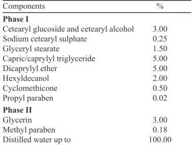
TABLE I- Composition of the O/W emulsion
Components %
Phase I
Cetearyl glucoside and cetearyl alcohol 3.00
Sodium cetearyl sulphate 0.25
Glyceryl stearate 1.50
Capric/caprylyl triglyceride 5.00
Dicaprylyl ether 5.00
Hexyldecanol 2.00
Cyclomethicone 0.50
Propyl paraben 0.02
Phase II
Glycerin 3.00
Methyl paraben 0.18
carefully removed with distilled water. The tissues were fixed to a support and submitted to removal of the SC by the tape stripping method using adhesive tapes (3M®). The tapes were transferred to a conical tube, 5 mL of methanol (extractor solvent) were added and the tubes mixed for 3 minutes. Extracted vitamin E was assayed by HPLC. The remaining tissue (epidermis + dermis) was cut into fragments and transferred to conical tubes. One mL of methanol was added and the tubes sonicated for 30 minutes. Contents were filtered through 0.45 mm pore membrane filters and the vitamin E content of the filtrates analyzed.
RESULTS AND DISCUSSION
Pharmaceutical and Cosmetic Technologies have dedicated special attention to the development and evaluation of formulations providing controlled release of active substance(s), taking into consideration that significant progress could be obtained by the use of pharmaceutical and cosmetic forms capable of modulating the release of active compounds and of localizing them at desired sites of action.
Micro/nanoparticles emerged as one of the most promising strategies to obtain controlled delivery systems (Berthold et al., 1998), They have been extensively used for oral, parenteral and topical administration, as well as to vehicle many compounds into the skin, including 5-fluorouracyl (Ghorab et al., 1990), α-tocopherol (Dingler et al., 1999), glycolic acid (Perugini et al., 2000), retinol (Jening et al., 2000), some allergenic substances (Brosse
et al., 2000) and others.
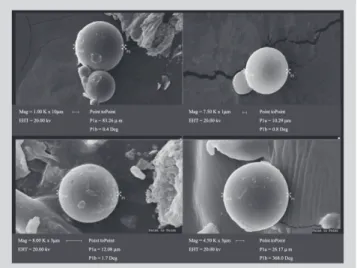
In the present study, CMC/chitosan microparticles presented diameters ranging between 2.726 and 7.654 mm (Table III). The analysis of the particle size distribution is important in skin penetration studies since depending on their size, it becomes possible to forecast the probability of these particles penetrating the stratum corneum or the hair follicles, in order to reach viable epidermis and dermis sites, where vitamin E will develop its biological function. SEM photomicrographs show that the micro-particles obtained in this work present spherical shapes
with a smooth and homogeneous surface and a non-aggregated distribution (Figure 1). Their morphology is of fundamental importance because it can directly influence the release and penetration of active substances from the encapsulated form into the skin.
The epidermal stratum corneum consists mainly of keratin, ceramides, cholesterol and free fatty acids. These lipids are distributed in a lamellar structure in the intercellular spaces (Kumpel et al., 1998; Dietz, Hameyer ,2003).
Madison et al., (1987) achieved the first visualization of the broad-narrow-broad arrangement of the stratum corneum intercellular lipid lamellae. It was recognized at the time that the broad lamellae in the stratum corneum, as well as the precursor lamellae in the lamellar granules, have the dimensions (approximately 5 nm) of a typical lipid bilayer, whereas, the broad-narrow-broad units were 13 nm wide. This 13 nm periodicity corresponds to the distance from the center of one pair of bilayers, to the center of the next pair (Kumpel et al., 1998).
Bronaught et al., 1982, in order to more completely characterize permeability relationships, measured the thickness of the stratum corneum, of the epidermis and of whole skin. He utilized frozen sections of the skin to overcome the well-known destructive effect of standard embedding and fixing procedures on the stratum corneum. Determination of hair follicle density, under the microscope, demonstrated the average diameter of the hair follicles of the human abdomen to be of 97 ± 3 mm, and of those of the dorsal area of the hairless mouse to be of 46 ± 1 mm. Based on this information and on our results of scanning electron microscopy and particle size distribution, we concluded that the particles under study should be able TABLE III – Particle size distribution of vitamin E
microparticles
Population Mean Diameter* Variation
(%) ± SD (µm) Coefficient (%)
10 0.948 ± 0.004 0.422
50-90 2.726- 7.654 ± 0.106 1.384
*The results represent the mean diameter of 5 determinations
FIGURE 1 - Photomicrography of CMC/chitosan
to penetrate into the human or mouse skin through hair follicles or to remain on the stratum corneum surface, delivering the active substances into the epidermis.
The Tables IV and V show the parameters of precision, accuracy, quantification limit and recovery obtained by HPLC method validation.
With the results obtained in the intra- and inter-assay, it was observed that the method chosen for the quantitative analyses of vitamin E presented precision, accuracy, sensitivity and selectivity adequate for the performance of the proposed studies.
Encapsulation yield was 81%, a satisfactory level, confirming the suitability of the complex coacervation method of preparation.
Physical stability studies showed that O/W and W/O emulsions were stable for a 2-month period, during which no changes like flocculation, cremeation or coalescence were observed, indicating that these emulsions can be used as vehicles for free and microencapsulated vitamin E.
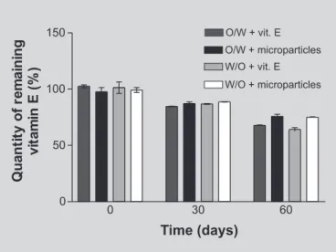
Alpha-tocopherol content in W/O and O/W emulsions stored at 5 or 25 oC remained practically constant for 60 days. Samples stored at 45 oC, for 60 days, showed a gradual decrease of vitamin E due to chemical degradation. A higher protection of vitamin E encapsulated in microparticles was noted, of 8.10% and 10.83% in O/W and W/O emulsions, respectively (Figure 2 and Table VI). Analysis by Student’s t test showed these results to be statistically different (p<0.05)
from those of formulations containing the free vitamin. Microparticles obtained using other polymers did not present significant stability for instable actives, (Rossler et al., 1994); collagen microparticles containing retinol incor-porated in hydrogels, did not show a statistically significant increase of stability compared to formulations containing free retinol. Microparticles had no influence on the chemical stability of retinol. Therefore in this respect, CMC/chitosan microparticles seem to be more efficient.
The pattern of release of vitamin E from CMC/ chitosan microparticles is shown on Figure 3. During the first stage of dissolution, a “burst effect” occurred which increased linearly for up to 12 hours, after which the vitamin concentration remained constant. Analysis of these results by linear regression data indicated that vitamin E release from microparticles, initially followed pseudo-zero order kinetics (Higuchi, 1963), stabilizing after 12 hours into a sustained release pattern.
TABLE IV – Precision, accuracy and quantification limit obtained by HPLC
Assay Theoretical Obtained C.V. A
concentration (µg/mL) concentration (µg/mL)
Inter-assay 0.5 00.4892 (± 0.0129) 2.6391 2.1677
50.0 50.1667 (± 1.0432) 2.0795 0.3333
Intra-assay 0.5 00.5091 (± 0.0354) 6.9500 1.8166
50.0 49.3481 (± 1.0418) 2.1111 1.3039
Quantification limit 0.20000 00000000.20069 (± 8.64565 . 103) 04.30801 00.34382
The results represent the mean and the standard deviation of 5 determinations; C.V. = Coefficient of Variation; A = Accuracy
TABLE V – Results obtained in the test of vitamin E recovery from O/W and W/O formulations by HPLC
Samples Vitamin E Recovery (%)*
O/W + vitamin E 102.510 ± 1.350
O/W + microparticles 97.730 ± 2.623
W/O + vitamin E 101.295 ± 3.596
W/O + microparticles 99.240 ± 2.220
The data represent the mean of 3 determinations with their respective standard deviation.
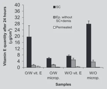
The results of retention studies presented on Figure 4 show the amounts of vitamin E retained in the stratum corneum, epidermis + dermis and permeated into the re-ceptor solution, after a 24 hour-experiment in diffusion cells. A higher availability of vitamin E incorporated into W/O emulsions was observed.
This result may be attributed to a higher thermo-dynamic activity in the W/O emulsions, since both the distribution coefficient and the concentration of the active substance depend not only on its total concentration, but also on its concentration in the solubilizing phase (Forsters et al., 1997). In discussing the smaller penetration of vitamin E from O/W emulsions, these authors attributed this fact to the formation of a lamellar layer, responsible for the viscosity of these formulations. Such lamellar layers would prevent the diffusion of vitamin E in the oily phase towards the skin.
The amounts of vitamin E permeated, quantified after 24 hours, showed a higher permeation in its free form from the O/W and W/O emulsions.
Wester et al. (1991) compared benzoyl peroxide absorption by the skin from a topical formulation containing the active substance dispersed or encapsulated in a system of microsponges (styrene/divinylbenzene) with a mean diameter of 25 µm. In vitro studies using human skin showed that after 24 h, smaller quantities of the active ingredient were recovered in the epidermis from the encapsulated form (1.1 ± 0.7% of the applied dose) in compared to the free form (5.2
± 4.1%). The same result occurred in the dermis. However, these results were statistically non-significant (p > 0.05). It was also observed that the amount permeating from the microsponge formulation was 3.5 ± 3.4% and that from the free form, 17.1 ± 15.7% .
Dingler et al. (1999) carried out the in vivo
administration of α-tocopherol and α-tocopherol acetate
to 20-30 year old volunteers, using dispersions of nanoparticles with mean diameters of 280 and 270 nm, respectively. After 30 minutes of contact with the skin, the excess of the formulation was eliminated and tape stripping carried out. The quantity of the two active ingredients that penetrated the stratum corneum from the encapsulated preparation was twice that from controls containing the vitamin in free form.
Perugini et al. (2000) evaluated the potential of three types of microparticulated systems, respectively: liposomes, liposomes modified by the addition of chitosan with diameters from 2-10 µm, and of 2-15 µm diameter chitosan microspheres, in the transport of glycolic acid, a small hydrophilic compound. Liposomes were able to TABLE VI – Percent of vitamin E remaining in formulations stored at 45 OC for 60 days
Time (days) O/W +vitamin E(%) O/W + microparticles(%) W/O +vitamin E(%) W/O + microparticles(%)
0 102.510 ± 1.350 97.730 ± 2.623 101.295 ± 3.596 99.240 ± 2.220
30 84.685 ± 0.285 87.155 ± 1.395 86.755 ± 0.375 88.655 ± 0.165
60 67.750 ± 0.430 75.855 ± 1.755 64.195 ± 1.555 75.030 ± 0.410
*Results represent means of 3 determinations with their respective standard deviations
FIGURE 3 - Vitamin E release from CMC/chitosan microparticles in phosphate buffer pH=7.2 containing 2 mM polysorbate 80. Bars represent standard derivations of means of six determinations.
delay the release of glycolic acid, and microspheres were unable to modulate active substance release even following crosslinking with glutaraldehyde, a compound causing a reduction of microsphere sizes.
In previous studies, Genta et al. (1997) were able to modulate the release of the acyclovir, a lypophilic drug, using chitosan microspheres with mean diameter ≤ 25 µm
for ocular administration.
Based on the experimental results obtained on the in vitro release and cutaneous penetration of vitamin E, as well as on literature data, it can be concluded that the general releasing properties of active substances from particulate systems are directly or indirectly related to the following factors: (i) The presence or absence of macro and micropores in the particulate system; microsponges (Wester
et al., 1991) and nylon particles (Parison, 1993) as typical examples of porous systems; (ii) particle size, with nanoparticles presenting a higher probability of penetrating the stratum corneum, while larger particles (microparticles, diameters > 1 µm) are retained on the skin surface or penetrate via hair follicles; (iii) intrinsic characteristics of delivery systems being directly related to the process and materials used for their manufacture; indirect factors related to (iv), the carrier into which the delivery system is incorporated; (v) intrinsic characteristics of the active substance to be encapsulated; and (vi) depending on the characteristics of active substance and carrier, different release behaviors may be expected, since after release from particulated systems, active substances have to diffuse through barrier(s) to reach their site(s) of action.
To conclude: in view of adequate morphology, particle size, sustained release profile and skin penetration behaviour, CMC/chitosan microparticles prepared by complex coacervation may become promising systems for the topical delivery of various therapeutically active compounds.
RESUMO
Estudo das características e propriedades da permeação in vitro de micropartículas de CMC/quitosana como
sitema de liberação cutânea para vitamina E
Micropartículas de carboximetilcelulose (CMC)/quitosana contendo vitamina E foram preparadas pelo método de coacervação complexa e seu uso potencial como um siste-ma de liberação tópico foi avaliado. Estudos da morfologia, da distribuição do tamanho de partículas, da eficiência de encapsulação, da estabilidade física e química e da liberação e permeação cutânea in vitro foram realiza-dos. As análises por Microscopia Eletrônica de Varredura mostraram que as partículas são esféricas, possuem uma superfície homogênea e ausência de agregados, com
diâ-metros na faixa de 2,7 a 7,6 μm. A eficiência de encap-sulação da vitamina E foi 81%. Os estudos de estabilidade química mostraram proteção da vitamina E encapsulada, sendo que a diferença em relação à quantidade de ativo re-manescente na emulsão O/A foi de 8,1% e na A/O, de 10,8%, após armazenamento a 45 °C por um período de 60 dias. O ensaio de liberação in vitro mostrou que 48% da vi-tamina E encapsulada, quantificada por CLAE, foram libe-radas em 24 horas de experimento. Nos estudos de retenção e permeação in vitro observou-se que a emulsão A/O pro-porcionou maior penetração da vitamina E tanto da forma livre como encapsulada.Os sistemas avaliados parecem ser promissores para veiculação de ativos em preparações de uso tópico.
UNITERMOS: Micropartículas. Quitosana. Vitamina E. Permeação cutânea in vitro
ACKNOWLEDGEMENTS
The authors wish to thank Cognis Brasil Ltda and CAPES for supporting this research.
REFERENCES
BERTHOLD, A.; CREMER, K.; KREUTER, J. Collagen microparticles: carriers for glucocorticosteroids., Eur. J. Pharm. Biopharm., Stuttgart, v. 45, p. 23-29, 1998.
BRONAUGH, R. L.; STEWART, R. F.; CONGDON, E. R. Methods for in vitro percutaneous absorption studies II. Animal models for human skin. Toxicol. Appl. Pharmacol., San Diego, v. 62, p. 481-488, 1982.
BROSSE, C.; SOUTIF, J. C.; BROSSE, J. C. Preparation of microcapsules for skin allergy testing by solvent evaporation process. J. Microencapsul., London, v. 17, p. 111-116, 2000.
CADWALLADER, D.E. Stability testing: its role in pre-formulation and pre-formulation of cosmetic products.
Cosmetics & Toiletries, Athens, v. 104, p. 87-102, 1989.
CALVO, P.; VILA-JATO, J.L.; ALONSO, M. J. Evaluation of cationic polymer-coated nanocapsules as ocular drug carriers. Int. J. Pharm., Amsterdam, v.153, p. 41-50, 1997.
CANNEL, J.S. Fundamentals of stability testing. Int. J. Cosmet. Sci., Oxford, v. 7, p. 291-303, 1985.
DIETZ, T., HAMEYER, P. How to formulate with ceramides – basic investigation. SÖFW – J., Angsburg, v. 129, p. 2-9, 2003.
DINGLER, A.; BLUM, R. P.; NIEHUS, H.; MULLER, R. H.; GOHLAS, S. Solid lipid nanoparticles (SLNTM/ LipopearlsTM) – a pharmaceutical and cosmetic carrier for the application of vitamin E in dermal products. J. Microencapsul., London, v. 16, p. 751-767, 1999.
EMBRIL, K.; NACHT, S. The Microsponge Delivery System (MDS): a topical delivery system with reduced irritancy incorporating multiple triggering mechanism for the release of actives. J. Microencapsul., London, v. 13, p. 575-588, 1996.
FORSTER, T. H .; JACKWERTH, B.; PITTERMANN, W.; RYBINSKI, W.; SCHMITT, M. Properties of emulsions.
Cosmet. Toiletries, Fairfield, v. 112, p. 73-82, 1997.
GENTA, I.; CONTI, B.; PERUGINI, P.; PAVANETTO, F.; SPADARO, A.; PUGLISI, G. Bioadhesive microsphere for ophthalmic administration of acyclovir. J. Pharm. Pharmacol., v. 49, 7p. 37-742, 1997.
GHORAB, M. M.; ZIA, H.; LUZZI, L. A. Preparation of controlled release anticancer agents, I. 5-Fluorouracil-ethyl cellulose microspheres. J. Microencapsul., London, v. 7, p. 447-454, 1990.
HANDBOOK OF VITAMINS. New York: Marcel, Dekker, Inc., 1991. 595 p.
HIGUCHI, T. Mechanism of sustained action medication. J. Pharm. Sci., Washington, v. 52, p. 1145-1149, 1963.
IDSON, B. Stabilitty testing of emulsions, I. Drug Cosmet. Ind., New York, v. 142, n. 1, p. 27-30, 1993a.
IDSON, B. Stability testing of emulsions, II. Drug Cosmet. Ind., New York, v. 142, n. 2, p. 38-43, 1993b.
JENNING, V.; SCHAFER-KORTING, M.; GOHLA, S.Vitamin A-loaded solid lipid nanoparticles for topical use: drug release properties. J. Control. Release, v. 66, p. 115-126, 2000.
KUEMPEL, D.; SWARTZENDRUBER, D.C.; SQUIER, C. A.; WERTZ, P.W. In vitro reconstitution of stratum corneum lipid lamellae. Biochim. Biophys. Acta, Amsterdam, v. 1372, p. 135-140, 1998.
KYDONEUS, A. F.; BERNER, B. Transdermal delivery of drugs, Boca Raton: CRC, 1987. v. 1, p. 3-16.
MADISON, K.C.; SWARTZENDRUBER, D.C.; WERTZ, P.W.; DOWNING, D.T. Presence of intact intercellular lipid lamellae in the upper layers of the stratum corneum.
J. Invest. Dermatol., New York, v. 88, p. 714-718, 1987.
MUZZARELLI, R. A. A. Chitin and the human body. In:INTERNATIONAL CONFERENCE OF THE EUROPEAN CHITIN SOCIETY, 1., Brest, 1995.
Proceedings. Brest: European Chitin Society, 1995. p. 448-461. (Advances in Chiting Science).
PARISON, V. Active delivery from nylon particles. Cosmet. Toiletries, Fairfield, v. 108, p. 97-100, 1993.
PERUGINI, P.; GENTA, I.; PAVANETTO, F.; CONTI, B.; SCALIA, S.; BARUFFINI, A. Study on glycolic acid delivery by liposomes and microspheres. Int. J. Pharm.,
Amsterdam, v. 196, p. 51-61, 2000.
ROSSLER, B.; KREUTER, J.; ROSS, G. Effect of collagen microparticles on the stability of retinol and its absoption into hairless mouse skin in vitro. Pharmazie,Berlin, v. 49, p. 175- 179, 1994.
TAMBURIC, S.; ABAMBA, G..; RYAN, J., 2000, Potencial umectante do α-tocoferol., Cosmet. Toiletries, Fairfield,
v. 12, p. 58-63, 2000.
THE MERCK INDEX; 12. ed. London: Whitehouse Station, 1996. Paginação irregular.
UNITED STATES. Food and Drug Administration. International Conference on Harmonization (ICH) Guidances. Guidance for Industry. Q1A(R2) Stability testing of new drug substances and products. Rockwill: FDA, 2003. Disponível em: <http://www.fda.gov/CbER/ gdlns/ichstab.htm>. Acesso em: 15 jul 2003.
WESTER, R. C.; PATEL, R.; NACHT, S.; LEYDEN, J.; MELENDRES, J.; MAIBACH, H. Controlled release of benzoil peroxide from a porous microsphere polymeric system can reduce topical irritancy. J. Am. Acad. Dermatol, St. Louis, v. 24, p. 720-726, 1991.