149
EXPRESSION OF MAJOR ECTONUCLEOTIDASES AFTER CORTICAL STAB BRAIN INJURY IN RATS: A REAL-TIME PCR STUDY
ANA PARABUCKI1, DANIJELA SAVIĆ1, DANIJELA LAKETA1,2, SANJA PEKOVIC1,
MIRJANA STOJILJKOVIĆ1, NADEZDA NEDELJKOVIĆ1,2 and IVANA BJELOBABA1
1 Department of Neurobiology, Institute for Biological Research “Siniša Stanković” University of Belgrade, 11000 Belgrade, Serbia
2 Institute of Physiology and Biochemistry, Faculty of Biology, University of Belgrade, 11000 Belgrade, Serbia Abstract - Ectonucleotidases are cell surface-located enzymes responsible for the extracellular degradation of nucleotides. hey are comprised of several protein families: ectonucleoside triphosphate diphosphohydrolases (E-NTPDase), ectonu-cleotide pyrophosphatase/phosphodiesterases (E-NPPases) and ecto-5’-nucleotidase. Previously we showed that cortical stab injury alters ectonucleotidase activities in the rat brain, but that the speciic enzymes responsible for these changes were not identiied. In this study we investigated the gene expression of the speciic ectonucleotidase enzymes, NTP-Dase1-3, NPP1-3 and ecto-5’-nucleotidase, two and seven days ater cortical stab injury in rats, using real-time PCR. Two days ater the injury we observed only one signiicant change: the downregulation in NTPDase2 mRNA expression. Our results indicate that traumatic brain injury induces signiicant upregulation of NTPDase1, NTPDase2and ecto-5’-nucle-otidasetranscripts, and the downregulation of NPP1, seven days ater the injury. hus, traumatic brain injury has diverse impacts on ectonucleotidases gene expression, which may be relected in the enzyme activities and extracellular nucleotide concentrations in the perilesional tissue.
Key Words:Ectonucleotidases, traumatic brain injury, NTPDase, NPP, ecto-5’-nucleotidase.
INTRODUCTION
Adenine nucleotides and nucleosides are ubiqui-tously present molecules. hey have a central role in energy metabolism and play important roles as extracellular messengers (Burnstock, 2013). Once released in the extracellular space, the extracellular actions of nucleotides and nucleosides are mediated through two types of purinergic receptors: recep-tors P2 and P1. P1 are receprecep-tors for nucleosides and are widely expressed in the central nervous system (CNS), and comprise four subtypes of G protein coupled receptors (A1, A2a, A2b and A3). P2 en-compasses two classes of ATP/ADP receptors: P2X
which are ion channels and P2Y which are G pro-tein coupled receptors. Several subclasses of P2X and P2Y receptors are present in the CNS (Ralevic and Burnstock, 1998).
ability to limit glutamate excitatory actions by acting on presynaptic A1 receptors (Burnstock et al., 2011). hus, the inactivation of nucleotides in the extracel-lular space represents an important control of neuro-transmitter-mediated signaling.
Several enzyme families, oten termed as ectonu-cleotidases, are responsible for nucleotide degrada-tion in the extracellular space. Major ectonucleoti-dases include ectonucleoside triphosphate diphos-phohydrolases (E-NTPDase), ectonucleotide pyro-phosphatase/phosphodiesterases (E-NPPases) and ecto-5’-nucleotidase (e-5NT) (Zimmermann, 1996).
E-NTPDases hydrolyze nucleoside triphosphates and diphosphates to nucleoside monophosphates. he irst three (NTPDase1-3) of this eight-member family are expressed in the brain (Zimmermann, 1996). E-NPPases use a wider palette of substrates; besides nucleoside triphosphates and diphosphates, they also hydrolyze dinucleoside polyphosphates, ADP ribose and NAD+. Of seven NPPs, at least three
(NPP1-3) are present in the mammalian brain (Cog-nato et al., 2008). Various nucleoside monophos-phates are hydrolyzed by e-5NT and this glycositol glycosylphosphatidylinositol (GPI)-bound protein is widely expressed in vertebrate brain (Zimmermann, 1992; Zimmermann et al., 1993).
herefore, ectonucleotidases terminate P2X-me-diated signaling by removing ATP, but their hydroly-sis products may activate other receptor types (i.e. ADP acts on P2Y, adenosine on P1 receptors).
he speciic roles of ectonucleotidases in neu-ropathological processes are becoming better de-scribed and recognized. Accordingly, ectonucleoti-dase enzyme activities and expression patterns are al-tered in diferent brain pathologies such as ischemia, epilepsy and experimental demyelination (for review see Deaglio and Robson, 2011; Amadio et al., 2011).
Our group focused on ectonucleotidase activities and protein expression ater brain injury. We charac-terized e-5NT (Bjelobaba et al., 2011) and NTPDase3 (Bjelobaba et al., 2010) responses in a somatosensory
cortical injury model. However, although we esti-mated changes in ATP and ADP hydrolyzing activity (Nedeljkovic et al., 2006; Bjelobaba et al., 2009), the speciic enzymes responsible for these changes were not identiied. he aim of this study was to estimate the changes in the mRNA expression proiles of the major ectonucleotidases, two and seven days ater the stab injury in rat somatosensory cortex.
MATERIALS AND METHODS
Animals
hree-month-old male Wistar rats weighing 250 ± 30 g at the time of surgery were used in this study. he animals were housed three per cage, with free access to food and water, and were subjected to a 12 h light/dark cycle,. All animals were treated in accord-ance with the principles from the Guide for Care and Use of Laboratory Animals (NIH publication No. 85-23), and the protocols were approved by the Belgrade University Animal Care and Use Committee. All ef-forts were made to minimize the number of used animals and their sufering.
Surgical procedure
lateral from the midline, and to a depth of 2 mm into the brain. he incision was closed with sutures. Rats submitted to sham injury were anesthetized, placed in the stereotaxic frame and subjected to the same surgical procedure, without causing further damage to the skull. he animals were placed in a heated room and monitored while recovering from anesthesia. Intact, age-matched animals were proc-essed as controls.
RNA isolation and gene expression analyses
Four rats from each group were used for gene ex-pression analysis. he animals were killed with an anesthetic overdose and transcardially perfused with cold saline. Immediately ater decapitation, the brains were quickly removed from the skull. From the injured (let) cortices, 2 mm sections around the center of lesion were dissected on ice and immedi-ately frozen in liquid nitrogen. he same size tissue explants were dissected from the let cortices of sham and intact controls. Tissue was stored at -80°C until processed using the TRIzol isolation method (Inv-itrogen, Grand Island, NY, USA) according to the manufacturer’s instructions. Ater isolation of total RNA and treatment with DNase (Fermentas, St. Le-on-Rot, Germany), RNA concentrations were meas-ured on an Eppendorf BioPhotometer Spectropho-tometer UV/VIS (Eppendorf, Wien, Austria), and the quality of RNA was checked on 1% agarose gel (Bioline, London, UK). RNA was reverse transcribed to cDNA using the High-Capacity cDNA Reverse Transcription Kit (Applied Biosystems, Carlsbad, CA, USA). Quantitative real-time PCR was conduct-ed using SYBR Green technology (Appliconduct-ed Biosys-tems, Carlsbad, CA, USA) and analyzed on AbiPrism 7000 (Applied Biosystems, Carlsbad, CA, USA) us-ing the followus-ing thermal proile parameters for each examined gene: 2 min at 50°C, 10 min at 95°C, 15 s at 95°C and 1 min at 60°C (the last two steps were repeated in 40 cycles).
Each reaction product was checked on agarose gels stained with ethidium bromide for speciicity of ampliication. Target gene expression levels were determined by the comparative 2^(-delta Ct)
quan-tiication method, using β-actin as a reference gene. Linear regressions analysis of the mean ampliication Ct values showed no efects of injury on the expres-sion of β-actin mRNA, and therefore justiied the use of β-actin as a suitable reference gene for the analysis of mRNA expression in this study. Used primers (Invitrogen, Grand Island, NY, USA) were designed in the free-access internet program “Primer 3” and primer sequences were as given in Table 1. β-actin (f) 5’-agattactgccctggctcct-3’, (r) 5’-acatctgct-ggaaggtggac-3’; NPPase1 (f) 5’-ccagaatcacatggcat-aattg-3’, (r) 5’-cggctgtcctttgtaccaca-3’; NPPase2 (f) 5’-gacagatgtggggaagtacga-3’, (r) 5’-tgcagaccacttgg-tagttgg-3’; NPPase3 (f) 5’-gcagaagacctttgggttga-3’, (r) 5’-caaataatggtttcgaatgtgg-3’; NTPDase1 (f) 5’-cccagctgaacagccattat-3’, (r) 5’-gatgaacagccctgt-gatga-3’; NTPDase2 (f) 5’-ggccaaagggctactctacc, (r) 5’-gttcctgacaggctgacgat-3’; NTPDase3 (f) 5’-acg-gttacagcaccaccttc, (r) 5’-acagctgtgggtcaccagtt-3’; e-5NT (f) 5’-caaatctgcctctggaaagc, (r) 5’-accttccagaag-gaccctgt-3’.
Statistical analysis
All data are shown as means ± SEM. Statistical signif-icance of diferences between the groups was deter-mined using one-way analysis of variance (ANOVA for repeated measures). P values less than 0.01 (P
<0.01) or 0.05 (P <0.05) were considered statistically signiicant.
RESULTS
Efects of CSI on ectonucleotidases gene expression
NTPDase1-3
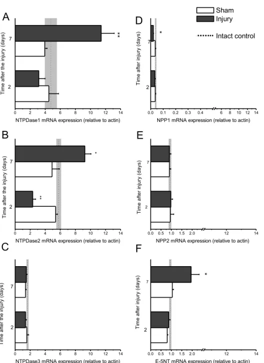
Fig. 1. Efect of CSI on ectonucleotidases mRNA expression. Expression of ectonucleotidases mRNA was measured by RTQ-PCR. he levels of each mRNA were normalized to those of the housekeeping gene β-actin. Ectonucleotidases mRNA relative expression values were compared to the corresponding control group and presented as mean ± SEM. A-C. he expression proile of NTPDase1-3 mRNA ater the CSI. (A) NTPDase1 mRNA expression is upregulated ~2 fold at seven days ater the injury, when compared to sham control (P
NTPDase3 mRNA expression did not change signii-cantly (Fig. 1C).
NPP1-3
NPP1 and NPP2 mRNA expression proiles are shown in Figs. 1D and 1E. NPP3 expression was very low and did not change in response to the injury (data not shown). NPP1 mRNA expression (Fig. 1D) was not afected by CSI ater two days; seven days af-ter the injury it was decreased to about half the level detected in the corresponding sham group (P <0.05). In contrast, NPP2 mRNA expression remained un-changed ater the injury (Fig. 1E).
Ecto-5-nucleotidase
he efect of CSI on E-5NT mRNA expression is shown in Fig. 1F. Although two days ater the injury E-5NT mRNA expression was not changed, it was signiicantly (by about 2 fold, P <0.01) upregulated ater seven days in comparison to the corresponding sham group.
Transcript Abundance
According to expression relative to β-actin, the most abundant ectonucleotidase transcripts in rat cortical tissue are the transcripts for NTPDase1 and NTP-Dase2, while the rest of the examined ectonucleoti-dases seem to be expressed at signiicantly lower lev-els. Comparison of transcript abundance in intact rat cortical tissue yields the sequence in the order NTP-Dase2 ≈ NTPDase1 >NTPDase3 > e-5NT ≥ NPP2 > NPP1 > NPP3.
DISCUSSION
his study was designed to investigate the expres-sion proiles of major ectonucleotidases ater cortical brain injury in rat, and is a continuation of our previ-ous work on ectonucleotidases in this injury model. We showed that the rates of ATP and ADP hydrolysis follow a biphasic pattern, with initial downregula-tion in the irst 24 hours ater the injury, and signii-cant upregulation 15 days ater injury (Nedeljkovic
et al., 2006). We hypothesized that the cause for the increase in ATPase/ADPase activity might be due to the upregulation of NTPDase1 at activated micro-glial cells (Nedeljkovic et al., 2006). Here we show that NTPDase2 may also contribute to the observed changes in nucleotides hydrolysis, at least for ATP hydrolysis. According to the results presented herein, transcripts for NTPDase1 and NTPDase2 are most abundant in the intact rat cortical tissue. Moreover, NTPDase2 mRNA displays a biphasic change in ex-pression, with a signiicant downregulation two days and pronounced upregulation seven days ater the injury. Interestingly, the time points selected for this study correspond to the onset (two days) and the peak of astrogliosis, and while NTPDase1 is associ-ated with microglia (Braun et al., 2000), NTPDase2 is usually assigned to astrocytes (Wink et al., 2006). It should be noted that implantation of NTPDase2-overexpressing C6 glioma cells has dramatic impact on tumor size increase and malignancy. herefore, it would be interesting to investigate the involvement of NTPDase2 in astrogliosis of diferent etiologies.
On the other hand, the expression of NTPDase3 does not seem to be altered by the injury. Our pre-vious indings of NTPDase3 low protein expression and absence of injury-induced changes in protein levels (Bjelobaba et al., 2010) also implied that this enzyme does not contribute much to the observed alterations of ATP/ADP hydrolyzing activities ater stab brain injury in rats (Nedeljkovic et al., 2006).
Compared to other ectonucleotidases, NPPases have been less thoroughly investigated in the CNS and related pathologies. We have investigated NPP1 protein distribution by immunohistochemistry, and in the rat cortex this enzyme seems to be expressed by neurons (Bjelobaba et al., 2006). In this study, we found that NPP1 gene expression is downregulated at seven days post-injury, which could be a relection of ongoing neuronal degeneration.
NPP2 protein in white matter astrocytes (Savaskan et al., 2007). However, the tissue sampled for our anal-yses did not contain any white matter, since in our model of stab injury, damage of white matter does not occur. Our results indicate that NPP2 transcript is the most abundant among the investigated NPPs, although NPP2 protein expression in the rat cortex seems to be very restricted and conined to layer IV (Savaskan et al., 2007).
It was already shown that the transcript of NPP3 is present in cortical tissue and in astrocytes of the developing rat brain (Cognato et al., 2008), but based on low level mRNA expression in real-time PCR analyses, we conclude that NPP3 does not contribute signiicantly to the control of extracellular nucleotide levels.
We speculate that the role of NPP enzymes might be more region-speciic. For instance, NPP1 enzyme exhibits the highest activity in the cerebellum, hy-pothalamus and hippocampus (Asensio et al., 2007), while NPP2 distribution is conined to white matter regions (Savaskan et al., 2007).
In our previously published papers, we have al-ready addressed e-5NT enzyme activity and protein expression in detail (Nedeljkovic et al., 2006; Bjeloba-ba et al., 2011). It should be noted that the observed mRNA expression proile of e-5NT corresponds well to the protein expression with near to control levels 2 days ater the injury and an increase seen at seven days post-injury. Nevertheless, the abundance of e-5NT mRNA is several times lower when compared to NTPDases1 and NTPDases 2 (Nedeljkovic et al., 2006). Based on immunohistochemical indings of astrocytic induction of e-5NT, upregulation in gene expression at seven days post-injury may also be as-signed to astrocytes.
In conclusion, our results suggest that the ma-jor ectonucleotidases are diferently challenged by cortical stab injury. NTPDase2 and NTPDase1 have the highest capacity for ATP hydrolysis, in physi-ological as well as pathphysi-ological conditions. NPP enzymes have a lower basal expression, but while
the expression of NPP2 is stable, injury induces a downregulation of NPP1. It seems plausible that injury-induced upregulation of NTPDases and e-5NT transcripts is due to astro/microgliosis and that these cells are more involved in regulating ex-tracellular nucleotide/nucleoside levels in patho-logical conditions.
Acknowledgments - his study was supported by the Serbian Ministry of Education, Science and Technology, Project No. III41014.
REFERENCES
Abbracchio M.P., Burnstock G., Verkhratsky A. and H. Zimmer-mann (2009). Purinergic signalling in the nervous system: an overview. Trends. Neurosci.32, 19-29.
Amadio S., Apolloni S., D’Ambrosi N. and C. Volonté (2011). Pu-rinergic signalling at the plasma membrane: a multipur-pose and multidirectional mode to deal with amyotrophic lateral sclerosis and multiple sclerosis. J. Neurochem.116, 796-805.
Asensio A.C., Rodríguez-Ferrer C.R, Castañeyra-Perdomo A., Oaknin S. and P. Rotllán (2007). Biochemical analysis of ecto-nucleotide pyrophosphatase phosphodiesterase ac-tivity in brain membranes indicates involvement of NPP1 isoenzyme in extracellular hydrolysis of diadenosine poly-phosphates in central nervous system. Neurochem. Int.
50(4), 581-90.
Bjelobaba I., Lavrnja I., Parabucki A., Stojkov D., Stojiljkovic M., Pekovic S. and Nedeljkovic N. (2010). he cortical stab in-jury induces beading of ibers expressing ecto-nucleoside triphosphate diphosphohydrolase 3. Neuroscience.170(1), 107-16.
Bjelobaba I., Parabucki A., Lavrnja I., Stojkov D., Dacic S., Pek-ovic S., Rakic L., StojiljkPek-ovic M. and N. Nedeljkovic (2011). Dynamic changes in the expression pattern of ecto-5’-nu-cleotidase in the rat model of cortical stab injury. J. Neuro-sci. Res.89(6), 862-73.
Bjelobaba I., Stojiljkovic M., Lavrnja I., Stojkov D., Pekovic S., Dacic S., Laketa D., Rakic L. and N. Nedeljkovic (2009). Regional changes in ectonucleotidase activity ater corti-cal stab injury in rat. Gen Physiol Biophys. 28, 32-8.
Braun N., Sévigny J., Robson S.C., Enjyoji K., Guckelberger O., Hammer K., Di Virgilio F. and H. Zimmermann (2000). Assignment of ecto-nucleoside triphosphate diphospho-hydrolase-1/cd39 expression to microglia and vasculature of the brain. Eur. J. Neurosci.12, 4357-4366.
Burnstock G. (2013). Introduction to purinergic signalling in the brain. Adv. Exp. Med. Biol.986, 1-12.
Burnstock G., Krügel U., Abbracchio M.P. and P. Illes (2011). Puri-nergic signalling: from normal behaviour to pathological brain function. Prog. Neurobiol.95(2), 229-74.
Burnstock, G. and G.E. Knight (2004). Cellular distribution and functions of P2 receptor subtypes in diferent systems. Int. Rev. Cytol. 240, 31-304
Cognato G. P., Czepielewski R.S., Sarkis J.J., Bogo M.R. and C.D. Bonan (2008). Expression mapping of ectonucleotide py-rophosphatase/phosphodiesterase 1-3 (E-NPP1-3) in dif-ferent brain structures during rat development. Int. J. Dev. Neurosci.26(6), 593-8.
Deaglio S. and S.C. Robson (2011). Ectonucleotidases as regula-tors of purinergic signaling in thrombosis, inlammation and immunity. Adv. Pharmacol.61, 01-32.
Nedeljkovic, N., Bjelobaba, I., Subasic, S., Lavrnja, I., Pekovic, S., Stojkov, D., Vjestica, A., Rakic, L. and M. Stojiljkovic
(2006). Upregulation of ectonucleotidase activity ater cortical stab injury in rat. Cell. Biol. Int.30, 541-546.
Ralevic V. and G. Burnstock (1998). Receptors for purines and pyrimidines. Pharmacol. Rev.50(3), 413-92.
Savaskan N.E., Rocha L., Kotter M.R., Baer A., Lubec G., van Meeteren L.A., Kishi Y., Aoki J., Moolenaar W.H., Nitsch R.
and A.U. Bräuer (2007). Autotaxin (NPP-2) in the brain: cell type-speciic expression and regulation during devel-opment and ater neurotrauma. Cell. Mol. Life. Sci. 64(2), 230-43.
Wink M.R., Braganhol E., Tamajusuku A.S., Lenz G., Zerbini L.F., Libermann T.A., Sévigny J., Battastini A.M. and S.C. Robson (2006). Nucleoside triphosphate diphosphohydro-lase-2 (NTPDase2/CD39L1) is the dominant ectonucle-otidase expressed by rat astrocytes. Neuroscience. 138(2), 421-32.
Zimmermann H. (1992). 5’-Nucleotidase: molecular structure and functional aspects. Biochem. J.285(2), 345-65
Zimmermann H., Vogel M. and U. Laube (1993). Hippocampal localization of 5’-nucleotidase as revealed by immunocy-tochemistry. Neuroscience.55(1), 105-12.
Zimmermann, H. (1996). Biochemistry, localization and func-tional roles of ecto-nucleotidases in the nervous system.