Defined Essential 8
™
Medium and Vitronectin
Efficiently Support Scalable Xeno-Free
Expansion of Human Induced Pluripotent
Stem Cells in Stirred Microcarrier Culture
Systems
Sara M. Badenes1, Tiago G. Fernandes1*, Cláudia S. M. Cordeiro1, Shayne Boucher2, David Kuninger2, Mohan C. Vemuri2, Maria Margarida Diogo2, Joaquim M. S. Cabral2
1Department of Bioengineering, and Institute for Bioengineering and Biosciences, Instituto Superior Técnico, Universidade de Lisboa, Lisboa, Portugal,2Thermo Fisher Scientific, Cell Biology, Life Sciences Solutions, Frederick, Maryland, United States of America
Abstract
Human induced pluripotent stem (hiPS) cell culture using Essential 8™xeno-free medium and the defined xeno-free matrix vitronectin was successfully implemented under adherent conditions. This matrix was able to support hiPS cell expansion either in coated plates or on polystyrene-coated microcarriers, while maintaining hiPS cell functionality and pluripotency. Importantly, scale-up of the microcarrier-based system was accomplished using a 50 mL spinner flask, under dynamic conditions. A three-level factorial design experiment was per-formed to identify optimal conditions in terms of a) initial cell density b) agitation speed, and c) to maximize cell yield in spinner flask cultures. A maximum cell yield of 3.5 is achieved by inoculating 55,000 cells/cm2of microcarrier surface area and using 44 rpm, which
gener-ates a cell density of 1.4x10⁶cells/mL after 10 days of culture. After dynamic culture, hiPS
cells maintained their typical morphology upon re-plating, exhibited pluripotency-associated marker expression as well as tri-lineage differentiation capability, which was verified by inducing their spontaneous differentiation through embryoid body formation, and subse-quent downstream differentiation to specific lineages such as neural and cardiac fates was successfully accomplished. In conclusion, a scalable, robust and cost-effective xeno-free culture system was successfully developed and implemented for the scale-up production of hiPS cells.
Introduction
Human induced pluripotent stem (hiPS) cells are capable of self renewing indefinitely, and to differentiate into all the cell types of the human body [1]. Because of these characteristics, anal-ogous to human embryonic stem (hES) cells, hiPS cells are promising sources for several bio-medical applications [2]. However, to fully realize the potential of hiPS cells for cellular OPEN ACCESS
Citation:Badenes SM, Fernandes TG, Cordeiro CSM, Boucher S, Kuninger D, Vemuri MC, et al. (2016) Defined Essential 8™Medium and Vitronectin Efficiently Support Scalable Xeno-Free Expansion of Human Induced Pluripotent Stem Cells in Stirred Microcarrier Culture Systems. PLoS ONE 11(3): e0151264. doi:10.1371/journal.pone.0151264
Editor:Vimal Selvaraj, Cornell University, UNITED STATES
Received:October 7, 2015
Accepted:February 19, 2016
Published:March 21, 2016
Copyright:© 2016 Badenes et al. This is an open access article distributed under the terms of the Creative Commons Attribution License, which permits unrestricted use, distribution, and reproduction in any medium, provided the original author and source are credited.
Data Availability Statement:All relevant data are within the paper and its Supporting Information files.
therapy, drug screening and disease modelling, the development of standardized and robust scalable processes to produce large numbers of these cells while maintaining their critical bio-logical functionality and safety are of prime importance.
Typically, hiPS cells are expanded using adherent static cell culture systems that cannot pro-vide a sufficient number of cells for downstream applications, presenting low cell yields and inherent variability of the culture process and of the final product. Translating cell culture from static plates to suspension systems is needed to achieve scalability of the process. Stirred bioreactors are an appropriate culture system for moderate large-scale cell production given their robustly controlled operation and well-established scale-up protocols [3,4,5]. Several methodologies for human pluripotent stem (hPS) cell culture in these systems have been implemented in the last few years, including cultivation of cells encapsulated typically inside hydrogels [6,7], adherent onto microcarriers [8,9], or as 3D aggregates in suspension [10,11]. Microcarrier technology confers distinct advantages as it provides homogeneous culture condi-tions to the cells, large surface areas for cell adhesion and growth [12,13] and importantly, a large surface/volume ratio. Also, microcarrier culture on fully controlled bioreactors allows monitoring and controlling of environmental parameters, and can be scaled up relatively easily. Nevertheless, despite recent progress on scalable microcarrier hPS cell suspension culture, most of the methods are based on the use of non-defined extracellular matrix (ECM) extracts, such as Matrigel™or Geltrex™, as surface for cell adherence on microcarriers [14,15,16], and commercially available serum-free media, such as mTeSR™and StemPro1[14,17,18], that con-tain animal-derived products.
Envisioning the bioprocess translation to Good Manufacturing Practice (GMP) standards, great efforts have been made towards the translation of scalable culture systems to chemically defined and xeno-free conditions. A completely defined medium, Essential 8™, that consists of only eight components, was recently developed [19,20,21], and several other studies have been reporting defined surfaces that support long-term hiPS cell culture, like vitronectin, laminin, fibronectin and various synthetic peptides [15,18,22,23]. Nevertheless, the use of Essential 8™
medium to support expansion of hiPS cells on microcarriers coated with defined substrates has never been reported.
To design a bioprocess to produce a biomedical product, it is of foremost importance to set up robust and reproducible production practices. Therefore, robust predictive strategies to evaluate process parameters that will impact culture output need to be developed. Rational design of experiments can provide a model to predict the culture output as a function of multi-ple culture parameters [24,25]. Therefore, in this work, we immulti-plemented a stirred culture sys-tem based on the use of vitronectin-coated microcarriers and Essential 8™medium for the scalable expansion of hiPS cells, using 50 mL spinner flasks. Importantly, a three-level factorial design model was used to identify the optimal conditions that maximize cell yield. Finally, given the potential applications of hiPS cells in differentiation and lineage specification studies, we investigated the differentiation capacity of hiPS cells cultured on microcarriers, under xeno-free chemically defined conditions, to cardiomyocytes and to neural progenitor cells.
Materials and Methods
Cells and microcarriers
Gibco™human induced pluripotent stem cell line used in this work was derived from CD34 + cells of healthy donors (Life Technologies). The hiPS cells were routinely cultured on Geltrex (1:60, Life Technologies) -coated 6-well plates in Essential 8 (E8) medium (Life Technologies), in a humidified 5% CO2incubator at 37°C. The medium was refreshed daily and cells were rou-tinely passaged at a split ratio of 1:4 using the EDTA method [26], when colonies reached 80%
cardiovascular applications (MITP-TB/ECE/0013/ 2013). The funders had no role in study design, data collection and analysis, decision to publish, or preparation of the manuscript. There are no current external funding sources for this study.
confluence. hiPS cells were adapted to Vitronectin (rhVTN-N, Life Technologies)–coated plates for two passages prior to inoculation onto microcarriers. Cells were routinely evaluated for karyotype abnormalities by conventional cytogenetics using the services of Genomed SA (Lisbon, Portugal).
Polystyrene microcarriers (Solohill Engineering, Inc.), with 360 cm2/g of superficial area, were used to support cell growth. Microcarriers were mixed during 1 h with Ethanol 70% (Sigma) at room temperature and washed 3 times with sterile phosphate-buffered saline (PBS). Coating of microcarriers was performed for 2 h at room temperature with Vitronectin in sterile PBS, using 0.5μg/cm2. Geltrex-coated microcarriers were used (coating: 0.25 mL/cm2of Gel-trex solution (1:60)) as a control. Prior to cell inoculation, microcarriers were incubated (at 37°C) for 30 min in culture medium.
Inoculation of hiPS cells on microcarriers
Inoculation as single cells. The protocol for the inoculation of hiPS cells on microcarriers as single cells was described recently [27].
Inoculation as clumps. Cells were incubated for 5 min with Cell Dissociation Buffer (Life Technologies) at room temperature, using the EDTA method [26]. Cells were then collected and inoculated on microcarriers with or without ROCK inhibitor (10μM, Y27632, from Stem-Gent) for the first 24 h of culture.
hiPS cell expansion on microcarriers
Static culture. The protocol for the screening of microcarriers for hiPS cell expansion under static culture in low-attachment 24-well plates (Corning Inc.) was recently published [27]. We used 3 cm2of microcarrier superficial area per well and cells were inoculated at an ini-tial density of 5x10⁴cells/cm2. Geltrex- and vitronectin-coated polystyrene microcarriers (GM and VtnM) were tested. 80% of E8 medium was changed daily for 5 days. The cell yield in total cell number was calculated as the ratioXday5/Xi, whereXday5is the number of viable cells,
attached to the microcarriers, at day 5, andXiis the number of cells inoculated at day 0.
Spinner flask culture. The expansion of hiPS cells in a microcarrier stirred suspension culture was performed in presiliconized (Sigmacote, Sigma) spinner flasks (StemSpanTM, Stem-Cell Technologies), with a working volume of 50 mL. The impeller was composed of a horizon-tal magnetic stir bar with a vertical paddle. Agitation was obtained by a magnetic stirrer platform (Variomag, Biosystem), which was placed inside a 5% CO2incubator at 37°C.
Cells were seeded as small clumps, at an initial density of 3, 5 or 7x10⁴cells/cm2, using a total of 1 g of coated polystyrene microcarriers (360 cm2/spinner) in 25 mL of E8 medium under static conditions, to promote cell-microcarrier contact. Medium was supplemented with ROCK inhibitor (10μM) for the first 24 h after inoculation. After 24 h, the medium was replaced and adjusted to 50 mL of fresh E8 medium. Subsequently, an intermittent stirring (3 min at 40 rpm every 2 h) was performed overnight to promote cell-cell and cell-microcarrier contact. Thereafter, the culture was continuously stirred at 30, 50 or 70 rpm and feeding was performed on a daily basis by replacing 80% of volume with fresh pre-warmed medium.
For spinner flask cultures, cell attachment efficiency to the microcarriers was calculated as the percentage ofXday1/Xi, whereXday1is the number of the viable cells attached to the
micro-carriers at day 1 of the culture andXiis the number of cells inoculated at day 0. The maximum
cell yield was calculated as the ratioXmax/Xi, whereXmaxis the maximum cell number, attached
to the microcarriers, achieved during the culture.
Technologies) at 37°C for 10 min, in the heater mixer set at 750 rpm. After dissociation by pipetting, the mixture was filtered through a 100μm mesh (cell strainer, from BD Biosciences) to remove the microcarriers. Cells were then centrifuged at 210gfor 5 min and viable and dead cells were determined by counting in a hemocytometer under optical microscope, using the trypan blue dye exclusion test.
Cell harvesting from the microcarriers and re-plating. The cell harvesting and re-plant-ing protocol at the end of the culture (static or dynamic) was performed as previously described [27]. Cells were resuspended in E8 medium supplemented with ROCK inhibitor (10μM) and then inoculated at a density of 5x10⁴cells/cm2of well area on GP.
Experimental design
The effects of two independent variables, initial cell density and agitation rate, on the cell yield were determined using a face-centered composite design (FC-CD) approach using STATIS-TICA software (StatSoft, Tulsa, OK). Each independent variable was evaluated at three differ-ent coded levels (low (−1), central (0) and high (+1)) as portrayed inS1 Tableand combined in a FC-CD design set up described as:
N¼2k pþ2kþC
0
whereNis the number of experiments,kis the number of variables (k= 2),pthe fractionaliza-tion number (in a full design,p= 0) andC0is the number of central points, that provides
esti-mation of the experimental error. Accordingly, a total of 12 [22−0+ (2×2) + 4] independent
experiments were performed.
The data was fitted to a full quadratic model (including linear and non-linear effects, plus two-way interaction) as follow:
Y ¼b0þb1X1þb11X1 2
þb2X2þb22X2 2
þb12X1X2
whereYis the response measured or dependent variable (cell yield),X1andX2are the two
independent variables,β0is the intersect;β1andβ2are the linear main effects,β11andβ22are the quadratic coefficients, andβ12is the coefficient for the second order interaction. The error
of the prediction was estimated from the error obtained by the genuine replicates performed on the central points of the matrix, done at least in 4 independent experiments [28]. The coeffi -cient of regression (R2) was also determined by the software.
Characterization of hiPS cells and derivatives
Flow cytometry. Cells were kept at 4°C in 2% (v/v) paraformaldehyde (PFA, from Sigma). For surface staining, approximately 5x10⁵cells were resuspended in 100μL of FACS buffer (3% (v/v) Fetal Bovine Serum (FBS, from Invitrogen) in PBS) with the diluted primary anti-body, and incubated for 15 min at room temperature in the dark. Cells were washed twice with PBS and resuspended in 300μL of PBS to be analysed by flow cytometry (FACSCalibur, Becton Dickinson). For negative controls, cells were incubated with the appropriate isotypes. For intra-cellular staining, the protocol used is described by Miranda et al. [29]. For the negative controls, cells were incubated only with 3% (v/v) Normal Goat Serum (NGS, from Sigma) in PBS. The CellQuest software (Becton Dickinson) was used for all acquisition/analyses.
Immunocytochemistry. For surface antigens, after removing the culture medium, cells were incubated for 30 min at 37°C in the presence of the primary antibodies diluted in medium. Cells were washed 3 times with PBS and incubated in the dark for 30 min, at 37°C, with the secondary antibodies. For intracellular staining, the protocol used is described by Miranda et al. [29]. Cells were examined using a fluorescence microscope (Leica DMI3000B/ Nikon Digital Camera Dxm1200F).
RT-PCR. RNA was isolated using PureLink1RNA Mini Kit (Life Technologies). cDNA was synthetized using 1μg of total RNA and the High Capacity cDNA Reverse Transcriptase kit (Life Technologies). StepOne QST Real-Time Polymerase Chain Reaction (RT-PCR) was performed using the TaqMan™Gene Expression Assay (Applied Biosystems) (S2 Table).
In vitrohiPS cell differentiation potential. hiPS cell differentiation potential was
evalu-atedin vitrovia embryoid body (EB) formation and spontaneous differentiation. Cells from a spinner flask culture were harvested and inoculated as single-cells in GP. At 80% confluence, cells were passaged with EDTA treatment to a 6-well low-attachment plate in EBs medium (DMEM with 20% (v/v) FBS, 1% (v/v) MEM-non essential amino acids, 1mM sodium pyru-vate, 0.1 mMβ–mercaptoethanol and 1% (v/v) Penicillin/Streptomycin, all from Invitrogen), supplement with ROCK inhibitor for the first 24 h. Medium was changed every 2 days for 4 weeks thereafter. EBs were then dissociated with trypsin 0.025% and cells inoculated in a 24-well plate coated with 4μg/mL laminin (StemGent) and 10μg/mL poly-D-lysine (Sigma). Medium was changed every 2 days for 1 week. Finally, cells were stained with anti-SOX17, TUJ1 andα-SMA antibodies.
Directed hiPS cell cardiomyocyte differentiation. The Gibco1
hPS cell Cardiomyocyte Differentiation Kit (Life Technologies) was used to induce cardiomyocyte differentiation of hiPS cells adherent to confluent microcarriers from a spinner flask culture (without cell har-vesting). Confluent microcarriers were placed in a 24-well low-attachment plate (3 cm2of microcarriers area/well) and the protocol was performed using manufacture’s instructions. Also, cells harvested from the microcarriers at the end of the spinner flask culture were inocu-lated as single-cells in GP, and at 80% confluence, cardiomyocyte differentiation was initiated. On both cases, at the end of the differentiation protocol, cells were stained for cTNT and OCT4 markers.
Neural induction by Dual-SMAD inhibition. Confluent microcarriers from a spinner flask culture were placed in 24-well low-attachment plate (3 cm2of microcarriers area/well).
N2B27 medium supplemented with 10μM of SB431542 (SB, Sigma) and 100 nM of
Statistical analysis
All data presented show n = 3 replicates, unless stated otherwise. Error bars represent the stan-dard error of the mean (SEM).
Results
Xeno-free surfaces for adherent hiPS cell culture in E8 medium
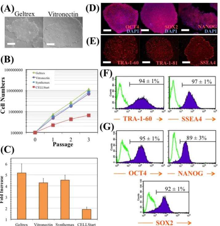
In order to select the best xeno-free substrate for expansion of hiPS cells in combination with E8 culture medium, different substrates were tested and compared. Since the ability of vitro-nectin (Vtn) surfaces to support long-term hiPS cell expansion in xeno-free E8 medium has been described in the literature [19], the model hiPS cell line was seeded onto Vtn and Geltrex surfaces and cultured in E8 medium. As shown inFig 1A, no significant differences were found in cell morphology between hiPS cells cultured on these two surfaces. On both cases, hiPS cells demonstrated a typical morphology of tightly packed colonies with defined borders and a high nucleus-to cytoplasm ratio.
Comparison studies were performed using different substrate surfaces for the adhesion, expansion and serial passaging of hiPS cells. Besides Geltrex and Vtn, CELLStart™(Life Tecnhologies) and Synthemax1(Corning Inc.) surfaces were also evaluated. As it can be seen inFig 1B, cell growth kinetics were similar when hiPS cells were cultured on all surfaces, except on CELLStart™surface. As shown inFig 1C, cells cultured on Geltrex surface pre-sented the highest fold increase (5.2±0.8), which could be expected due to its complex and rich protein composition [30]. However, this non-defined ECM extract surface may be a source of xenogeneic risk. In the same figure, it is shown a similar cell fold expansion when culturing the cells on Vtn (4.3±0.4) and Synthemax1(4.5±0.5) surfaces. Although
Synthe-max1
is a chemically-synthesized substrate [31], Vtn is more cost-effective adhesion-pro-moting reagent [22] as was evaluated in the literature [30]. hiPS cells were then cultured during four consecutive passages on Vtn surface and immunofluorescence microscopy was performed to evaluate the expression of the intracellular and extracellular pluripotency markers OCT4, SOX2 and NANOG (with the corresponding DAPI stains of the nuclei), and TRA-1-60, TRA-1-81 and SSEA4, respectively (Fig 1D and 1E). The fluorescence images indicated that hiPS cells can be maintained in their undifferentiated state on Vtn surface. Moreover, flow cytometry analysis revealed consistently high expression levels of pluripotent markers TRA-1-60 (94±1%), SSEA4 (97±1%), OCT4 (95±1%), NANOG (89±3%) and SOX2 (92±1%) (Fig 1F and 1G). Finally, it was also verified that hiPS cells consistently displayed a normal karyotype (46 XX) after four passages on Vtn-coated tissue culture plates, in E8 medium (data not shown). In conclusion, the combination of Vtn surfaces and E8 medium support robust and long-term culture of undifferentiated hiPS cells under adherent static conditions.
hiPS cell expansion on vitronectin-coated microcarriers: inoculation
strategy
After demonstrating that Vtn could support the long-term culture of hiPS cells in xeno-free E8 medium, this matrix was used to coat polystyrene microcarriers and then to implement a scal-able culture. Vtn-coated microcarriers were inoculated with 5x10⁴cells/cm2in low attachment 24-well plates.
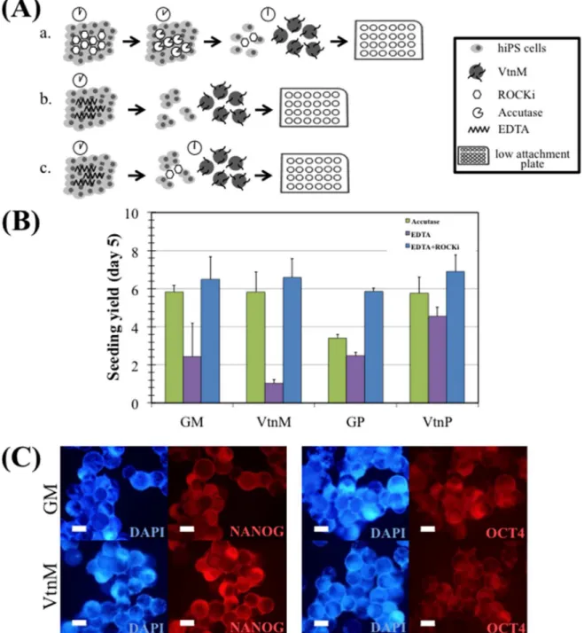
treatment and inoculated as cell clumps; and in strategy (c) cells were dissociated with EDTA treatment and were inoculated as cell clumps, in the presence of ROCK inhibitor for 24 h.
Fig 1. hiPS cell maintenance in E8 medium and vitronectin.(A) Colonies cultured with E8 medium in Geltrex (left panel), and in vitronectin (right panel). Scale bars—100μm. (B) Cumulative fold increase in total cell numbers over four passages with E8 medium in Geltrex (green), in vitronectin (purple), in
Synthemax1
(blue) or in CELLStart™(red). (C) Average fold increase per passage. The fold increase was 5.2 for cells growing in Geltrex, 4.3 for vitronectin, 4.5 for Synthemax1and 1.9 for CELLStart
™. Error bars represent SEM (standard error of the mean). Four passages and two independent experiments were performed. (D) Immunostaining of colonies cultured with E8 medium in vitronectin. Colonies were stained for pluripotency markers OCT4, SOX2, and NANOG, and nuclei counterstained with DAPI. Scale bars—100μm. (E) Immunostaining of colonies cultured with E8 medium in vitronectin. Colonies were
stained for pluripotency surface markers TRA-1-60, TRA-1-81, and SSEA4. Scale bars—100μm. (F) and (G) Flow cytometry analysis of pluripotency surface
markers (TRA-1-60 and SSEA4) (F) and transcription factors (OCT4, NANOG, SOX2) (G) in cells expanded with E8 medium and vitronectin.
When hiPS cells were treated with EDTA for 3 minutes, this resulted in the formation of small clumps that were able to survive. However, due to the considerable size of these clumps (8–12 cells) cells tend to grow as aggregates rather than attaching onto microcar-riers. Consequently, the time of incubation with EDTA was increased to 5 min in order to
Fig 2. hiPS cell expansion on microcarriers in E8 medium, under static conditions.(A) Three different inoculation strategies: a. accutase, b. EDTA and c. EDTA+ROCKi were compared. (B) Seeding yield after 5 days of culture of hiPS cells on Geltrex coated polystyrene microcarriers (GM) and plate (GP), vitronectin coated polystyrene microcarriers (VtnM) and plate (VtnP), using the three different inoculation strategies: accutase (green), EDTA (purple) and EDTA+ROCKi (blue). Cells were inoculated at a 5x10⁴cells/cm2density. Error bars represent SEM (standard error of the mean). Three replicate wells were performed for each condition. (C) Immunostaining of hiPS cells cultured on microcarriers with E8 medium at day 5. Cells were stained for pluripotency markers NANOG (left panel) and OCT4 (right panel), and nuclei counterstained with DAPI. Scale bars–100μm.
obtain smaller clumps (3–6 cells) and allow cell adhesion to the microcarriers. Since higher incubation time with EDTA had to be performed in the microcarrier-based culture, the addition of ROCK inhibitor for the first 24 h of culture was considered beneficial for smaller clump survival.
InFig 2B, it can be observed the cell yield (seeMaterials and Methods) for the three dif-ferent inoculation strategies, after 5 days of static culture of hiPS cells on Vtn-coated poly-styrene microcarriers (VtnM) and plate (VtnP). Cell expansion on Geltrex-coated
polystyrene microcarriers (GM) and plate (GP) were evaluated as a control. The highest cell yields were obtained using the inoculation strategy (c), and results with VtnM (5.8±1.0 for strategy (a), 1.0±0.2 for strategy (b) and 6.6±1.0 for strategy (c)) were similar to the ones obtained with GM. Moreover, for strategy (c), the culture on microcarriers showed similar yields to the static culture on plate (6.4±0.7). The inclusion of the small molecule Y27632 (ROCK inhibitor) has already been reported [19,32] to improve initial cell survival and to support high clonal efficiency. Therefore, cell-microcarrier adhesion efficiency was improved and higher cell yields were obtained in this case. Importantly, as presented inFig 2C, after 5 days of culture, hiPS cells cultured on VtnM and GM, stained positively for NANOG and OCT4 pluripotency intracellular markers. Considering these results, strategy (c) was chosen for scaling-up the microcarrier-based culture.
Optimization of hiPS cells expansion on a scalable stirred spinner flask
culture by a face-centered composite design
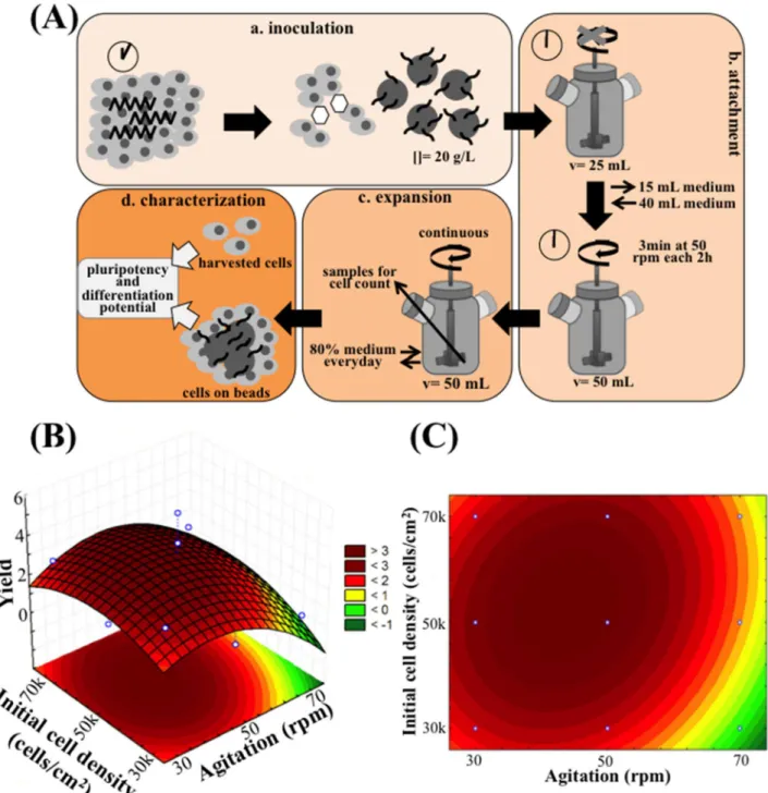
The next step was to implement a dynamic microcarrier-based system in 50 mL-spinner flasks, envisaging the scalability of the expansion process. The protocol followed for the expansion experiments in the spinner flask is presented inFig 3A, and is composed of four steps. On step (a) cells were inoculated in the spinner flask, using the EDTA/ROCKi method, in VtnM (20 g/ L, corresponding to 360 cm2of superficial area) and using half of the working volume (25 mL E8 medium supplemented with ROCK inhibitor). On Step (b), attachment of the cells to the VtnM is initiated. This step corresponds to the initial 2 days of culture. The spinner flask was operated under static conditions during the first 24 h, which is a critical period for the success of the culture that depends on cell attachment efficiency. In our experiments, attachment effi-ciencies of hiPS cells to VtnM were very similar, 32.5±0.9%, for inoculations with different ini-tial cell densities. Step (b) also involved the second day of culture when ROCK inhibitor was removed from the medium, working volume was established at 50 mL and spinner flask was operated at an intermittent agitation (3 min at 50 rpm every 2 h) to maximize cell and cell-microcarrier interactions. Step (c) corresponds to the period of cell expansion, which started with the initiation of the exponential growth phase at day 3 and ceased when the maximum cell yield was attained, between days 7 and 11, depending on culture conditions. During this step, the spinner flask was operated under a continuous agitation, 80% of the medium was changed everyday and samples were taken each day for cell counting. The final step (d) involved the characterization of the hiPS cells cultured on VtnM in the stirred spinner flask, through analysis of their pluripotency state and their differentiation potential by flow cytome-try and immunocytochemiscytome-try.
agitation speed, values were 30, 50 and 70 rpm, which were selected taking into consideration the operating conditions already reported for dynamic microcarrier cultures with hESC and mESC [12,33,34,35]. The equation that describes the quadratic model obtained for cell yield
Fig 3. Scaling-up hiPS cell expansion in E8 medium to a 50 mL spinner flask, under dynamic conditions.(A) The procedure consists in 4 steps: a. EDTA+ROCKi inoculation strategy into 20 g/L VtnM; b. Attachment is performed in half of the total volume in static conditions for 24 h (in the presence of ROCKi), following by intermittent stirring overnight to allow cell- microcarrier interaction; c. Expansion is performed with a specific continuous agitation with 80% E8 medium change everyday; and d. At the end of the culture, cell pluripotency and differentiation potential are evaluated. Quadratic model relating initial cell density and agitation speed with cell yield during spinner flask culture of human iPS cells: 3D representation (B) and 2D heat map (C) are shown. For initial cell density: 30 000 cells/cm2(
−1 level)Initial Cell Density70 000 cells/cm2(1 level); for agitation speed: 30 rpm (−1 level)Agitation Speed70 rpm (1 level).
response is:
Yield¼3
:298 1:610X1 2:408X1 2
þ0
:677X2 1:448X2 2
þ0
:700X1X2
whereX1is the agitation rate andX2is the initial cell density.
The second order polynomial generated for cell yield in a spinner flask culture does not fully describe the expected experimental data (R2= 0.479), which could be anticipated due to the inherent variability of this cell culture system. Nevertheless, based on the regression model, response surface plot and 2D heat plot were established as shown inFig 3B and 3C. The opti-mal conditions predicted by the model to reach a maximum cell yield of 3.5 were 55,000 cells/ cm2for the initial cell density and 44 rpm for the agitation rate.
hiPS cell expansion under optimized dynamic culture conditions
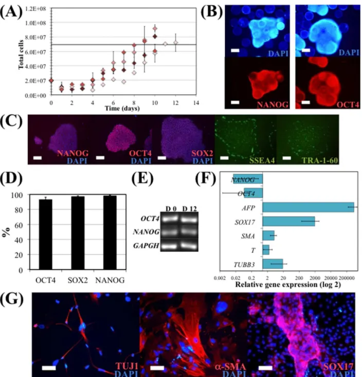
In order to verify the validity of the proposed model, several runs of hiPS cell expansion in spinner flask were performed under the optimum conditions given by the FC-CD, which were an initial cell density of 55,000 cells/cm2and an agitation rate of 44 rpm. Maximum cell num-bers achieved in these experimental culture runs were compared with the model-predicted value of the maximum cell number achieved in a culture under the optimal conditions (Fig 4A). Predicted and experimental results for maximum cell yield are similar, 3.5 and 4.0±0.4, respectively. Interestingly, the values of the maximum cell yield obtained in the experimental culture runs were all above the value predicted by the model. Therefore, although the inherent variability of hiPS cell expansion on microcarriers under dynamic conditions, in a spinner flask, the model obtained by the FC-CD proved to be a useful approximation for this culture system.
hiPS cells expanded under these optimal conditions were then evaluated for their pluripo-tency and undifferentiated state. It was confirmed that hiPS cells growing attached onto VtnM-coated microcarriers in a spinner flask retained their pluripotency characteristics, since these cells presented NANOG and OCT4 expression, as detected by immunocytochemical analysis. Also, the cells maintained their capacity to form typical undifferentiated colonies when har-vested from microcarriers and re-plated on GP, since they stained positively for the pluripo-tency markers NANOG, OCT4, SOX2, SSEA4 and TRA-1-60 (Fig 4C).
Pluripotency maintenance was demonstrated by flow cytometry analysis. As shown inFig 4D, more than 93% of the cells were positive for the pluripotency markers NANOG, SOX2 and OCT4 after 12 days of culture. mRNA was isolated from hiPS cells at day 0 and at the end of the spinner flask culture (day 12) in order to assess for the expression of the hiPS cell markers
OCT4andNANOG, by RT-PCR (Fig 4E). It was confirmed that cells cultured in spinner flasks maintained gene expression of the pluripotency markers. Furthermore, hiPS cells collected at the end of a spinner flask culture retained a normal karyotype (46 XX).
Fig 4. hiPS cell expansion in E8 medium, in a 50 mL spinner flask.EDTA clump inoculation was performed using 55,000 cells/cm2and a continuous agitation at 44 rpm (optimal values obtained solving the quadratic model). (A) Total cell numbers during the 10 days expansion. Maximum cell yield obtained solving the quadratic model is represented by the black line. Error bars represent SEM (standard error of the mean) of duplicate samples. (B) Immunostaining of hiPS cells cultured on microcarriers in the spinner flask with E8 medium at day 10. Cells were stained for pluripotency markers NANOG (left panel) and OCT4 (right panel), and nuclei counterstained with DAPI. Scale bars–100μm. (C) Immunostaining of hiPS cells harvested from microcarriers after 10 days expansion in spinner flask and re-plated on GP. Cells were stained after 3 days for intracellular pluripotency markers NANOG, OCT4 and SOX2, and nuclei counterstained with DAPI; and for surface markers SSEA4 and TRA-1-60. Scale bars–100μm. (D) Flow cytometry analysis of hiPS cells harvested from
microcarriers after 10 days expansion in spinner flask. Cells were stained for OCT4 and NANOG. (E) mRNA was isolated from hiPSC at day 0 and at the end of the spinner flask culture (day 12) on microcarriers, and the undifferentiated hiPSC marker transcripts (OCT4andNANOG) were analysed by RT-PCR. (F) Quantitative RT-PCR analysis of spontaneous differentiated EB of hiPSC cultured in spinner flask. The relative expression of each gene was measured against the same gene prior to differentiation. (G) Immunostaining showing the formation of cells expressing SOX17 (endoderm), TUJ1 (ectoderm) andα
-SMA (mesoderm) after EB formation and spontaneous differentiation assay with hiPSC cultured in spinner flask. Nuclei were counterstained with DAPI. Scale bar: 50μm.
immunostainning of the differentiated cells re-plated on laminin/poly-D-lysine-coated well plates (Fig 4G).
Differentiation potential of hiPS cells cultured in dynamic conditions
Two different experimental settings were performed in order to evaluate the differentiation potential of hiPS cells after expansion under dynamic conditions in the spinner flask: directed cardiomyocyte (CM) differentiation and commitment to neural progenitor (NP) cells. Both directed differentiation protocols were performed by a) plating microcarriers with hiPS cells in low-attachment plates, and b) plating hiPS cells harvested from microcarriers on GP.
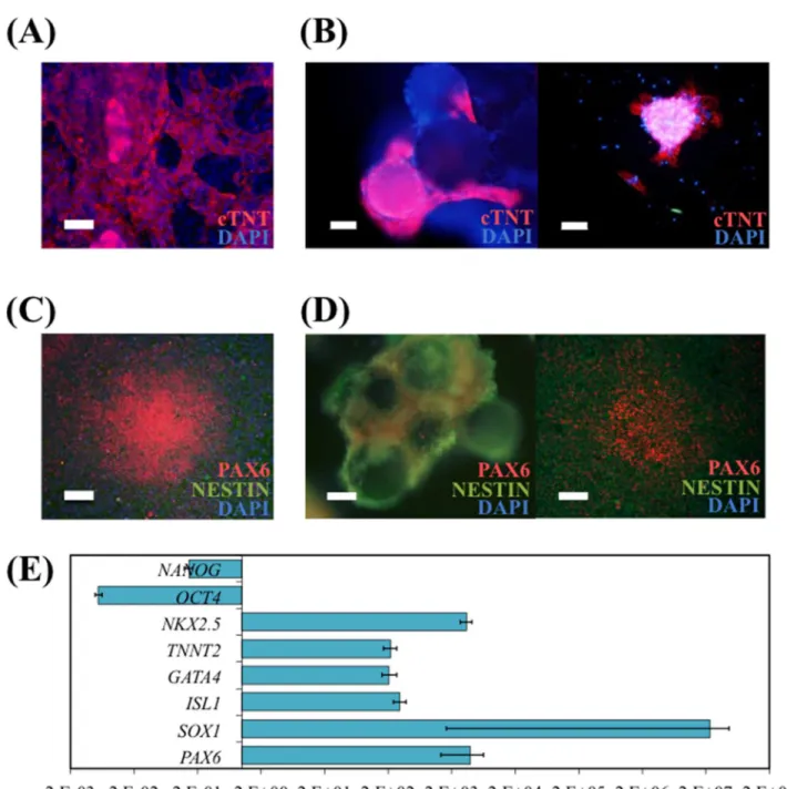
Directed differentiation of hiPS cells into CM was performed after the spinner flask culture. Spontaneous contracting regions in GP (S1 Video) and beating cell-VtnM aggregates (S2 Video) on low-attachment plate were observed at day 10 of differentiation. CM induction was confirmed at day 16 by immunocytochemistry analysis (Fig 5A and 5B) since cTNT+cells were obtained both on GP and on VtnM. Also, upon re-plating of CM obtained on VtnM onto GP, after the differentiation protocol, it was possible to observe the presence of contracting colonies (S3 Video) that stained positively for the cardiac marker cTNT.
NP cells were also obtained from spinner flask-expanded hiPS cells by dual inhibition of SMAD signaling [36]. After a 12 day-differentiation protocol on GP and on VtnM with hiPS cells cultured in spinner flask, immunocytochemical analysis showed strong expression of the early neural differentiation markers PAX6 and NESTIN, whereas the expression of the pluripo-tency marker OCT4 was not observed (Fig 5C and 5D). Also, NP cells that were obtained on VtnM, were re-plated on GP after the differentiation protocol and after 4 days it was observed neuroepithelial cells arranged in neural rosette structures, which expressed PAX6 and NESTIN markers.
InFig 5Eare presented the relative gene expressions obtained by quantitative RT-PCR after cardiac and neural differentiation. It was demonstrated an increase of transcription levels of representative genes of cardiac markers (early markersISL1andGATA4and late markers
TNNT2andNKX2.5) or increase of transcription levels of representative genes of neural pro-genitor markers (PAX6andSOX1), while there was a decrease in pluripotency marker gene expression (OCT4andNANOG).
Discussion
Biomedical applications of stem cell-derived products depend on the availability of large num-bers of cells, or their differentiated progeny. However, developing GMP-compliant scalable and efficient process for stem cell production, namely hiPS cell expansion, followed by directed differentiation into progenitor cells and then fully mature cells, is still a challenge.
Fig 5. hiPS cells cultured in spinner flasks with E8 medium and VtnM retain their differentiation potential.Cardiomyocyte (CM) differentiation was performed using Life Technologies Cardiomyocyte Differentiation Kit (A) of hiPS cells harvested from microcarriers and re-plated on GP. Immunostaining depicting CM for the cTNT marker. Nuclei were counterstained with DAPI. Scale bar–100μm; and (B) of hiPS cells on VtnM in static conditions in a low attachment plate. Immunostaining for the cTNT marker depicting CM on microcarriers (left panel) and plated CM on GP after differentiation protocol (right panel). Nuclei were counterstained with DAPI. Scale bars–50μm. Neural induction by Dual-SMAD inhibition (C) of hiPS cells harvested from microcarriers and re-plated on GP. Immunostaining depicting neural progenitor (NP) cells for the neuroectoderm markers PAX6 and NESTIN. Nuclei were counterstained with DAPI. Scale-bar: 100μm; and (D) of hiPS cells on VtnB in static conditions in a low attachment plate. Immunostaining for the neuroectoderm markers
PAX6 and NESTIN depicting NP cells on microcarriers (left panel) and plated NP cells on GP after differentiation protocol (right panel). Nuclei were counterstained with DAPI. Scale bars–50μm. (E) Quantitative RT-PCR analysis of differentiated hiPSC cultured in spinner flask, subject to cardiomyocytes or neural differentiation. The relative expression of each gene was measured against the same gene prior to differentiation.
Although TeSR™medium has been used for hPS cell expansion in the complete absence of animal proteins, the inclusion of human serum albumin (HSA) and human-sourced matrix proteins makes the production process expensive and impractical for scale-up. Recently, basic components of hES cell and iPS cell culture were re-optimized in the absence of BSA andβ -mercaptoethanol (BME, a toxic component in the absence of BSA) and a completely defined medium, E8 (eight components, including DMEM/F12), was developed [19]. E8 medium reduces process cost and simplifies quality control, being a promising medium for studying specific signaling pathways in self-renewal and differentiation, due to its simple composition. Therefore, the use of E8 medium may facilitate the hiPS cell research transfer to the clinic. In the present work, it was demonstrated that Vtn-coated surface combined with E8 medium can support hiPS cell expansion and serial passaging in tissue culture plates, while cells maintain their undifferentiated and pluripotent states. In parallel, with the development of cell culture medium, a consistent xeno-free dissociation method is also important. Conventionally, hPS cells are passaged as aggregates, using and enzymatic treatment, but this process is always accompanied with an excessive cell death. It was reported recently that, after a specific EDTA treatment, hiPS cells could be partially dissociated to generate small aggregates (3 to 5 cells) that survived [26] and attached to a GP within minutes, spread in 2 h and presented a colony-like morphology in 24 h. These results were confirmed in our work since we demonstrated that our hiPS cell model retained stable proliferation and pluripotency markers after growth on Vtn surface combined with E8 medium, for four consecutive passages, using EDTA treatment.
We have also demonstrated that by coating polystyrene microcarriers with Vtn substrate, the model hiPS cell line was effectively expanded in a suspension culture in static conditions. The optimization for the inoculation protocol indicated that higher cell yields are obtained when cells are inoculated as small clumps (3–6 cells) using EDTA treatment (5 min), in the presence of ROCK inhibitor, for the first 24 h of culture. The cell yields achieved in the micro-carrier suspension culture were comparable to the ones obtained when cells were cultured in tissue culture plates.
growth curves that correspond to the 70 rpm cultures (S1 Fig), it was interesting to notice that cell expansion only occurred when inoculating at the highest density (7x10⁴cells/cm2) and still in this case the culture presented a larger lag phase. In relation to cell inoculation densities, the initial cell number will affect the maximum yield of the culture due to the lack of critical auto-crine signals in low density cultures or the buildup of toxic metabolites in high densities cul-tures [25].
An optimal response was achieved indicating that the optimal conditions were 44 rpm for agitation rate and 55,000 cells/cm2for initial cell density, which corresponded to an expected yield of 3.5 that was validated and confirmed experimentally. This means that with the culture system implemented, a maximum hiPS cell density of 1.4x10⁶cells/ml could be obtained. Importantly, cells cultured in this system maintain their pluripotency state and presented a normal karyotype. hiPS cells harvested from microcarriers at the end of the spinner flask cul-ture were able to differentiate into derivatives of the three embryonic germ layers through EB formation and spontaneous differentiation.
Envisioning the incorporation of both expansion and differentiation steps in an integrated bioprocess, the use of microcarrier technology to directly generate hiPS cell-derived- NP cells and CM without harvesting the cells after the expansion period was evaluated, using serum-free and xeno-serum-free conditions. We were able to efficiently differentiate hiPS cells attached to VtnM, which were previously cultured in a stirred spinner flask, to a) NESTIN+and PAX6+ cells after 12 days of neural commitment protocol [36] and to b) clusters of beating cells after 10 days of CM differentiation (using ready-to-use media, Life Technologies). It has been already reported the generation of NP cells using this technology [47], however the system involved the use of Matrigel™to coat the microcarriers. Also, CM were recently generated [48] using a differentiation protocol based on modulators of the Wnt signaling, nonetheless it involves the use of murine laminin to coat the microcarriers.
Conclusion
In conclusion, a scalable and efficient bioprocess for hiPS cell expansion using xeno-free and defined conditions was developed and optimized, in order to generate larger numbers of hiPS cells, needed for clinical, drug discovery and industrial applications. Importantly, this work paves the way towards the development of strategies for the scalable integrated expansion and directed differentiation to specific lineages, (for example, neural and cardiac) of hiPS cells under defined xeno-free conditions.
Supporting Information
S1 Fig. hiPSC (Gibco hiPSC line) expansion in E8 medium, in a 50 mL spinner flask.
Growth curves in terms of total cell numbers during expansion at a continuous agitation of 70 rpm. EDTA clumps inoculation was performed using 30,000 (green), 50,000 (purple) and 70,000 (blue) cells/cm2.
(TIF)
S1 Table. Three-level face-centered composite design (FC-CD).Coded levels and correspon-dent values of each variable (cell density and agitation rate) of the FC-CD and experimental values of the maximum yield of the spinner flask culture for each condition of the FC-CD. (DOCX)
S2 Table. TaqMan™assays used for quantitative real time PCR analysis.
S1 Video. Human iPSCs (Gibco hiPSC line) cultured in spinner flasks with E8 medium and VtnB retain their differentiation potential.Cardiomyocyte differentiation was performed using Life Technologies Cardiomyocytes Differentiation Kit by plating hiPS cells harvested from microcarriers in GP. Spontaneous contracting regions on GP were observed at day 10 of differentiation.
(MOV)
S2 Video. Human iPSCs (Gibco hiPSC line) cultured in spinner flasks with E8 medium and VtnB retain their differentiation potential.Cardiomyocyte differentiation was performed using Life Technologies Cardiomyocytes Differentiation Kit by plating microcarriers with hiPS cells in low-attachment plates. Beating cell-VtnM aggregates in low-attachment plate were observed at day 10 of differentiation.
(MOV)
S3 Video. Human iPSCs (Gibco hiPSC line) cultured in spinner flasks with E8 medium and VtnB retain their differentiation potential.CM, obtained on VtnM after 10 days of differen-tiation using Life Technologies Cardiomyocytes Differendifferen-tiation Kit, were re-plated onto GP and it was observed the presence of contracting colonies.
(MOV)
Author Contributions
Conceived and designed the experiments: SMB TGF DK MCV MMD JMSC. Performed the experiments: SMB TGF CSC. Analyzed the data: SMB TGF DK MCV MMD JMSC. Contrib-uted reagents/materials/analysis tools: SB DK MCV JMSC. Wrote the paper: SMB TGF SB MCV MMD JMSC.
References
1. Takahashi K, Tanabe K, Ohnuki M, Narita M, Ichisaka T, Tomoda K, et al. (2007) Induction of pluripo-tent stem cells from adult human fibroblasts by defined factors. Cell 131: 861–872. PMID:18035408
2. Yamanaka S (2009) A fresh look at iPS cells. Cell 137: 13–17. doi:10.1016/j.cell.2009.03.034PMID: 19345179
3. Want AJ, Nienow AW, Hewitt CJ, Coopman K (2012) Large-scale expansion and exploitation of pluripo-tent stem cells for regenerative medicine purposes: beyond the T flask. Regen Med 7: 71–84.
4. Fan YJ, Wu JC, Ashok P, Hsiung M, Tzanakakis ES (2015) Production of Human Pluripotent Stem Cell Therapeutics under Defined Xeno-free Conditions: Progress and Challenges. Stem Cell Reviews and Reports 11: 96–109. doi:10.1007/s12015-014-9544-xPMID:25077810
5. Rodrigues CA, Fernandes TG, Diogo MM, da Silva CL, Cabral JM (2011) Stem cell cultivation in biore-actors. Biotechnol Adv 29: 815–829. doi:10.1016/j.biotechadv.2011.06.009PMID:21726624
6. Jing DH, Parikh A, Tzanakakis ES (2010) Cardiac Cell Generation From Encapsulated Embryonic Stem Cells in Static and Scalable Culture Systems. Cell Transplantation 19: 1397–1412. doi:10.3727/ 096368910X513955PMID:20587137
7. Serra M, Correia C, Malpique R, Brito C, Jensen J, Bjorquist P, et al. (2011) Microencapsulation Tech-nology: A Powerful Tool for Integrating Expansion and Cryopreservation of Human Embryonic Stem Cells. PLoS One 6.
8. Nie Y, Bergendahl V, Hei DJ, Jones JM, Palecek SP (2009) Scalable Culture and Cryopreservation of Human Embryonic Stem Cells on Microcarriers. Biotechnology Progress 25: 20–31. doi:10.1002/btpr. 110PMID:19197994
9. Chen AKL, Chen XL, Choo ABH, Reuveny S, Oh SKW (2011) Critical microcarrier properties affecting the expansion of undifferentiated human embryonic stem cells. Stem Cell Research 7: 97–111. doi:10. 1016/j.scr.2011.04.007PMID:21763618
11. Olmer R, Haase A, Merkert S, Cui W, Palecek J, Ran C, et al. (2010) Long term expansion of undiffer-entiated human iPS and ES cells in suspension culture using a defined medium. Stem Cell Research 5: 51–64. doi:10.1016/j.scr.2010.03.005PMID:20478754
12. Phillips BW, Horne R, Lay TS, Rust WL, Teck TT, Crook JM (2008) Attachment and growth of human embryonic stem cells on microcarriers. J Biotechnol 138: 24–32. doi:10.1016/j.jbiotec.2008.07.1997 PMID:18771697
13. Storm MP, Orchard CB, Bone HK, Chaudhuri JB, Welham MJ (2010) Three-dimensional culture sys-tems for the expansion of pluripotent embryonic stem cells. Biotechnol Bioeng 107: 683–695. doi:10. 1002/bit.22850PMID:20589846
14. Oh SK, Chen AK, Mok Y, Chen X, Lim UM, Chin A et al. (2009) Long-term microcarrier suspension cul-tures of human embryonic stem cells. Stem Cell Res 2: 219–230. doi:10.1016/j.scr.2009.02.005 PMID:19393590
15. Melkoumian Z, Weber JL, Weber DM, Fadeev AG, Zhou Y, Dolley-Sonneville P, et al. (2010) Synthetic peptide-acrylate surfaces for long-term self-renewal and cardiomyocyte differentiation of human embry-onic stem cells. Nat Biotechnol 28: 606–610. doi:10.1038/nbt.1629PMID:20512120
16. Ludwig TE, Levenstein ME, Jones JM, Berggren WT, Mitchen ER, Frane JL, et al. (2006) Derivation of human embryonic stem cells in defined conditions. Nat Biotechnol 24: 185–187. PMID:16388305
17. Chen VC, Couture SM, Ye J, Lin Z, Hua G, Huang HI, et al. (2012) Scalable GMP compliant suspension culture system for human ES cells. Stem Cell Res 8: 388–402. doi:10.1016/j.scr.2012.02.001PMID: 22459095
18. Heng BC, Li J, Chen AK, Reuveny S, Cool SM, Birch WR, et al. (2012) Translating human embryonic stem cells from 2-dimensional to 3-dimensional cultures in a defined medium on laminin- and vitronec-tin-coated surfaces. Stem Cells Dev 21: 1701–1715. doi:10.1089/scd.2011.0509PMID:22034857
19. Chen G, Gulbranson DR, Hou Z, Bolin JM, Ruotti V, Probasco MD, et al. (2011) Chemically defined conditions for human iPSC derivation and culture. Nat Methods 8: 424–429. doi:10.1038/nmeth.1593 PMID:21478862
20. Wang Y, Chou BK, Dowey S, He C, Gerecht S, Cheng L (2013) Scalable expansion of human induced pluripotent stem cells in the defined xeno-free E8 medium under adherent and suspension culture con-ditions. Stem Cell Res 11: 1103–1116. doi:10.1016/j.scr.2013.07.011PMID:23973800
21. Lei Y, Schaffer DV (2013) A fully defined and scalable 3D culture system for human pluripotent stem cell expansion and differentiation. Proc Natl Acad Sci U S A 110: E5039–5048. doi:10.1073/pnas. 1309408110PMID:24248365
22. Braam SR, Zeinstra L, Litjens S, Ward-van Oostwaard D, van den Brink S, van Laake L, et al. (2008) Recombinant vitronectin is a functionally defined substrate that supports human embryonic stem cell self-renewal via alphavbeta5 integrin. Stem Cells 26: 2257–2265. doi:10.1634/stemcells.2008-0291 PMID:18599809
23. Barbosa HS, Fernandes TG, Dias TP, Diogo MM, Cabral JM (2012) New insights into the mechanisms of embryonic stem cell self-renewal under hypoxia: a multifactorial analysis approach. PLoS One 7: e38963. doi:10.1371/journal.pone.0038963PMID:22701736
24. Kirouac DC, Zandstra PW (2008) The systematic production of cells for cell therapies. Cell Stem Cell 3: 369–381. doi:10.1016/j.stem.2008.09.001PMID:18940729
25. Hunt MM, Meng G, Rancourt DE, Gates ID, Kallos MS (2014) Factorial experimental design for the cul-ture of human embryonic stem cells as aggregates in stirred suspension bioreactors reveals the poten-tial for interaction effects between bioprocess parameters. Tissue Eng Part C Methods 20: 76–89. doi: 10.1089/ten.TEC.2013.0040PMID:23668683
26. Beers J, Gulbranson DR, George N, Siniscalchi LI, Jones J, Thomson JA, et al. (2012) Passaging and colony expansion of human pluripotent stem cells by enzyme-free dissociation in chemically defined culture conditions. Nat Protoc 7: 2029–2040. doi:10.1038/nprot.2012.130PMID:23099485
27. Badenes SM, Fernandes TG, Rodrigues CAV, Diogo MM, Cabral JSM (2015) Scalable expansion of human-induced pluripotent stem cells in xeno-free microcarriers. Methods Mol Biol 1283: 23–29. doi: 10.1007/7651_2014_106PMID:25108454
28. Marinho PA, Chailangkarn T, Muotri AR (2015) Systematic optimization of human pluripotent stem cells media using Design of Experiments. Sci Rep 5: 9834. doi:10.1038/srep09834PMID:25940691 29. Miranda CC, Fernandes TG, Pascoal JF, Haupt S, Brustle O, Cabral JM, et al. (2015) Spatial and
tem-poral control of cell aggregation efficiently directs human pluripotent stem cells towards neural commit-ment. Biotechnol J 10: 1612–1624. doi:10.1002/biot.201400846PMID:25866360
31. Jin S, Yao H, Weber JL, Melkoumian ZK, Ye K (2012) A synthetic, xeno-free peptide surface for expan-sion and directed differentiation of human induced pluripotent stem cells. PLoS One 7: e50880. doi:10. 1371/journal.pone.0050880PMID:23226418
32. Watanabe K, Ueno M, Kamiya D, Nishiyama A, Matsumura M, Wataya T, et al. (2007) A ROCK inhibitor permits survival of dissociated human embryonic stem cells. Nat Biotechnol 25: 681–686. PMID: 17529971
33. Lock LT, Tzanakakis ES (2009) Expansion and differentiation of human embryonic stem cells to endo-derm progeny in a microcarrier stirred-suspension culture. Tissue Eng Part A 15: 2051–2063. doi:10. 1089/ten.tea.2008.0455PMID:19196140
34. Marinho PA, Vareschini DT, Gomes IC, Paulsen Bda S, Furtado DR, Castilho Ldos R, et al. (2013) Xeno-free production of human embryonic stem cells in stirred microcarrier systems using a novel ani-mal/human-component-free medium. Tissue Eng Part C Methods 19: 146–155. doi:10.1089/ten.TEC. 2012.0141PMID:22834864
35. Fernandes AM, Marinho PA, Sartore RC, Paulsen BS, Mariante RM, Castilho LR, et al. (2009) Suc-cessful scale-up of human embryonic stem cell production in a stirred microcarrier culture system. Braz J Med Biol Res 42: 515–522. PMID:19448900
36. Chambers SM, Fasano CA, Papapetrou EP, Tomishima M, Sadelain M, Studer L (2009) Highly efficient neural conversion of human ES and iPS cells by dual inhibition of SMAD signaling. Nat Biotechnol 27: 275–280. doi:10.1038/nbt.1529PMID:19252484
37. Martin MJ, Muotri A, Gage F, Varki A (2005) Human embryonic stem cells express an immunogenic nonhuman sialic acid. Nature Medicine 11: 228–232. PMID:15685172
38. Carlson J, Garg R, Compton SR, Zeiss C, Uchio E (2008) Poliomyelitis in SCID Mice Following Injection of Basement Membrane Matrix Contaminated with Lactate Dehydrogenase-elevating Virus. Journal of the American Association for Laboratory Animal Science 47: 80–81.
39. Rodin S, Domogatskaya A, Strom S, Hansson EM, Chien KR, Inzunza J, et al. (2010) Long-term self-renewal of human pluripotent stem cells on human recombinant laminin-511. Nature Biotechnology 28: 611–U102. doi:10.1038/nbt.1620PMID:20512123
40. Miyazaki T, Futaki S, Hasegawa K, Kawasaki M, Sanzen N, Hayashi M, et al. (2008) Recombinant human laminin isoforms can support the undifferentiated growth of human embryonic stem cells. Bio-chemical and Biophysical Research Communications 375: 27–32. doi:10.1016/j.bbrc.2008.07.111 PMID:18675790
41. Li JA, Bardy J, Yap LYW, Chen A, Nurcombe V, Cool SM, et al. (2010) Impact of vitronectin concentra-tion and surface properties on the stable propagaconcentra-tion of human embryonic stem cells. Biointerphases 5: Fa132–Fa142. doi:10.1116/1.3525804PMID:21171706
42. Singh P, Carraher C, Schwarzbauer JE (2010) Assembly of Fibronectin Extracellular Matrix. Annual Review of Cell and Developmental Biology, Vol 26 26: 397–419.
43. Yamasaki S, Taguchi Y, Shimamoto A, Mukasa H, Tahara H (2014) Generation of Human Induced Plu-ripotent Stem (iPS) Cells in Serum- and Feeder-Free Defined Culture and TGF-beta 1 Regulation of Pluripotency (vol 9, e87151, 2014). PLoS One 9.
44. Nagata S (1975) Mixing: Principles and Applications. New York: Wiley. pp. 24–32.
45. Stathopoulos NA, Hellums JD (1985) Shear stress effects on human embryonic kidney cells in Vitro. Biotechnol Bioeng 27: 1021–1026. PMID:18553772
46. Cormier JT, zur Nieden NI, Rancourt DE, Kallos MS (2006) Expansion of undifferentiated murine embryonic stem cells as aggregates in suspension culture bioreactors. Tissue Eng 12: 3233–3245. PMID:17518637
47. Bardy J, Chen AK, Lim YM, Wu S, Wei S, Weiping H, et al. (2013) Microcarrier suspension cultures for high-density expansion and differentiation of human pluripotent stem cells to neural progenitor cells. Tissue Eng Part C Methods 19: 166–180. doi:10.1089/ten.TEC.2012.0146PMID:22834957