ABSTRACT | Purposes: To develop an efficient and xeno-free standard eye-derived induced pluripotent stem cell repro-gramming protocol for use during induced pluripotent stem cell-based cell therapies in treating retinal degenerative diseases and to compare the relative effectiveness of both animal- and non-animal-derived culture systems in the generation of induced pluripotent stem cells. Methods: Primary cultured human pterygium fibroblasts and human Tenon’s capsule fibroblasts were induced to induced pluripotent stem cells using a non-in-tegrated virus under two xeno-free systems; as part of this study, a traditional non-xeno-free reprogramming system was also assessed. Induced pluripotent stem cell clones were selected and counted by live staining. Reprogramming efficiencies were evaluated between the fibroblasts and among different culture systems. In a series of experiments, such as PCR and immunofluorescence staining, the induced pluripotent stem cells were characterized. Results: Human pterygium fibro-blast- and human Tenon’s capsule fibrofibro-blast-derived induced pluripotent stem cells were successfully established using different reprogramming systems, under which they exhibited properties of induced pluripotent stem cells. Reprogramming efficiencies of induced pluripotent stem cells using the cell therapy system, the traditional system, and the E6/E8 system were 0.014%, 0.028%, and 0.001%, respectively, and those of human pterygium fibroblast- and human Tenon’s capsule fibroblast-derived induced pluripotent stem cells-using the aforementioned systems-were 0.018% and 0.017%, respectively.
Conclusions: Sendai virus facilitates induced pluripotent stem cell reprogramming of ocular fibroblasts-both human pterygium
and human Tenon’s capsule fibroblasts being safe and efficient for induced pluripotent stem cell reprogramming. Although the reprogramming efficiencies of ocular-derived induced pluripo-tent stem cells under xeno-free conditions were not superior to those observed using the traditional reprogramming system, the cell therapy system reprogramming system is a good option when induced pluripotent stem cells are to be induced under xeno-free conditions.
Keywords: Induced pluripotent stem cells; Sendai virus; Fibro-blasts, cellular reprogramming techniques
RESUMO | Objetivos: Desenvolver um protocolo padrão, eficiente e xeno-livre, para a reprogramação de células-tronco pluripotentes induzidas, que possa ser usado durante as terapias de células-tronco pluripotentes induzidas para o tratamento de doenças degenerativas da retina, e comparar a eficácia relativa de sistemas de cultivo de origem animal e de origem não animal na geração de células-tronco pluripotentes induzidas. Métodos:
Cultivos primários de fibroblastos de pterígio humano e de fibroblastos da cápsula de Tenon humanos foram induzidos a células-tronco pluripotentes induzidas usando um vírus não integrado sob dois sistemas xeno-livres; um sistema tradicional de reprogramação não xeno-livre também foi avaliado como parte deste estudo. Os clones de células-tronco pluripotentes induzidas foram selecionados e contados por coloração de células vivas. As eficiências de reprogramação foram avaliadas entre os diferentes fibroblastos e entre os diferentes sistemas de cultivo. Uma série de experimentos, como o PCR e a coloração por imunofluorescência, foram conduzidos para caracterizar as células-tronco pluripotentes induzidas. Resultados: Célu-las-tronco pluripotentes induzidas derivadas de fibroblastos de pterígio humano e fibroblastos da cápsula de Tenon humanos foram estabelecidas com sucesso sob diferentes sistemas de reprogramação e exibiram propriedades de células-tronco pluripotentes induzidas. As eficiências de reprogramação das células-tronco pluripotentes induzidas usando o sistema de terapia celular, o sistema tradicional e o sistema E6/E8 foram 0,014, 0,028% e 0,001%, respectivamente. Além disso, as efi-ciências de reprogramação de células-tronco pluripotentes
Induction of pluripotent stem cells by reprogramming
human ocular fibroblasts under xeno-free conditions
Indução de células-tronco pluripotentes pela reprogramação de
fibroblastos oculares humanos sob condições xeno-livre
Yunfan Xiong1, Ying Liu1, Jian Ge1
1. State Key Laboratory of Ophthalmology, Zhongshan Ophthalmic Center, Sun Yat-sen University, Guangzhou 510060, China.
Submitted for publication: January 28, 2018 Accepted for publication: June 22, 2018
Funding: This study was supported by National Natural Science Foundation of China (Grant Nos. 81371007, 81430009 and 81170846).
Disclosure of potential conflicts of interest: None of the authors have any potential conflicts of interest to disclose.
Corresponding author: Jian Ge
State Key Laboratory of Ophthalmology. Zhongshan Ophthalmic Center Sun Yat-sen University - Guangzhou, Guangdong - 510060 China E-mail: [email protected]/ [email protected]
induzidas derivadas de fibroblastos de pterígio humano e de fibroblastos da cápsula de Tenon humanos usando todos os sistemas acima foram de 0,018% e 0,017%, respectivamente.
Conclusões: O vírus Sendai pode ser usado para facilitar a reprogramação de fibroblastos oculares pelas células-tronco pluripotentes induzidas. Tanto os fibroblastos de pterígio hu-mano quanto os fibroblastos da cápsula de Tenon huhu-manos são seguros e eficientes para a reprogramação de células-tronco pluripotentes induzidas. Embora as eficiências de reprogramação das células-tronco pluripotentes induzidas de origem ocular sob condições xeno-livres não tenham sido superiores às eficiências observadas para o sistema tradicional de reprogramação, o sistema de reprogramação sistema de terapia celular é uma boa opção para a indução de células-tronco pluripotentes induzidas sob condições xeno-livres.
Descritores: Células-tronco pluripotentes induzidas; Vírus Sendai; Fibroblastos; Técnicas de reprogramação celular
INTRODUCTION
In 2007, Takahashi et al. successfully reprogrammed human cells into induced pluripotent stem cells (iPSCs)(1).
iPSCs can differentiate into various types of terminal cells harboring the same genetic make-up as donor pa-tients. iPSCs can not only be used for in vitro studies
per-taining to the pathogenesis of human diseases but also are capable of unlimited in vitro proliferation-the latter
characteristic suggesting their potential use in targeted cell transplantation therapies for human patients(2,3).
However, before iPSC technology can be adapted for large-scale clinical applications, at least two basic condi-tions need to be satisfied: first, the induction efficiency of iPSCs needs to be sufficiently high and second, the abso-lute safety of patients during transplantation procedures must be ensured(4). The traditional iPSC culture system
contains various animal-derived components, including animal-derived bFGF and bovine serum. In the traditional method of culturing iPSCs, an animal-sourced feeder cell layer, such as mouse embryonic fibroblast cells (MEFCs)(1),
is also required to maintain pluripotency and self-renewal capacity. However, animal-derived components can cau-se zoonotic dicau-seacau-ses introduced by pathogens such as viruses, bacteria, and prions(5). In addition, non-human
sialic acids secreted by feeder cells, when absorbed by stem cells, can cause immunogenicity(6,7). This phenomenon
can cause immune rejection during cell transplantation. To avoid these safety issues, animal-derived components in traditional iPSC culture medium can be replaced by cor-responding human or recombinant source components. Various non-animal-derived culture systems (xeno-free culture systems) comprising various alternative
com-ponents, including the E6/E8 and CTS systems can be used(8). As part of the present analysis, the relative
effec-tiveness of animal- and non-animal-de rived culture sys-tems in the generation of iPSCs was compared.
The use of iPSCs for the treatment of human degene-rative eye diseases was the specific interest in the present study. Deng et al.(9) have previously reported a
relatively simple method of generating patient-tailored iPSCs from human Tenon’s capsule fibroblasts (HTFs). The resultant cells represented a potentially invaluable resource for the treatment of multifarious ocular dege-nerative diseases. In a separate study conducted by Qiu et al.(10), a detailed protocol was reported for the
gene-ration of iPSCs from human lens epithelial cells isolated from cataract patients. The development of protocols for the generation of iPSCs from cells of ocular ori-gin-considering that pluripotent cells harbor epigenetic me mory characteristics that underpin successful trans-formation into healthy target cells(11)-carries even more
significance. In the present study, new strategies for the generation of iPSCs from human eye-derived fibroblasts following Sendai virus transfection were investigated.
Because a transfection strategy that involved the use of a viral vector was employed, it was important to point out the concerns accompanying this approach. Indeed, the integration of viral DNA into donor cell genomes may result in the following serious consequences: (1) viral DNA, by interfering with critical genes, can lead to cell death and (2) viral DNA integration, by affecting tumor suppressor gene expression, can result in tumo-rigenesis(12,13). Sendai virus, an ssRNA virus, replicates
in the cytoplasm of transfected cells, without synthe-sizing DNA or integrating into the host cell genome(14).
Compared with other transfection vectors, Sendai virus exhibits a high transfection efficiency and is not patho-genic to humans(15). In conjunction with animal-free
cul-ture systems, Sendai virus can be used to generate iPSCs under xeno-free conditions(16,17). The aim of this study
was to compare the generation of iPSCs from various ocular fibroblast cell types using both xeno-free and conventional culture conditions. Ultimately the results of this study may aid in the development of strategies underpinning the successful generation of potentially therapeutic iPSCs.
METHODS
Materials
Center eye bank were the primary cells used in this study. Tissues from seven pterygium excisions were collected. In addition, seven Tenon’s capsule tissue samples were collected from patients or from the Zhongshan Eye Center eye bank. All protocols were approved by the Ethics Committee at Zhongshan Eye Center. All patients signed consent forms prior to the initiation of the study. All animals were provided by the Experimental Animal Center of Sun Yat-sen University.
iPSCs were generated from human eye-derived fi-broblasts following Sendai virus transfection, and the obtained cells were allowed to proliferate in culture. All reagents, feeder layers, the CytoTune™-iPS 2.0 Sendai Reprogramming Kit, the Essential-6/Essential-8 (E6/E8) culture system, and the Cell Therapy System (CTS) culture system were purchased from Life Technologies (USA). The E6/E8 and CTS culture systems met all appropriate standards associated with xeno-free conditions(8). The
culture medium, basic fibroblast growth factor (bFGF), and GlutaMAX were all human-derived or were human recombinant proteins. Instead, of MEFCs, Vitronectin and CELLstart derived from humans were used.
Experimental design
In this study, the following two human ocular fibroblast cell types were used: HTFs and human pterygium fibro-blasts (HPFs). Both of these cell types were cultured and transfected with Sendai virus in both a traditional cultu-re system (containing animal-derived components) and under xeno-free conditions (E6/E8 and CTS). Subsequently, various human eye fibroblast types were compared under different culture systems in terms of their reprogramming efficiencies and their capacity to generate iPSCs.
The cell culture systems were as follows: system I com-prised conventional stem cell culture medium (containing animal-derived components) + an MEFC feeder layer; system II comprised E6/E8 medium + feeder-free cultu-re conditions (Vitronectin-coated dishes); and system III comprised CTS + feeder-free culture conditions (CELLStart-coated dishes). iPSC reprogramming was performed using the CytoTune™-iPS 2.0 Sendai Repro-gramming Kit. ReproRepro-gramming efficiency was assessed by live staining. iPSC clones were selected, propagated, and subsequently characterized.
Primary cultures of human ocular fibroblasts
Human pterygium tissues or human Tenon’s capsule tissues were obtained in operating rooms under sterile
conditions. Once retrieved, the tissues were immediate-ly placed in a cell culture chamber, repeatedimmediate-ly washed with 100 U/mL penicillin and streptomycin in aseptic PBS solution, and cut into 1 × 1 mm tissue blocks using ophthalmic micro-scissors in a 60-mm tissue culture dish. Next, the cells were incubated with Dulbecco’s Modified Eagle Medium (DMEM) supplemented with 10% human serum in a cell incubator at 37°C. After 2-4 weeks, the cells became confluent near the edges of the tissue pieces. Next, the cells underwent first passage and were subse-quently passaged every 2 days. Cell growth curves were calculated using a hemocytometer. The cell doubling time was calculated and cells exhibiting vigorous growth were selected for iPSC reprogramming.
Reprogramming of human eye-derived iPSCs
Preparation of the cell feeder layer or alternative feeder-free cell culture
System I: BALB/c mouse embryos (E13.5) were used to obtain MEFs. MEF passages 3-5 were treated with 10 µg/ml mitomycin and used to prepare the cell feeder layer. System II: diluted vitronectin was added to the corresponding culture dishes to achieve a final concen-tration of 1.0 µg/cm2. Culture vessels (with vitronectin)
were maintained at room temperature for 1 h and im-mediately used. System III: Diluted CELLStart was added to the corresponding culture dishes to achieve a concen-tration of 0.078 mL/cm2. The cultures were subsequently
placed in a tissue culture incubator at 37°C for 2 h and immediately used.
Sendai virus transfection of human ocular fibroblasts to generate human eye-derived iPSCs
HTF/HPF passages 3 or 4 were seeded in 6-well plates for approximately 2 days. When cell numbers reached 2-3 × 10⁵/well, reprogramming was initiated using the
E6/E8 medium containing essential 6/essential 8 medium, 2% essential 8 supplement, and 10 ng/ml recombinant human bFGF; for system III: CTS medium containing KnockOut™ DMEM CTS™ supplemented with 15% KnockOut™ SR XenoFree CTS™, 1% KnockOut™ SR GF Cocktail CTS™, 1% β -mercaptoethanol, 1% GlutaMAX™ -I CTS™, and 20 ng/ml recombinant human bFGF). From day 9, cultures were examined on a daily basis under a microscope for the presence of cell clones. When the cell clones had grown large enough (after approximately 3-4 weeks), morphological characteristics were assessed and live staining was performed prior to the propagation of clones. Cells were allowed to further proliferate to obtain a sufficient number of cells for iPSC characterization.
Live staining to evaluate the reprogramming efficiency of different human ocular fibroblasts under different culture conditions
Three or four weeks after Sendai virus transfection, TRA-1-60 antibody was added into the culture medium for 1 h at 37°C. Culture medium containing secondary an-tibody was subsequently added in the dark for 1 h at 37°C. Cultures were observed under a fluorescence microsco-pe, and cell clones exhibiting fluorescent staining were marked at the bottom of the culture dish using a marker pen. The numbers of positively stained clones derived from HTFs/HPFs were counted and compared between different culture conditions. The clone formation rate was calculated (clone formation rate= number of positive clones of live cells in the culture dish/the number of HPFs or HTFs inoculated on the 8th day after transfection).
iPSC characterization
iPSC characterization included iPSC pluripotency gene detection by PCR, alkaline phosphatase staining, im-munofluorescence staining, flow cytometry, karyotype analysis, and in vivo (teratoma formation) and in vitro
(embryoid body formation) differentiation tests.
PCR was performed to test the relative expression of
OCT3/4, NANOG, and SOX2, and alkaline phosphatase
staining, immunofluorescence staining, and flow cyto-metry were performed to test specific markers of iPSCs. After conducting the teratoma formation test, iPSC sus-pensions were injected into the leg muscles of NOD-SCID mice (2 × 10⁵-2 × 10⁶ cells/each position).
Statistical analysis
Donor cell growth curve fitting, donor age, and cell doubling time comparisons for the different groups
of proliferating cells were performed using SPSS 19.0 software.
RESULTS
Primary cultures of human ocular fibroblasts
Fibroblasts were successfully obtained from six human pterygium tissues-HPF1-HPF6-and five human Tenon’s capsule tissues-HTF1-HTF5. SPSS software was used to generate cell growth curves and to calculate the cell doubling time, thereby facilitating an assessment of the cells’ ability to proliferate. Subsequently, cell lines HPF1-5 and HTF1, 3, and 5, which had shorter doubling times, were selected for further experiments and iPSC reprogramming.
Reprogramming using sendai virus in different systems to generate iPSCs
Clones became visible on the 12th day after Sendai virus transfection. Live cell staining and clonal selection were initiated after 3-6 weeks. Only a limited number of clones were generated using the E6/E8 system, and they stopped growing from the beginning of the third week; further, there was no improvement in cellular growth after switching to E8 culture medium. A large number of clones were generated using both the CTS and con-ventional systems, and the associated clones were also larger than those generated using the E6/E8 system. If a cell line was observed not to contain any TRA-1-60-po-sitive clone or clones that were too small after 6 weeks, the selection process was terminated. TRA-1-60-positive clones were selected for iPSCs.
Evaluation of iPSC reprogramming efficiency
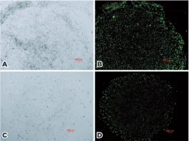
Figure 1 shows the imaging results for TRA-1-60 live cell staining. These images were obtained before and after staining using an inverted microscope and a fluorescence microscope. The expression of TRA-1-60 indicates suc-cessful reprogramming of the cell clones.
TRA-1-60-positive clones were harvested from the transfected fibroblasts under the three reprogramming systems. Table 1 shows the reprogramming efficiency of each culture system, and table 2 shows the reprogramming efficiency of the two types of fibroblasts.
iPSC characterization
Table 1. Comparison of iPSC reprogramming efficiency in different culture systems
Culture systems Number of transfected fibroblasts (×105) Number of TRA-1-60 positive clones Reprogramming efficiency (%)
Traditional system 14.050 390 0.028
E6/E8 system 07.025 008 0.001
CTS system 07.025 100 0.014
CTS= cell therapy system; iPSCs= induced pluripotent stem cells.
Table 2. Comparison of iPSC reprogramming efficiency from different ocular fibroblasts
Transfected fibroblasts Number of transfected fibroblasts (×105) Number of TRA-1-60 positive clones Reprogramming efficiency (%)
HPFs 16.1 297 0.018
HTFs 12.0 201 0.017
iPSCs= induced pluripotent stem cells; HPFs= human pterygium fibroblasts; HTFs= human Tenon’s capsule fibroblasts.
systems is shown in figure 2. The relative expression of
OCT3/4, NANOG, and SOX2 was also assessed in putative
iPSCs. HTF was used as a negative control, and ES was used as a positive control. n-HTF1-iPSC and n-HPF2-iPSC represent HTF1- and HPF2-derived iPSCs obtained using the conventional culture system, respectively. c-HTF1-iPSC and c-HPF2-iPSC represent HTF1- and HPF2-derived iPSCs obtained from the CTS culture system, respecti-vely (Figure 3). The results demonstrated that the ex-pression of endogenous pluripotent genes in iPSCs was >200-fold higher than that in HTFs and was similar to the expression in ES cells.
Immunofluorescence staining demonstrated that iPSCs derived from HPFs and HTFs in the CTS system (CTS-HPF/ HTF-iPSC) expressed not only the characteristic cell
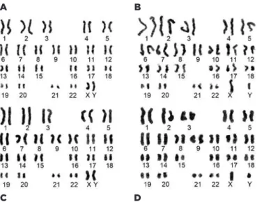
surface markers of human pluripotent stem cells, in-cluding TRA-1-60, TRA-1-81, and SSEA-4, but also the intracellular marker proteins Nanog, SOX2, and OCT4 (Figure 4). Importantly, HTF/HPF-iPSCs obtained by re-programming using the conventional and CTS systems exhibited normal human karyotypes (Figure 5).
Both in vivo and in vitro differentiation tests were conducted to further characterize iPSCs. First, NOD-SCID immunodeficient mice were injected with CTS-HPF/ HTF-iPSC suspensions. Four to six weeks after injection, tumors were observed. Ten weeks after injection, the teratomas were retrieved and histological examination was performed. Endoderm, mesoderm, and ectoderm layers were observed, indicating the pluripotency of the iPSCs. After the culturing of CTS-HPF/HTF-iPSCs in iPSC medium without bFGF for 2-3 weeks, embryoid bodies were observed (Figure 6).
DISCUSSION
Effects of donor cell tissue on iPSC generation
Both iPSCs and ES cells are pluripotent stem cells. Thus, iPSCs should not, in theory, carry tissue-specific markers unique to terminally differentiated cells, the reprogramming process that encompasses the transition of adult cells to iPSCs thus resulting in the removal of tissue-specific markers. However, the latter transition apparently does not always occur. Many studies have revealed that iPSCs tend to carry specific epigenetic characteristics associated with their donor cells, a phe-nomenon known as epigenetic memory(11,18). Further
stu-dies have shown that these residual epigenetic markers prevent iPSCs from differentiating into cells other than those normally associated with the donor cell linea-ges(19). It follows that iPSCs reprogrammed from retinal Figure 1. TRA-1-60 live cell staining. (A) Clonal morphology of HTF1-dervied
cells under the CTS system; (B) live staining imaging result of (A); (C) clonal morphology of HPF5-dervied cells under the conventional culture system; (d) live staining imaging result of (C). Magnification, 50-100×
A B
Figure 3. Endogenous pluripotent gene expression in HPF/HTF-iPSCs.
donor cells rather than other organs may be optimal for the study of cortical blindness in ophthalmopathy. In 2012, our group was the first to report a case of successful reprogramming of human Tenon’s capsule-derived fi-broblasts into iPSCs(9).
Effect of iPSC proliferation on reprogramming efficiency
In 2008, Park et al. reported that SV40LT and hTERT, two anti-aging factors, improve the reprogramming efficiency of iPSCs(20). In 2011, Ruiz et al. also reported
that cell proliferation, in the early stages of iPSC deve-lopment, is highly associated with reprogramming effi-ciency; cells exhibiting greater levels of proliferation were reported to be more likely to form iPSC clones(21).
Figure 4. Immunofluorescence staining of CTS-HPF2-iPSCs (400×).
For this reason, as part of this study-prior to further pro-cessing-we assessed the growth dynamics of transfected cells. Cortical blindness is common among elderly pa-tients, and aging will inevitably reduce the efficiency of iPSC induction(22,23). Pterygium, a wing-like pathological
tissue formed following the abnormal growth of fibrous vascular tissue under the conjunctiva, is an eye disease occurring primarily in the elderly. This tissue type, rich in fibroblasts, exhibits a strong capacity to proliferate. HPFs and HTFs are both eye-derived fibroblasts. Our study demonstrates that HPF-derived iPSCs do not exhibit abnormal karyotypes, although the HPFCs were obtained from pathological tissue. Moreover, HPFs obtained from elderly patients-in a similar way to HPFs from young patients-were efficient at reprogramming. Our results therefore suggest that HPFs might represent a viable choice in the generation of iPSCs for elderly patients.
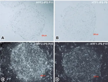
Figure 2. iPSC morphology (100×). (A), (B) iPSCs cultured under the CTS system; (C), (D) iPSCs induced using a MEF cell feeder layer.
D B
The expression levels of OCT3/4, NANOG, and SOX2
were assessed in putative iPSCs to further characterize the reprogrammed cells using HTF as a negative control and ES as a positive control. In maintaining the pluripo-tency of iPSCs, OCT3/4, Nanog, and SOX2 are impor-tant transcriptional regulators(24-26). Similar expression
profiles were observed for these markers in cells that
underwent varying reprogramming regimens. This is further evidence for the efficacy of the clonal selection strategies utilized in the study, resulting in the genera-tion of fully reprogrammed iPSCs. Upon analysis of the reprogramming efficiencies, the CTS system was signifi-cantly more effective than the E6/E8 system, while both systems were less effective than the traditional control system. However, it is likely that reprogramming condi-tions could be further optimized to negate the observed differences in reprogramming efficiency.
iPSC clone selection
Fully reprogrammed iPSCs exhibit morphological and molecular characteristics similar to those of ESCs. Previous studies have been performed mainly by selec-ting clones that exhibit typical ESC clonal morphology. However, since many partially reprogrammed cells can grow into clones with morphologies similar to those of typical ESC clones, visual assessment of iPSC clones is relatively subjective and has a high false-positive rate. Advances in molecular technology have ushered in live staining procedures to select iPSC clones by identifying signature proteins that are known to be specific to PSCs. iPSC clones are identifiable either colorimetrically or by fluorescence, and the staining process does not adversely affect the observed cells. After a short time, the color or fluorescence automatically disappears, allowing for
conti-Figure 5. Karyotype analysis. (A) HPF2-derived iPSCs from the CTS sys-tem; (B) HTF1-derived iPSCs from the CTS syssys-tem; (C) HPF2-derived iPSCs from the conventional system; (D) HTF1-derived iPSCs from the conventional system.
B
D C
A
Figure 6. In vitro and in vivo differentiation tests. (A), (B), (C) show the histology of CTS-HTF1-iPSC-derived terato-mas. (D), (E) show the formation of embryoid bodies. (a) Adenoid tissue (endoderm); (B) muscle tissue (mesoderm); (C) neural tube (ectoderm). (D) 15 days after CTS-HPF2-iPSC suspension cultures, embryoid bodies formed; (E) 15 days after CTS-HTF1-iPSC suspension cultures, embryoid bodies formed.
A
D E
nued passaging of the cells. The emergence of this method has greatly enhanced the efficacy of iPSC clonal selection.
In 2011, Ramirez et al. reported that TRA-1-60, as a PSC marker, is more specific than other markers(27). Hence
in this study, a TRA-1-60 live cell staining method was used in combination with morphological assessment to select iPSC clones. Our results revealed the success of this strategy for clonal selection.
REFERENCES
1. Takahashi K, Tanabe K, Ohnuki M, Narita M, Ichisaka T, Tomoda K, et al. Induction of pluripotent stem cells from adult human fibroblasts by defined factors. Cell. 2007;131(5):861-72.
2. Trounson A, Shepard KA, DeWitt ND. Human disease modeling with induced pluripotent stem cells. Curr Opin Genet Dev. 2012; 22(5):509-16.
3. Ramsden CM, Powner MB, Carr AJ, Smart MJ, da Cruz L, Coffey PJ. Stem cells in retinal regeneration: past, present and future. Deve-lopment. 2013;140(12):2576-85.
4. Borooah S, Phillips MJ, Bilican B, Wright AF, Wilmut I, Chandran S, et al. Using human induced pluripotent stem cells to treat retinal disease. Prog Retin Eye Res. 2013;37:163-81.
5. De Sousa PA, Galea G, Turner M. The road to providing human embryo stem cells for therapeutic use: the UK experience. Repro-duction. 2006;132(5):681-9.
6. Martin MJ, Muotri A, Gage F, Varki A. Human embryonic stem cells express an immunogenic nonhuman sialic acid. Nat Med. 2005;11(2):228-32.
7. Heiskanen A, Satomaa T, Tiitinen S, Laitinen A, Mannelin S, Impola U et al. N-glycolylneuraminic acid xenoantigen contamination of human embryonic and mesenchymal stem cells is substantially reversible. Stem Cells. 2007;25(1):197-202.
8. Chen G, Gulbranson DR, Hou Z, Bolin JM, Ruotti V, Probasco MD et al. Chemically defined conditions for human iPSC derivation and culture. Nat Methods. 2011;8(5):424-9.
9. Deng F, Hu H, Chen M, Sun X, Liu X, Dong Z, et al. Generation of induced pluripotent stem cells from human Tenon’s capsule fibroblasts. Mol Vis. 2012;18:2871-81.
10. Qiu X, Yang J, Liu T, Jiang Y, Le Q, Lu Y. Efficient generation of lens progenitor cells from cataract patient-specific induced pluripotent stem cells. PLoS One. 2012;7(3):e32612.
11. Kim K, Doi A, Wen B, Ng K, Zhao R, Cahan P, et al. Epigenetic me-mory in induced pluripotent stem cells. Nature. 2010;467(7313): 285-90.
12. Harui A, Suzuki S, Kochanek S, Mitani K. Frequency and stability of chromosomal integration of adenovirus vectors. J Virol. 1999; 73(7):6141-6.
13. Okita K, Ichisaka T, Yamanaka S. Generation of germline-competent induced pluripotent stem cells. Nature. 2007;448(7151):313-7.
14. Lamb RA, Parks GD. Paramyxoviridae: the viruses and their re-plication. In: Fields BN, Knipe DM, Howley PM, editors. Fields Virology. Volume 1. 6th ed. Philadelphia (PA): Lippincott, Williams & Wilkins; 2013. p. 957-95.
15. Tokusumi T, Iida A, Hirata T, Kato A, Nagai Y, Hasegawa M. Re-combinant Sendai viruses expressing different levels of a foreign reporter gene. Virus Res. 2002;86(1-2):33-8.
16. Macarthur CC, Fontes A, Ravinder N, Kuninger D, Kaur J, Bailey M, et al. Generation of human-induced pluripotent stem cells by a nonintegrating RNA Sendai virus vector in feeder-free or xeno-free conditions. Stem Cells Int. 2012;2012:564612.
17. Kele M, Day K, Rönnholm H, Schuster J, Dahl N, Falk A. Gene-ration of human iPS cell line CTL07-II from human fibroblasts, under defined and xeno-free conditions. Stem Cell Res (Amst). 2016;17(3):474-8.
18. Doi A, Park IH, Wen B, Murakami P, Aryee MJ, Irizarry R, et al. Differential methylation of tissue- and cancer-specific CpG island shores distinguishes human induced pluripotent stem cells, em-bryonic stem cells and fibroblasts. Nat Genet. 2009;41(12):1350-3. 19. Kim K, Zhao R, Doi A, Ng K, Unternaehrer J, Cahan P, et al.
Donor cell type can influence the epigenome and differentia-tion potential of human induced pluripotent stem cells. Nat Bio technol. 2011;29(12):1117-9.Erratum in Nat Biotechnol. 2012 Jan;30(1):112.
20. Park IH, Lerou PH, Zhao R, Huo H, Daley GQ. Generation of hu-man-induced pluripotent stem cells. Nat Protoc. 2008;3(7):1180-6. 21. Ruiz S, Panopoulos AD, Herrerías A, Bissig KD, Lutz M, Berggren
WT, et al. A high proliferation rate is required for cell reprogramming and maintenance of human embryonic stem cell identity. Curr Biol. 2011;21(1):45-52.
22. Marión RM, Strati K, Li H, Murga M, Blanco R, Ortega S, et al. A p53-mediated DNA damage response limits reprogramming to en-sure iPS cell genomic integrity. Nature. 2009;460(7259):1149-53. 23. Li H, Collado M, Villasante A, Strati K, Ortega S, Cañamero M,
et al. The Ink4/Arf locus is a barrier for iPS cell reprogramming. Nature. 2009 Aug;460(7259):1136-9.
24. Nichols J, Zevnik B, Anastassiadis K, Niwa H, Klewe-Nebenius D, Chambers I et al. Formation of pluripotent stem cells in the mam-malian embryo depends on the POU transcription factor Oct4. Cell. 1998;95(3):379-91.
25. Avilion AA, Nicolis SK, Pevny LH, Perez L, Vivian N, Lovell-Badge R. Multipotent cell lineages in early mouse development depend on SOX2 function. Genes Dev. 2003;17(1):126-40.
26. Kalmar T, Lim C, Hayward P, Muñoz-Descalzo S, Nichols J, Garcia-Ojalvo J, et al. Regulated fluctuations in nanog expression mediate cell fate decisions in embryonic stem cells. PLoS Biol. 2009; 7(7):e1000149.