Orthodontic retreatment of a Class III patient
with significant midline asymmetry and bilateral
posterior crossbite
Ademir R. Brunetto1
Posterior crossbite might cause serious long-term functional problems if not early treated. Nevertheless, in older patients, treatment might include palatal expansion in order to correct such malocclusion. In view of the above, this article aims at reporting late correction of bilateral posterior crossbite associated with Angle Class III malocclusion, right subdivision, with consequent midline shift (good skeletal pattern). The case was presented to the Brazilian Board of Orthodontics and Dentofacial Orthopedics (BBO), with DI equal to or greater than 10, as a requirement for the title of certified by the BBO.
Keywords:Angle Class III malocclusion. Dental asymmetry. Posterior crossbite.
How to cite this article: Brunetto AR. Orthodontic retreatment of a Class III patient with significant midline asymmetry and bilateral posterior cross-bite. Dental Press J Orthod. 2015 Jan-Feb;20(1):118-26. DOI: http://dx.doi. org/10.1590/2176-9451.20.1.118-126.bbo
Submitted: September 2, 2014 - Revised and accepted: October 1, 2014
» Patients displayed in this article previously approved the use of their facial and intraoral photographs.
Contact address: Ademir Roberto Brunetto
Avenida Sete de Setembro, 4456 - Centro, Curitiba / PR, CEP: 80.250-210 — Brazil — E-mail: [email protected]
» The author reports no commercial, proprietary or financial interest in the prod-ucts or companies described in this article.
1 Specialist in Orthodontics and Facial Orthopedics, University of California.
Certified by the Brazilian Board of Orthodontics and Facial Orthopedics (BBO). Former director of the Brazilian Board of Orthodontics and Facial Orthopedics (BBO).
* Case report, DI 20, approved by the Brazilian Board of Orthodontics and
Dentofacial Orthopedics (BBO).
DOI: http://dx.doi.org/10.1590/2176-9451.20.1.118-126.bbo
A mordida cruzada posterior pode causar sérios problemas funcionais em longo prazo, se não tratada precocemente. Porém, em alguns pacientes com idade mais avançada, ainda é possível realizar a disjunção palatina para corrigir essa condição. Assim sendo, o objetivo do presente caso é relatar a correção tardia de uma mordida cruzada posterior bilateral, associada a uma má oclusão de Classe III de Angle, subdivisão direita, com consequente desvios de linhas médias dentá-rias (bom padrão esquelético). Esse caso foi apresentado à Diretoria do Board Brasileiro de Ortodontia e Ortopedia Facial (BBO), se enquadrando na categoria IGC igual ou superior a 10, como requisito para revalidação do título de Diplomado.
Figure 1 - Initial facial and intraoral photographs.
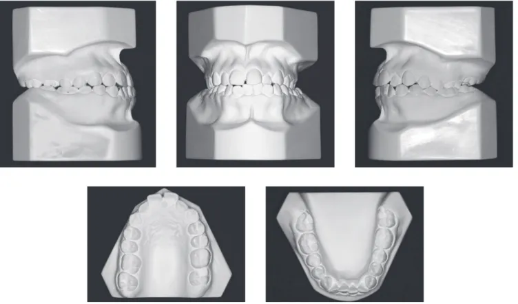
of 3.5-mm space and asymmetry (right canine and irst molar more mesially placed). In occlusion, Angle Class III malocclusion, right subdivision, was found in asso-ciation with bilateral posterior and #22 crossbite. The patient also presented right, 2-mm upper midline shit and let, 2-mm lower midline shit, which resulted in an anesthetic smile. A 2-mm overbite and 1-mm overjet were also found on teeth #21 and 42 (Figs 1 and 2).
Lateral cephalogram and cephalometric tracing re-vealed Class I skeletal pattern (ANB = 1o), mild
max-illary retrognathism (SNA = 80o) and vertical growth
pattern (SN. GoGn = 37o and Y-axis = 56o). From a
dental point of view, upper incisors were slightly pro-clined and protruded, while lower incisors were prop-erly positioned (Fig 3 and Table 1). Facial asymmetry was evident in frontal radiograph which revealed mild
INTRODUCTION
Facial analysis revealed patient’s concave proile with everted, slightly forward lower lip in comparison to up-per lip, and increased nasolabial angle. In frontal view, she presented mild asymmetry with chin shit to the let associated with slightly increased lower third and passive labial seal. Her smile was signiicantly and esthetically compromised mainly due to diastema between incisors, lack of coincidence between dental and facial midlines, and large buccal corridor (Fig 1).
Figure 2 - Initial casts.
Figure 3 - Initial lateral cephalogram (A) and cephalometric tracing (B).
Figure 4 - Initial frontal cephalometric radiograph revealing mild iatrogenesis.
Figure 5 - Initial panoramic radiograph.
mandibular deviation to the let due to asymmetric con-dyle growth (Fig 4). Panoramic radiograph raised the initial possibility of ankylosis of #16, which was further denied by infraocclusion. Tooth #27 was absent and had been replaced, in position, by #28 which, in turn, was also in infraocclusion. The patient was in good peri-odontal health and had #18 missing (Fig 5).
TREATMENT PLAN
Taking patient’s most signiicant problems into ac-count, treatment plan included palatal expansion with Haas appliance initially activated 2/4 of a turn a day fol-lowed by 1/4 a day until overcorrection was achieved. Ater a 120-day retention period, ixed appliance was mounted on the upper and mandibular arches with 0.022 x 0.028-in preadjusted, Edgewise slots. Initial alignment and leveling were performed by means of the following sequence of NiTi archwires: 0.012-in, 0.016-in, 0.016 x 0.022-in and 0.017 x 0.025-in on both up-per and mandibular arches. Subsequently, mechanics with ¼-in intermaxillary Class III elastics (200 g/force) associated with jig and NiTi springs was applied on one side. Once dental and facial midlines were coinciding, the inishing phase was started with ideal 0.019 x 0.025-in sta0.025-inless archwires, with l0.025-ingual torque applied to up-per anterior teeth and buccal torque applied to second molars. The patient was then referred to lower third molars extraction.
Alternative treatment plan included the use of mini-implants in the upper let and lower right hemi-arches for canine distalization so as to achieve arch symmetry and midline coincidence. Another option would in-clude surgical maxillary advancement for sagittal dis-crepancy correction. Taking the cost-beneit relation-ship of treatment planning into account, we initially opted for conservative treatment.
TREATMENT PROGRESS
Palatal expansion achieved satisfactory results, with mid palatal suture opening without excess compensa-tory proclination of upper posterior teeth. Once the expansion appliance was rendered stable and a 120-day retention period had passed, the sequence of archwires established at initial treatment planning was followed. Tooth #16, initially under suspicion of ankylosis, re-sponded well to orthodontic forces. Patient’s compli-ance was satisfactory with regard to the use of elastics, thereby increasing the possibilities of achieving accept-able occlusion.
RESULTS
At smiling, the patient evinced a decreased buccal cor-ridor which, together with midline coincidence, also led to signiicant esthetic changes (Fig 6).
Upper dental arch analysis revealed satisfactory alignment achieved as a result of increased arch circum-ference that, in turn, led to expansion. Teeth #16 and 28, previously under infraocclusion, were duly aligned. Right canine underwent mesialization which improved intra-arch anteroposterior symmetry. In the mandibular arch, satisfactory alignment and leveling were achieved by means of torque correction in lower posterior teeth as a result of palatal expansion. Right canine distaliza-tion achieved by means of Class III elastics on one side also aided to create space necessary to correct model discrepancy. In terms of function, molar and canine
relationship was achieved on both sides, with ideal lat-eral and anterior disocclusion. In addition to midline coincidence, overjet and overbite were within normal standards (Figs 6 and 7).
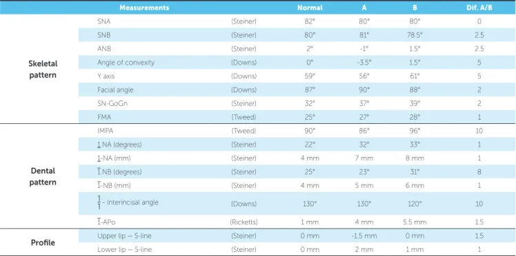
Cephalometry revealed decreased SNB and, as a consequence, ANB angle due to slight clockwise rota-tion of the mandible evinced by an increase of 2o in the
Sn.GoGn angle. Upper incisors were slightly proclined, unlike lower incisors in which proclination was much more signiicant. There was upper lip protrusion and lower lip setback, both of which were responsible to es-tablish facial proile balance (Fig 8, Tab 1). In addition, there was signiicant change in the distance between up-per molars (an increase of 5 mm) proving greater palatal opening in the posterior region. Total cephalometric
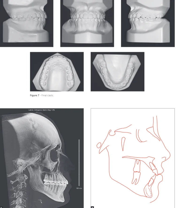
Figure 7 - Final casts.
Figure 8 - Final lateral cephalogram (A) and cephalometric tracing (B).
Figure 9 - Total (A) and partial (B) initial (black) and final (red) cephalometric tracings superimposition.
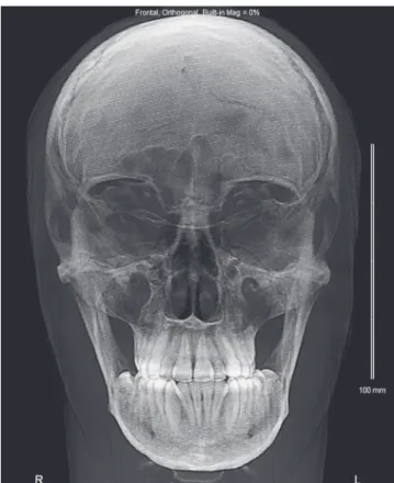
Figure 10 - Final frontal cephalometric radiograph.
Figure 11 - Final panoramic radiograph.
Table 1 - Initial (A) and final (B) cephalometric values.
Table 2 - Model measurements.
Measurements Normal A B Dif. A/B
Skeletal pattern
SNA (Steiner) 82° 80° 80° 0
SNB (Steiner) 80° 81° 78.5° 2.5
ANB (Steiner) 2° -1° 1.5° 2.5
Angle of convexity (Downs) 0° -3.5° 1.5° 5
Y axis (Downs) 59° 56° 61° 5
Facial angle (Downs) 87° 90° 88° 2
SN-GoGn (Steiner) 32° 37° 39° 2
FMA (Tweed) 25° 27° 28° 1
Dental pattern
IMPA (Tweed) 90° 86° 96° 10
1.NA (degrees) (Steiner) 22° 32° 33° 1
1-NA (mm) (Steiner) 4 mm 7 mm 8 mm 1
1.NB (degrees) (Steiner) 25° 23° 31° 8
1-NB (mm) (Steiner) 4 mm 5 mm 6 mm 1
1
1- Interincisal angle (Downs) 130° 130° 120° 10
1-APo (Ricketts) 1 mm 4 mm 5.5 mm 1.5
Profile Upper lip — S-line (Steiner) 0 mm -1.5 mm 0 mm 1.5
Lower lip — S-line (Steiner) 0 mm 2 mm 1 mm 1
superimposition evinced clockwise rotation of the mandible and posterior displacement of the lower lip. Partial superimposition of maxillary segments revealed signiicant posterior anchorage loss (on the right side) and slight extrusion and proclination of incisors. Con-versely, partial superimposition of mandibular segments revealed slight distalization of molars (on the right side) and proclination of incisors (Fig 9).
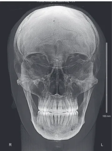
Frontal cephalogram showed that mandibular shit to the let remained stable ater maxillary expansion, while midlines were corrected so as to coincide with the me-dian sagittal plane (Fig 10). Panoramic radiograph not only revealed good periodontal health ater orthodontic movement, but also satisfactory root parallelism in both the maxilla and mandible. Tooth #28 was positioned slightly upward and distally, which is considered ideal; however, it should undergo natural movement, includ-ing more physiological movements, so as to be better positioned in line of occlusion (Fig 11).
FINAL CONSIDERATIONS
Maxillary expansion is ideally performed when the mid palatal suture is not yet mature and does not present interdigitation, which occurs before patients achieve puber-tal growth spurt.1-4 The younger the patient is, the greater
the orthopedic component and the smaller the chances of relapse. Nevertheless, in some cases, patients have already achieved this stage of growth, but might as well satisfactorily respond to forces applied to the palatal suture despite great-er bone density and intgreat-erdigitation. Capelloza Filho et al5
found a success rate of 81.5% in expansion of adult patients. On the other hand, the technique might produce more se-vere deleterious efects in adults than in children and adoles-cents, including edema, buccal clinical attachment loss and occlusal plane instability.5,6 Thus, the procedure needs to be
carefully performed and strictly followed by a professional. Nevertheless, it should always be considered, since it poten-tially prevents surgery (LeFort I osteotomy).7
There is also some discussion on the amount of anterior and posterior opening and in which proportion they occur. In the case reported herein, we noticed greater opening in the posterior region, which was evinced by an increase of 5 mm in the distance between molars despite signiicant an-chorage loss of #16. This inding is in accordance with most studies.8 The literature also reports clockwise rotation of
the mandible as a consequence of palatal expansion, which
Measurements A B Dif. A/B
Distance between upper canines 37 38 1
Distance between lower canines 27 29 2
Distance between upper molars 46 51 5
1. Wertz RA. Skeletal and dental changes accompanying rapid midpalatal suture opening. Am J Orthod. 1970;58(1):41-66.
2. Silva Filho OG, Lara TS, Silva HC, Bertoz FA. Post expansion evaluation of the midpalatal suture in children submitted to rapid palatal expansion: a CT study. J Clin Pediatr Dent. 2006;31(2):142-8.
3. Ghoneima A, Abdel-Fattah E, Hartsield J, El-Bedwehi A, Kamel A, Kula K.
Efects of rapid maxillary expansion on the cranial and circummaxillary sutures. Am J Orthod Dentofacial Orthop. 2011;140(4):510-9. 4. Lione R, Ballanti F, Franchi L, Baccetti T, Cozza P. Treatment and
posttreatment skeletal efects of rapid maxillary expansion studied with low-dose computed tomography in growing subjects. Am J Orthod Dentofacial Orthop. 2008;134(3):389-92.
5. Capelozza Filho L, Cardoso Neto J, Silva Filho OG, Ursi WJ.
Non-surgically assisted rapid maxillary expansion in adults. Int J Adult Orthodon Orthognath Surg. 1996;11:57-66; discussion 67-70.
6. Graber LW, Vanarsdall R. Orthodontics: current principles and techniques. Saint Louis: Mosby; 1994.
7. Albuquerque RR, Eto LF. Previsibilidade de sucesso na disjunção palatina avaliada pelo estágio de maturação esquelética: estudo piloto. Rev Dental Press Ortod Ortop Facial. 2006;11(2):74-83.
8. Lione R, Franchi L, Cozza P. Does rapid maxillary expansion induce
adverse efects in growing subjects? Angle Orthod. 2013;83(1):172-82.
9. Akkaya S, Lorenzon S, Ucem TT. A comparison of sagittal and vertical
efects between bonded rapid and slow maxillary expansion procedures. Eur J Orthod. 1999;21(2):175-80.
10. Chung CH, Font B. Skeletal and dental changes in the sagittal, vertical, and transverse dimensions after rapid palatal expansion. Am J Orthod Dentofacial Orthop. 2004;126(5):569-75.
11. Cozza P, Giancotti A, Petrosino A. Rapid palatal expansion in mixed dentition using a modiied expander: a cephalometric investigation. J Orthod. 2001;28(2):129-34.
REFERENCES
12. Cozza P, Baccetti T, Franchi L, De Tofol L, McNamara JA, Jr. Mandibular changes produced by functional appliances in Class II malocclusion: a systematic review. Am J Orthod Dentofacial Orthop. 2006;129(5):599. e1-12; discussion e1-6.
13. Wertz R, Dreskin M. Midpalatal suture opening: a normative study. Am J Orthod. 1977;71(4):367-81.
14. Lucato AS, Boeck EM, Vedovello SAS, Pereira Neto JS, Mangnani MBBA. Sliding Jig: confeção e mecanimo de ação. Rev Clín Ortod Dental Press. 2003 Dez-2004 Jan;2(6):10-7.
15. Moscardini MS. O “Sliding-Jig” na prática ortodôntica. Rev Clín Ortod Dental Press. 2010;9(2):59-64.
16. Krukemeyer AM, Arruda AO, Inglehart MR. Pain and orthodontic treatment. Angle Orthod. 2009;79(6):1175-81.
17. King GJ, Fischlschweiger W. The efect of force magnitude on extractable bone resorptive activity and cemental cratering in orthodontic tooth movement. J Dent Res. 1982;61(6):775-9.
18. Darendeliler MA, Kharbanda OP, Chan EK, Srivicharnkul P, Rex T, Swain MV, et al. Root resorption and its association with alterations in physical properties, mineral contents and resorption craters in human premolars following application of light and heavy controlled orthodontic forces. Orthod Craniofac Res. 2004;7(2):79-97.
19. Segal GR, Schifman PH, Tuncay OC. Meta analysis of the treatment-related factors of external apical root resorption. Orthod Craniofac Res. 2004;7(2):71-8.
20. Fox N. Longer orthodontic treatment may result in greater external apical root resorption. Evid Based Dent. 2005;6(1):21.
is caused due to extrusion of palatal cusps in anchorage teeth.9,10,11 In the case reported herein, we found clockwise
rotation of the mandible leading to slight increase in lower facial height; however, without causing signiicant esthetic damage to the face and, therefore, being clinically irrel-evant.12 Additionally, such mandibular movement is likely
to recede in the long-term.13
We opted for mechanics with Class III elastics and jig/springs on one side due to opposite shit of upper and lower midlines. When applied to one side only, this mechanics not only promotes mesialization of upper posterior teeth, but also distalization of lower posterior teeth (highly evident in partial cephalometric superim-position).14,15 Movement of the maxilla and mandible in
opposite directions favors simultaneous correction of dental midlines, causing them to coincide with the fa-cial midline. However, long-term use of intermaxillary elastics on one side should be avoided, as it may lead to imbalance of stomatognathic system muscles and, as a result, temporomandibular disorders, migraine or local pain. Control should be ongoing, and should any of the
aforementioned factors be identiied, mechanics should be immediately removed. Despite being a common fact, symptoms should never be underestimated by the ortho-dontist, as it might hinder treatment assessment.16
In addition to patient’s advanced age, previous use of ixed orthodontic appliance was also considered a com-plicating factor. Whenever teeth, periodontal ligament and alveolar bone have already been subject to non-physiological forces, additional care should be taken, particularly with regard to the magnitude of orthodontic forces applied. Consensus has been reached on the fact that treatment duration and magnitude of forces play an important role in triggering orthodontically induced root resorption.17-20 In the present study, total treatment time
(36 months) increased due to patient’s absence, despite cooperation on the use of intermaxillary elastics.